. 2001 Aug 14;98(17):9630-5.
doi: 10.1073/pnas.181341498.

HIF-1alpha binding to VHL is regulated by stimulus-sensitive proline hydroxylation

Affiliations

HIF-1alpha binding to VHL is regulated by stimulus-sensitive proline hydroxylation

F Yu et al. Proc Natl Acad Sci U S A. .

Erratum in

  • Proc Natl Acad Sci U S A 2001 Dec 4;98(25):14744

Abstract

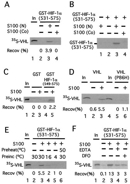
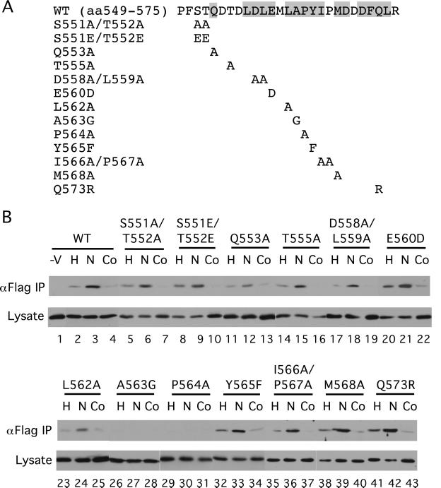
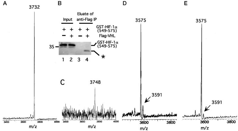
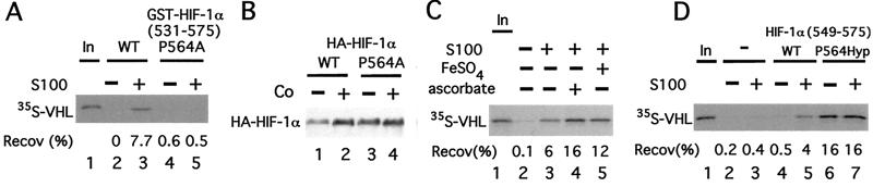
Hypoxia-inducible factor-1alpha (HIF-1alpha) is a global transcriptional regulator of the hypoxic response. Under normoxic conditions, HIF-1alpha is recognized by the von Hippel-Lindau tumor-suppressor protein (VHL), a component of an E3 ubiquitin ligase complex. This interaction thereby promotes the rapid degradation of HIF-1alpha. Under hypoxic conditions, HIF-1alpha is stabilized. We have previously shown that VHL binds in a hypoxia-sensitive manner to a 27-aa segment of HIF-1alpha, and that this regulation depends on a posttranslational modification of HIF-1alpha. Through a combination of in vivo coimmunoprecipitation assays using VHL and a panel of point mutants of HIF-1alpha in this region, as well as MS and in vitro binding assays, we now provide evidence that this modification, which occurs under normoxic conditions, is hydroxylation of Pro-564 of HIF-1alpha. The data furthermore show that this proline hydroxylation is the primary regulator of VHL binding.


Figures

Figure 1
Figure 1
A cytoplasmic activity modifies HIF-1α to promote VHL binding. (A) GST-HIF-1α (531) prebound to GSH agarose was preincubated in the absence or presence of 50 μg of cytoplasmic extract (S100) obtained from normoxic (N)- or cobalt (Co)-treated HeLa S3 cells for 1 h at 30°C and washed. Then the resins were incubated with 35S-VHL for 1 h at 4°C, washed, eluted, and the eluates then were examined by SDS/PAGE and autoradiography. The position of 35S-VHL is indicated. (B) Far Western analysis. GST-HIF-1α (531) prebound to GSH agarose was preincubated in the absence or presence of 50 μg of cytoplasmic extract (S100) obtained from N- or Co-treated HeLa S3 cells for 1 h at 30°C. The resin was washed, eluted, and subjected to SDS/PAGE. Subsequently, far Western blotting, using in vitro-translated 35S-labeled VHL, was performed. The position of GST-HIF-1α (531) is indicated. (CF) GST or GST-HIF-1α (549) (C) or GST-HIF-1α (531) (DF) prebound to GSH agarose was preincubated in the absence or presence of 90 μg of cytoplasmic extract (S100) obtained from normoxic HeLa S3 cells for 1 h at 30°C (DF) or at 16°C or 4°C (E) and washed. The resins then were incubated with 35S-VHL (CF) or 35S-VHL P86H (D) for 1 h at 4°C, washed, eluted, and the eluates then were examined by SDS/PAGE and autoradiography. In E, S100 was preheated at 50°C for 10 min before the preincubation in one sample. In F, either 1 mM EDTA or 100 μM desferrioxamine (DFO) was included in the preincubation, as indicated. “In” designates 10% of the input in A and CF. The recoveries (Recov) of 35S-VHL (in %) are indicated.
Figure 2
Figure 2
The in vivo interaction with VHL is altered by select HIF-1α mutants. (A) The sequence of WT HIF-1α residues 549–575 is shown at the top with conserved residues (see ref. 11) indicated by shading. Mutants are indicated below. (B) COS-1 cells were cotransfected with an expression vector for Flag-VHL and those for GAL4-HIF-1α (531) with the indicated mutations. In a control transfection (−V), the Flag-VHL vector was omitted. Twenty-four hours after transfection, all cells were treated with Cbz-LLL, and cells were additionally exposed for 2.5 h to 1% O2 (H) or 100 μM cobalt chloride (Co), or were maintained under normoxic conditions (N). Cells were lysed, the Flag-VHL was immunoprecipitated with anti-Flag Abs, and then the immunoprecipitates were analyzed for the presence of the GAL4-fusion proteins by immunoblotting with anti-GAL4 Abs (top rows). Aliquots of the whole-cell extracts were also analyzed by immunoblotting with anti-GAL4 Abs (bottom rows). Shown are representative results of three to four independent experiments.
Figure 3
Figure 3
Cytoplasmic extracts induce a change in the mass of HIF-1α (549). (A) GST-HIF-1α (549) was cleaved with Factor Xa protease to release HIF-1α (549), which then was subjected to MALDI-TOF MS. The amu of the main peak, 3732, is indicated. (B and C) GST-HIF-1α (549) (20 μg) was treated with S100 (810 μg) from normoxic HeLa S3 cells for 1 h at 30°C. Then in vitro-translated Flag-VHL (100 μl) was added for 1 h at 4°C, and the Flag-VHL–GST-HIF-1α (549) complex was immunoprecipitated by using 10 μl of anti-Flag (M2) agarose (Sigma). The immunoprecipitates were treated with Factor Xa protease to cleave the GST moiety, followed by treatment (with no intervening washes) with 40 μg of Flag peptide to elute the Flag-VHL–HIF-1α (549) peptide complex. (B) Aliquots of GST-HIF-1α (549) before immunoprecipitation (lanes 1 and 2) and the eluate after immunoprecipitation (lanes 3 and 4) were subjected to SDS/PAGE and immunoblotting by using anti-GST Abs (lanes 2 and 4, respectively). Samples from a control reaction prepared by using a mock in vitro translation reaction instead of the in vitro-translated Flag-VHL are shown (lanes 1 and 3). The asterisk denotes slower migrating GST species obtained after Factor Xa proteolysis that represents GST lacking HIF-1α (549). Position of molecular weight marker is shown to the left. “Input” represents 1% of the total GST-HIF-1α (549). (C) The eluate was subjected to MALDI-TOF MS. The amu of the most prominent peak, 3748, is indicated. (D and E) COS-1 cells were transfected with pcDNA3-FlagGAL4-HIF-1α (550). Twenty-four hours after transfection, all cells were treated with Cbz-LLL, and cells were either maintained under normoxic conditions (D) or exposed for 2.5 h to 100 μM cobalt chloride (E). Cells were lysed, the FlagGAL4-HIF-1α (550) was immunoprecipitated with anti-Flag Abs, and then the immunoprecipitates were treated with Factor Xa protease. The cleaved HIF-1α (550) peptide was subjected to MALDI-TOF MS. The amu of two peaks at 3575 and 3591 are indicated.
Figure 4
Figure 4
Hydroxylation of Proline-564 of HIF-1α promotes its binding to VHL. (A) GST-HIF-1α (531) or GST-HIF-1α (531) P564A prebound to GSH agarose was preincubated in the absence or presence of 90 μg of cytoplasmic extract (S100) obtained from normoxic HeLa S3 cells for 1 h at 30°C and washed. Then the resins were incubated with 35S-VHL for 1 h at 4°C, washed, eluted, and the eluates then were examined by SDS/PAGE and autoradiography. (B) COS-1 cells were cotransfected with pcDNA3-FlagVHL and either pcDNA3-HA-HIF-1α WT or pcDNA3-HA-HIF-1α P564A. Twenty-four hours after transfection, some cells were stimulated for 2.5 h with 100 μM cobalt chloride. Cells were lysed, and aliquots of the lysate were examined by immunoblotting with anti-HA Abs. The position of HA-HIF-1α is indicated. (C) GST-HIF-1α (531) prebound to GSH agarose was preincubated in the absence or presence of 90 μg of HeLa cytoplasmic extract (S100) supplemented with either 100 μM FeSO4 or 5 mM ascorbic acid, as indicated, for 1 h at 30°C, and washed. Then the resins were incubated with 35S-VHL for 1 h at 4°C, washed, eluted, and the eluates then were examined by SDS/PAGE and autoradiography. (D) Agarose (SulfoLink) was either mock-treated or coupled to peptides corresponding to HIF-1α (549) or HIF-1α (549) P564Hyp. The resins (10 μl) were preincubated in the absence or presence of 90 μg of cytoplasmic extract (S100) obtained from normoxic HeLa S3 cells for 1 h at 30°C, and washed. Then the resins were incubated with 35S-VHL for 1 h at 4°C, washed, eluted, and the eluates then were examined by SDS/PAGE and autoradiography. In A and CD, the positions of 35S-VHL and its recovery (Recov; in %) are indicated. “In” designates 10% of the input.

References

    1. Semenza G L. Curr Opin Cell Biol. 2001;13:167–171. - PubMed
    1. Maxwell P H, Wiesener M S, Chang G W, Clifford S C, Vaux E C, Cockman M E, Wykoff C C, Pugh C W, Maher E R, Ratcliffe P J. Nature (London) 1999;399:271–275. - PubMed
    1. Huang L E, Gu J, Schau M, Bunn H F. Proc Natl Acad Sci USA. 1998;95:7987–7992. - PMC - PubMed
    1. Cockman M E, Masson N, Mole D R, Jaakkola P, Chang G W, Clifford S C, Maher E R, Pugh C W, Ratcliffe P J, Maxwell P H. J Biol Chem. 2000;275:25733–25741. - PubMed
    1. Ohh M, Park C W, Ivan M, Hoffman M A, Kim T Y, Huang L E, Pavletich N, Chau V, Kaelin W G. Nat Cell Biol. 2000;2:423–427. - PubMed

Publication types

MeSH terms

LinkOut - more resources