

Presence and distribution of sensory nerve fibers in human peritoneal adhesions
- PMID: 11505072
- PMCID: PMC1422013
- DOI: 10.1097/00000658-200108000-00016
Presence and distribution of sensory nerve fibers in human peritoneal adhesions
Abstract
Objective: To assess the distribution and type of nerve fibers present in human peritoneal adhesions and to relate data on location and size of nerves with estimated age and with clinical parameters such as reports of chronic pelvic pain.
Summary background data: Peritoneal adhesions are implicated in the cause of chronic abdominopelvic pain, and many patients are relieved of their symptoms after adhesiolysis. Adhesions are thought to cause pain indirectly by restricting organ motion, thus stretching and pulling smooth muscle of adjacent viscera or the abdominal wall. However, in mapping studies using microlaparoscopic techniques, 80% of patients with pelvic adhesions reported tenderness when these structures were probed, an observation suggesting that adhesions themselves are capable of generating pain stimuli.
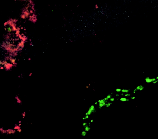
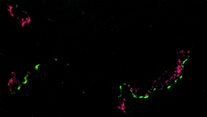
Methods: Human peritoneal adhesions were collected from 25 patients undergoing laparotomy, 20 of whom reported chronic pelvic pain. Tissue samples were prepared for histologic, immunohistochemical, and ultrastructural analysis. Nerve fibers were characterized using antibodies against several neuronal markers, including those expressed by sensory nerve fibers. In addition, the distribution of nerve fibers, their orientation, and their association with blood vessels were investigated by acetylcholinesterase histochemistry and dual immunolocalization.
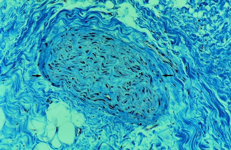
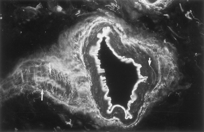
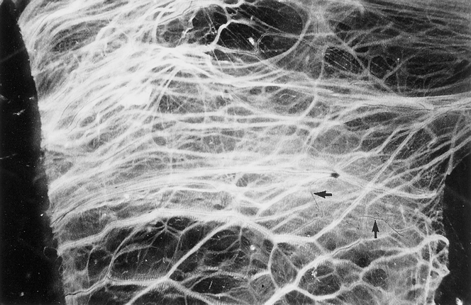
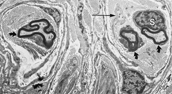
Results: Nerve fibers, identified histologically, ultrastructurally, and immunohistochemically, were present in all the peritoneal adhesions examined. The location of the adhesion, its size, and its estimated age did not influence the type of nerve fibers found. Further, fibers expressing the sensory neuronal markers calcitonin gene-related protein and substance P were present in all adhesions irrespective of reports of chronic abdominopelvic pain. The nerves comprised both myelinated and nonmyelinated axons and were often, but not invariably, associated with blood vessels.
Conclusions: This study provides the first direct evidence for the presence of sensory nerve fibers in human peritoneal adhesions, suggesting that these structures may be capable of conducting pain after appropriate stimulation.
Figures
References
-
- DeCherney AH, Mezer HC. The nature of posttuboplasty pelvic adhesions as determined by early and late laparoscopy. Fertil Steril 1984; 41: 643–666. - PubMed
-
- Tulandi T, Collins JA, Burrows E, et al. Treatment-dependent and treatment-independent pregnancy among women with periadnexal adhesions. Am J Obstet Gynecol 1990; 162: 354–357. - PubMed
-
- Stricker B, Blanco J, Fox HE. The gynecologic contribution to intestinal obstruction in females. J Am Coll Surg 1994; 178: 617–620. - PubMed
-
- Kresch AJ, Seifer DB, Sachs LB, et al. Laparoscopy in 100 women with chronic pelvic pain. Obstet Gynaecol 1984; 64: 672–674. - PubMed
Publication types
MeSH terms
Substances
LinkOut - more resources
Full Text Sources
Other Literature Sources
Medical
