

Recombinant GABA(C) receptors expressed in rat hippocampal neurons after infection with an adenovirus containing the human rho1 subunit
- PMID: 11507165
- PMCID: PMC2278778
- DOI: 10.1111/j.1469-7793.2001.00145.x
Recombinant GABA(C) receptors expressed in rat hippocampal neurons after infection with an adenovirus containing the human rho1 subunit
Abstract
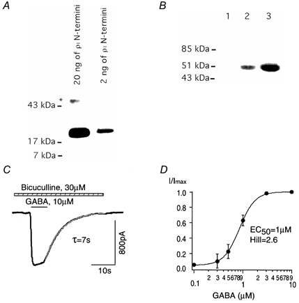
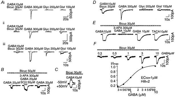
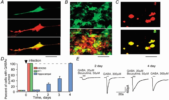
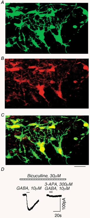
1. A recombinant adenovirus was generated with the human rho1 GABA(C) receptor subunit (adeno-rho). Patch-clamp and antibody staining were employed to confirm functional expression of recombinant rho1 receptors after infection of human embryonic kidney cells (HEK293 cell line), human embryonic retinal cells (911 cell line), dissociated rat hippocampal neurons and cultured rat hippocampal slices. 2. Standard whole-cell recording and Western blot analysis using rho1 GABA(C) receptor antibodies revealed that recombinant rho1 receptors were expressed in HEK293 and 911 cells after adeno-rho infection and exhibited properties similar to those of rho1 receptors after standard transfection. 3. Cultured rat hippocampal neurons (postnatal day (P)3-P5) did not show a native GABA(C)-like current. After adeno-rho infection, however, a GABA(C)-like current appeared in 70-90 % of the neurons. 4. Five days after infection, expression of GABA(C) receptors in hippocampal neurons significantly decreased native GABA(A) receptor currents from 1200 +/- 300 to 150 +/- 70 pA (n = 10). The native glutamate-activated current was unchanged. 5. Hippocampal slices (P8) did not show a native GABA(C)-like current, although recombinant rho1 receptors could be expressed in cultured hippocampal slices after adeno-rho infection. 6. These data indicate that an adenovirus can be used to express recombinant GABA(C) receptors in hippocampal neurons. This finding could represent an important step towards the gene therapy of CNS receptor-related diseases.
Figures
Similar articles
-
Kinetic properties of GABA rho1 homomeric receptors expressed in HEK293 cells.Biophys J. 2006 Sep 15;91(6):2155-62. doi: 10.1529/biophysj.106.085431. Epub 2006 Jun 23. Biophys J. 2006. PMID: 16798806 Free PMC article.
-
Suppression of neuronal hyperexcitability and associated delayed neuronal death by adenoviral expression of GABA(C) receptors.J Neurosci. 2001 May 15;21(10):3419-28. doi: 10.1523/JNEUROSCI.21-10-03419.2001. J Neurosci. 2001. PMID: 11331372 Free PMC article.
-
GABA(C) rho(1) subunits form functional receptors but not functional synapses in hippocampal neurons.J Neurophysiol. 2001 Nov;86(5):2605-15. doi: 10.1152/jn.2001.86.5.2605. J Neurophysiol. 2001. PMID: 11698546
-
Physiological and pharmacological characterization of glutamate and GABA receptors on carp retinal neurons.Prog Brain Res. 2001;131:277-93. doi: 10.1016/s0079-6123(01)31023-3. Prog Brain Res. 2001. PMID: 11420948 Review. No abstract available.
-
Neurochemicals for the investigation of GABA(C) receptors.Neurochem Res. 2010 Dec;35(12):1970-7. doi: 10.1007/s11064-010-0271-7. Epub 2010 Oct 21. Neurochem Res. 2010. PMID: 20963487 Review.
Cited by
-
An Update on GABAρ Receptors.Curr Neuropharmacol. 2010 Dec;8(4):422-33. doi: 10.2174/157015910793358141. Curr Neuropharmacol. 2010. PMID: 21629448 Free PMC article.
-
Inhibition of neuronal degenerin/epithelial Na+ channels by the multiple sclerosis drug 4-aminopyridine.J Biol Chem. 2013 Mar 29;288(13):9418-27. doi: 10.1074/jbc.M112.449413. Epub 2013 Feb 12. J Biol Chem. 2013. PMID: 23404498 Free PMC article.
-
Kinetic properties of GABA rho1 homomeric receptors expressed in HEK293 cells.Biophys J. 2006 Sep 15;91(6):2155-62. doi: 10.1529/biophysj.106.085431. Epub 2006 Jun 23. Biophys J. 2006. PMID: 16798806 Free PMC article.
References
-
- Amin J, Dickerson I M, Weiss D S. The agonist binding site of the γ-aminobutyric acid type A channel is not formed by the extracellular cysteine loop. Molecular Pharmacology. 1994;45:317–323. - PubMed
-
- Amin J, Weiss D S. Homomeric rho 1 GABA channels: activation properties and domains. Receptors and Channels. 1994;2:227–236. - PubMed
-
- Boue-Grabot E, Roudbaraki M, Bascles L, Tramu G, Bloch B, Garret M. Expression of GABA receptor rho subunits in rat brain. Journal of Neurochemistry. 1998;70:899–907. - PubMed
-
- Cossart R, Dinocourt C, Hirsch J, Merchan-Perez A, Felipe J, Esclapez M, Bernard C, Ben-Ari Y. Dendritic but not somatic GABAergic inhibition is decreased in experimental epilepsy. Nature Neuroscience. 2001;4:52–62. - PubMed
Publication types
MeSH terms
Substances
Grants and funding
LinkOut - more resources
Full Text Sources
Other Literature Sources
Miscellaneous