. 2001 Sep;75(18):8556-68.
doi: 10.1128/jvi.75.18.8556-8568.2001.

An Epstein-Barr virus isolated from a lymphoblastoid cell line has a 16-kilobase-pair deletion which includes gp350 and the Epstein-Barr virus nuclear antigen 3A

Affiliations

An Epstein-Barr virus isolated from a lymphoblastoid cell line has a 16-kilobase-pair deletion which includes gp350 and the Epstein-Barr virus nuclear antigen 3A

W Lee et al. J Virol. 2001 Sep.

Abstract

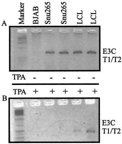
Epstein-Barr virus (EBV) is associated with human cancers, including nasopharyngeal carcinoma, Burkitt's lymphoma, gastric carcinoma and, somewhat controversially, breast carcinoma. EBV infects and efficiently transforms human primary B lymphocytes in vitro. A number of EBV-encoded genes are critical for EBV-mediated transformation of human B lymphocytes. In this study we show that an EBV-infected lymphoblastoid cell line obtained from the spontaneous outgrowth of B cells from a leukemia patient contains a deletion, which involves a region of approximately 16 kbp. This deletion encodes major EBV genes involved in both infection and transformation of human primary B lymphocytes and includes the glycoprotein gp350, the entire open reading frame of EBNA3A, and the amino-terminal region of EBNA3B. A fusion protein created by this deletion, which lies between the BMRF1 early antigen and the EBNA3B latent antigen, is truncated immediately downstream of the junction 21 amino acids into the region of the EBNA3B sequence, which is out of frame with respect to the EBNA3B protein sequence, and indicates that EBNA3B is not expressed. The fusion is from EBV coordinate 80299 within the BMRF1 sequence to coordinate 90998 in the EBNA3B sequence. Additionally, we have shown that there is no detectable induction in viral replication observed when SNU-265 is treated with phorbol esters, and no transformants were detected when supernatant is used to infect primary B lymphocytes after 8 weeks in culture. Therefore, we have identified an EBV genome with a major deletion in critical genes involved in mediating EBV infection and the transformation of human primary B lymphocytes that is incompetent for replication of this naturally occurring EBV isolate.


Figures

FIG. 1
FIG. 1
Schematic illustration showing a linear EBV genome with the relative positions of the EBNA2, -3A, -3B, and -3C and gp350 genes. These genes were analyzed by PCR to determine the genotypes and deletion ends of the SNU-265 genome. The respective gene-specific primers employed are indicated over each ORF (denoted by open bars) by a combination of names and arrowheads. Vertical lines and filled bars indicate the BamHI recognition sites and the terminal repeats, respectively. Also shown are the replication origin of viral episome, OriP, and the position of the EBNA1 and LMP1 genes (2, 13, 14).
FIG. 2
FIG. 2
PCR analyses of the EBNA2, -3A, -3B, and -3C genes in SNU-265 to determine the allelic types of each of these EBNA genes. Aliquots of cell DNA samples obtained from SNU-265, as well as control DNAs from B95–8, AG986, SNU-1103, and BJAB, were subjected to 40 cycles of PCR amplication, using primers that were specific for EBNA2 (E2-1–E2-2) (A), EBNA3B (B1-B2) (B), EBNA3A (A1-A2) (C), and EBNA3C (C1-C2) (D). For the primer sequences, see Table 1. Aliquots of PCR products were resolved on a 2.5% ME-agarose gel and visualized by ethidium bromide staining. Note that the SNU-265 genome is type 1 for EBNA2, -3B, and -3C and has no signal for the EBNA3A gene compared to the prototypic B95–8 type I genome and the AG876 prototypic type 2 genome. BJAB was used as an EBV-negative control in this assay.
FIG. 3
FIG. 3
Determination of the sensitivity of the PCR analysis in the detection of the EBNA3A gene. To estimate sensitivity levels of our PCR method, 104 IB4 cells, which contained four integrated EBV genomes per cell, were serially diluted with 104 BJAB cells, an EBV-negative BL cell line, and subjected to PCR amplification. An aliquot of each dilution was amplified at the same time to determine the approximate number of copies of EBV genomes that can be detected using this primer set for EBNA3A. (A) Amplified products were separated on a 2.5% ME-agarose gel and visualized by ethidium bromide staining. (B) This gel was then transferred to a nylon membrane and Southern blotted using an EcoRI K probe for the EBNA3A gene. Signals were visualized by exposure to X-ray film. By Southern blot, a weak signal was seen in the dilution of 10−4, suggesting that approximately four copies of the genome can be detected by this PCR analysis.
FIG. 4
FIG. 4
Southern blotting analysis of the SNU-265 genome using the HindIII E or EcoRI G2+F fragments as probes to determine the position of the SNU-265 deletion. (A) Map of the genomic region analyzed, indicating the fragments generated by EcoRI digestion (top) and those generated by BamHI digestion (bottom) from a region of the EBV genome cloned as a SalI C fragment (75,601- to 105,296-bp EBV coordinates based on the B95-8 genome). The solid lines above show the EcoRI G2+F (76,596- to 91,421-bp) and HindE (91,821- to 102,891-bp) fragments used as 32P-labeled probes in panels B and C. (B) Results of Southern blot analysis of the SNU-265 genome digested with EcoRI (R) and BamHI (B) probed with 32P-labeled EcoRI G2+F fragments. The fragments shown on the left of the panel are the BamHI fragments detected and those on the right of the bands on the panel are the EcoRI fragments. In panel B, the leftmost panel is a shorter exposure of the blot to clearly show the B95–8 bands used as controls in this analysis. The right panel is a longer exposure to detect the EBV genomes in the SNU-265 and SNU-1103 with a smaller number of EBV genome copies. The closed circle in the EcoRI digest of SNU-265 and the asterisk in the BamHI digest of SNU-265 indicate the new fusion bands created by the deletion, respectively. Note that the EcoRI G2+F fragments and the BamHI L, M, O and S fragments are missing in SNU-265. The BamHI a fragment is detected. Analysis using the Hind E probe also shows the fusion fragments on EcoRI and BamHI digestion, suggesting fusion of the EcoRI B fragment and the BamHI E fragment, respectively, with the left end in the BamHI a region (53).
FIG. 5
FIG. 5
Southern blotting assay of SNU-265 using KpnI subfragments of EcoRI G2 as probes to roughly map the 5′ endpoint of the deletion. (A) The nylon membrane used in Fig. 4 was stripped and reprobed with subfragments in the EcoRI G2 region. (B) Southern blot with the 4-kb KpnI fragment showing the BamHI O and a fragments but not BamHI M, which migrates above BamHI O, indicated by the asterisk. The solid circle in the EcoRI-digested lane suggests a fusion of the EcoRI G2 and B fragments creating a larger migrating band. (C) Southern blot using a probe of 1.6 kb from the BamHI M fragment. As shown, no signal was seen in the SNU-265 EcoRI- and BamHI-digested lanes. However, the EcoRI G2 and BamHI M fragments are clearly seen in the B95-8 and SNU-1103 control lanes. These results indicate that the region on BamHI M is deleted in the SNU-265 genome.
FIG. 6
FIG. 6
PCR analysis to amplify the SNU-265 deletion-specific junction. (A) 265DF1 forward primer with the EBV coordinates and 265DR1 reverse primer with the EBV coordinates, according to the B95-8 strain. An ∼1.3-kb fragment was the expected PCR-amplified product, whereas the corresponding wild-type region is approximately 16 kb and is not amplified with the PCR analysis described here. (B) PCR-amplified product from SNU-265 genomic DNA, with no signal in the B95–8 lane as expected. This finding suggests that this product is specific for the SNU-265 genome.
FIG. 7
FIG. 7
Sequencing analysis of the SNU-265 deletion-specific junction. The PCR-amplified product in Fig. 6 was cloned as a BamHI and EcoRI insert into pBluescript SK(+) and sequenced using the T7 Sequenase sequencing kit from Amersham. (A) Sequence reaction products obtained from the signals of the 33P-labeled ends of the terminated products. The arrow on the left shows the position of the junction from the BMRF1 reading frame, and the remaining sequence was specific for the EBNA3B reading frame. (B) Illustration of the fusion product created by this deletion with the EBV coordinates shown in panel A as reference points.
FIG. 8
FIG. 8
Western blot analysis to determine the major latent gene products expressed by the SNU-265 LCL. (A) Western blot of cell lysates from SNU-265, two EBV-positive cell lines (B95-8 and LCL1), and an EBV-negative control cell line using a human polyclonal serum which recognizes all of the major EBNA proteins for the type 1 EBV strains. The lines on the right indicates the positions of the EBNA3 proteins migrating approximately 160 kDa and the EBNA2 and EBNA1 signals, which are approximately 80 to 90 kDa. (B) Same blot as in panel A but stripped and reprobed with EBNA3C-specific monoclonal antibody A10. This blot indicates that the signal expressed in the SNU-265 cell line is an EBNA3C-specific signal. (C) Further stripping and reprobing of the blot with another monoclonal antibody specific for LMP1, S12, shows the expression of LMP1 in SNU-265.
FIG. 9
FIG. 9
PCR analysis of phorbol ester-treated SNU-265 shows no observable induction in virus production. (A) PCR analysis from uninduced DNA lysates prepared from SNU-265, an LCL as a positive control, and BJAB as a negative control prepared from 50,000 cells. (B) PCR analysis of the virus progeny produced from the same cells as in panel A but induced with phorbol ester for 4 days. No signal was seen with SNU-265, whereas a signal was seen from both LCL lanes.

References

    1. Aman P, Lewin N, Nordstrom M, Klein G. EBV-activation of human B-lymphocytes. Curr Top Microbiol Immunol. 1986;132:266–271. - PubMed
    1. Baer R, Banker A, Biggin M, Deininger P, Farrell P, Gibson T, Hatful G, Satchwell S, Seguin C, Tuffnell P, Barrell B. DNA sequence and expression of the B95–8 Epstein-Barr virus genome. Nature. 1984;310:207–211. - PubMed
    1. Bister K, Yamamoto N, zur Hausen H. Differential inducibility of Epstein-Barr virus in cloned, non-producer Raji cells. Int J Cancer. 1979;23:818–825. - PubMed
    1. Cahir-McFarland E D, Davidson D M, Schauer S L, Duong J, Kieff E. NF-κB inhibition causes spontaneous apoptosis in Epstein-Barr virus-transformed lymphoblastoid cells. Proc Natl Acad Sci USA. 2000;97:6055–6060. - PMC - PubMed
    1. Calender A, Billaud M, Aubry J P, Banchereau J, Vuillaume M, Lenoir G M. Epstein-Barr virus (EBV) induces expression of B-cell activation markers on in vitro infection of EBV-negative B-lymphoma cells. Proc Natl Acad Sci USA. 1987;84:8060–8064. - PMC - PubMed

Publication types

MeSH terms