Comparative Study
. 2001 Mar;33(3):149-58.
doi: 10.1023/a:1017991925821.

A comparison of caveolae and caveolin-1 to folate receptor alpha in retina and retinal pigment epithelium

Affiliations
Comparative Study

A comparison of caveolae and caveolin-1 to folate receptor alpha in retina and retinal pigment epithelium

C C Bridges et al. Histochem J. 2001 Mar.

Abstract

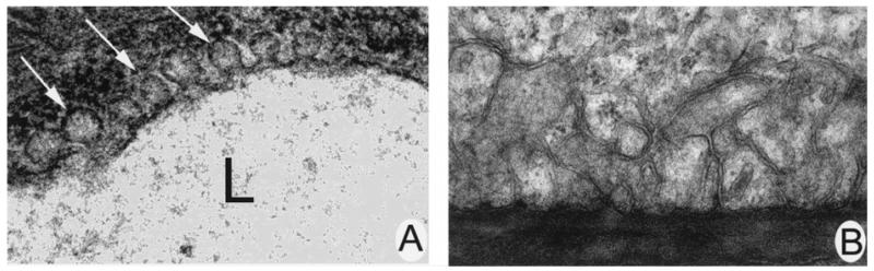
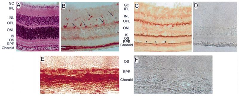
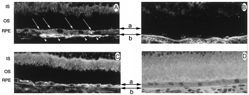
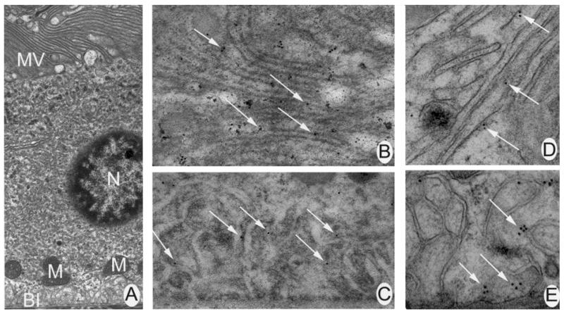
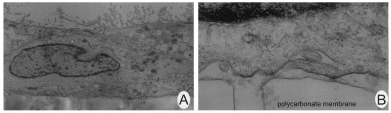
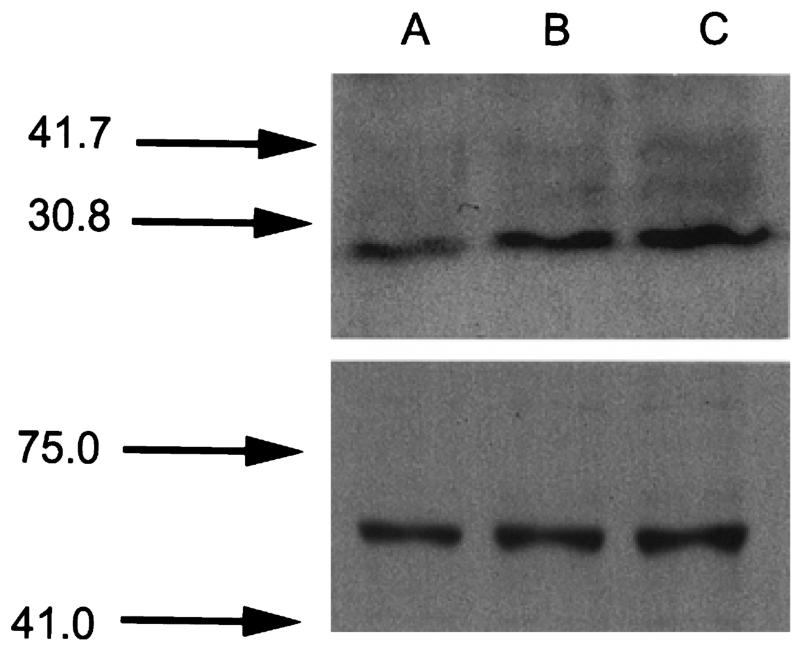
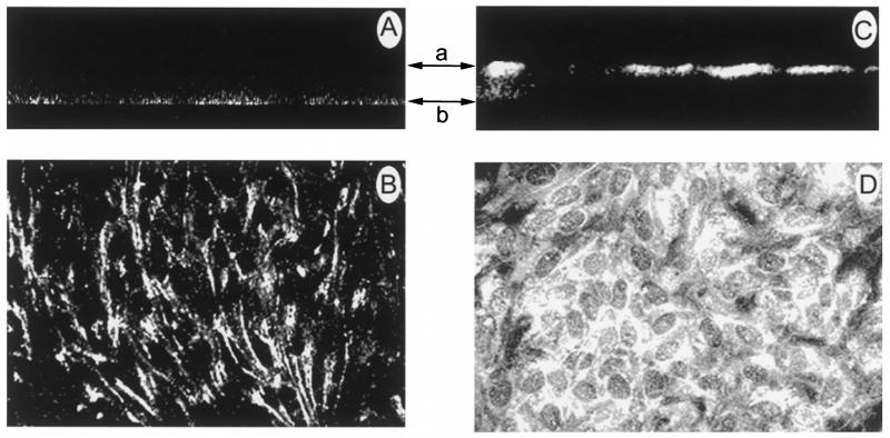
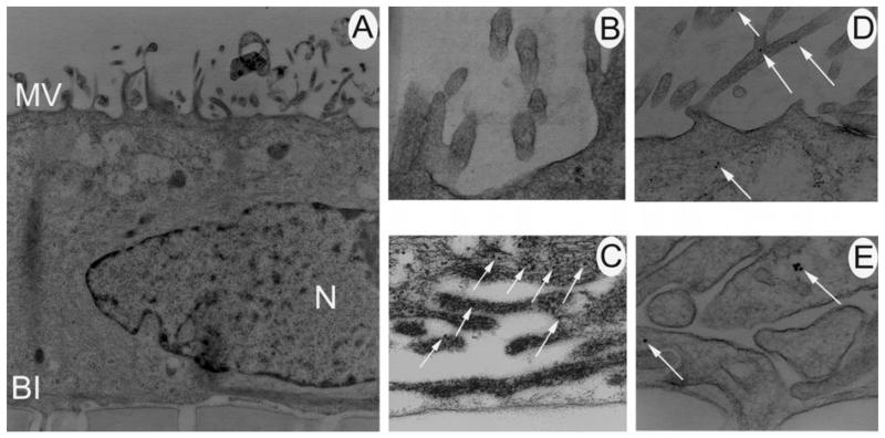
Caveolae are flask-shaped membrane invaginations present in most mammalian cells. They are distinguished by the presence of a striated coat composed of the protein, caveolin. Caveolae have been implicated in numerous cellular processes, including potocytosis in which caveolae are hypothesized to co-localize with folate receptor alpha and participate in folate uptake. Our laboratory has recently localized folate receptor alpha to the basolateral surface of the retinal pigment epithelium (RPE). It is present also in many other cells of the retina. In the present study, we asked whether caveolae were present in the RPE, and if so, whether their pattern of distribution was similar to folate receptor alpha. We also examined the distribution pattern of caveolin-1, which can be a marker of caveolae. Extensive electron microscopical analysis revealed caveolae associated with endothelial cells. However, none were detected in intact or cultured RPE. Laser scanning confocal microscopical analysis of intact RPE localized caveolin-1 to the apical and basal surfaces, a distribution unlike folate receptor alpha. Western analysis confirmed the presence of caveolin-1 in cultured RPE cells and laser scanning confocal microscopy localized the protein to the basal plasma membrane of the RPE, a distribution like that of folate receptor alpha. This distribution was confirmed by electron microscopic immunolocalization. The lack of caveolae in the RPE suggests that these structures may not be essential for folate internalization in the RPE.


Figures

Figure 1
Figure 1
Electron microscopical analysis of caveolae in intact retina. (A) Electron micrograph of retinal endothelial cells with caveolae (arrows) present (60,000×). (B) Electron micrograph of representative section of basal infoldings of a RPE cell. No caveolae were identified (27,500×).
Figure 2
Figure 2
Light immunohistochemical analysis of caveolin-1 in mouse retina. (A) Haematoxylin- and eosin-stained cryosection of mouse retina shown for comparison (400×). (B) Cryosection of mouse retina labelled with an antibody against caveolin-1 (400×). The dark reddish-brown staining is representative of caveolin-1 localization. Arrows point to blood vessels. (C) Cryosection of mouse retina labelled with antibody against folate receptor α (400×). Arrowheads point to RPE. (D) Cryosection of mouse retina labelled with normal rabbit serum in place of the primary antibody (400×). No immunoreactivity was observed. (E) High magnification of the RPE–choroid complex of a mouse retina labelled with an antibody against caveolin-1. (F) High magnification of the RPE–choroid complex of a mouse retina incubated with normal rabbit serum in place of the primary antibody. No immunoreactivity was observed (400×). Abbreviations: GC, ganglion cells; IPL, inner plexiform layer; INL, inner nuclear layer; OPL, outer plexiform layer; ONL, outer nuclear layer; IS, inner segments of the photoreceptor cells; OS, outer segments of the photoreceptor cells; RPE, retinal pigment epithelium.
Figure 3
Figure 3
Laser scanning confocal microscopic localization of caveolin-1 and folate receptor α in intact mouse RPE. (A) High magnification of mouse retina incubated with an antibody against caveolin-1. The apical and basal plasma membranes of the RPE are represented by the top (a) and bottom (b) two-headed arrows, respectively. Caveolin-1 is present on the apical (arrows) and basal (arrowheads) plasmalemmal surfaces of the RPE cell. (B) High magnification of mouse retina incubated with an antibody against the folate receptor α. Note the intense bands of fluorescence on the basolateral membrane of RPE, but no immunoreactivity in the apical region. (C) High magnification of mouse retina incubated with an antibody against the Na+,K+-ATPase. As expected, an intense band of fluorescent labelling was observed on the apical surface of the RPE monolayer. (D) Haematoxylin and eosin-stained section of mouse retina shown for comparison (630×). Abbreviations: IS, inner segments; OS, outer segments; RPE, retinal pigment epithelium.
Figure 4
Figure 4
Electron microscopical immunohistochemical analysis of caveolin-1 in mammalian retina. (A) Mouse RPE depicting apical microvilli (AM) and basal infoldings (BI). The nucleus (N) and mitochondria (M) are also visible (7700×). Panel A serves as a comparison for panels B–E, which are higher magnifications of the apical and basal plasma membranes of a RPE cell. (B) Higher magnification of apical microvilli labelled with an antibody against caveolin-1. Arrows point to gold particles indicative of positive labelling (27,500×). (C) Higher magnification of the basal infoldings of RPE labelled with an antibody against caveolin-1. Arrows point to gold particles (35,500×). (D) Higher magnification of apical microvilli labelled with antibody against the Na+,K+-ATPase (60,000×). Gold particles were abundant. (E) Higher magnification of the basolateral infoldings of a RPE cell labelled with an antibody against folate receptor α (60,000×). Gold particles were present across the membrane.
Figure 5
Figure 5
Electron microscopical analysis of caveolae in cultured human ARPE-19 cells. (A) Electron micrograph of a cultured human ARPE-19 cell grown on a permeable membrane support (4600×). (B) High magnification of a representative basal section of an ARPE-19 cell showing no caveolae (46,000×).
Figure 6
Figure 6
Western analysis of caveolin-1 in cultured human ARPE-19 cells. Proteins extracted from ARPE-19 cells were subjected to SDS-PAGE and immunoblotted as described in the text. The upper panel shows an immunoblot probed with an antibody to detect caveolin-1. The lower panel shows an immunoblot probed with an antibody to detect a protein known to be present in the RPE, reduced-folate transporter-1. Lanes A–C represent increasing protein loading amounts of 10, 20, and 30 μg, repectively.
Figure 7
Figure 7
Laser scanning confocal microscope analysis of caveolin-1 distribution in cultured human ARPE-19 cells. Panels A and C are sections of cells shown in a vertical dimension (z, y). The upper and lower arrows represent the apical and basolateral surfaces, respectively. Panels B and D are sections of cells shown in a horizontal dimension (x, y). Panels A and B show cells incubated with an antibody against caveolin-1. The vertical scan (panel A) revealed a basolateral distribution for caveolin-1. A ring-like fluorescence pattern was observed in the horizontal scan (panel B), suggestive of a basolateral distribution. As a positive control, cells were incubated with antibody against the Na+,K+-ATPase. The apical band of fluorescence in the vertical scan (panel C) and the dome-like fluorescence pattern in the horizontal scan (panel D) suggests an apical distribution for the protein (630×).
Figure 8
Figure 8
Electron microscopical analysis of caveolin-1 in cultured human ARPE-19 cells. (A) Micrograph of an ARPE-19 cell depicting apical microvilli (AM) and basal infoldings (BI). The nucleus (N) can also be seen (7700×). Panel A serves as a comparison for panels B–E, which are higher magnifications of the apical and basal plasma membranes of a RPE cell. (B) Higher magnification of apical microvilli labelled with an antibody against caveolin-1. No gold particles were observed (60,000×). (C) Higher magnification of the basal infoldings of RPE labelled with an antibody against caveolin-1. Arrows point to gold particles (35,500×). (D) Higher magnification of apical microvilli labelled with antibody against the Na+,K+-ATPase. Gold particles were abundant (46,000×). (E) Higher magnification of the basolateral infoldings of A RPE cells labelled with an antibody against folate receptor α. Gold particles were present across the membrane (46,000×).

References

    1. Anderson RG, Kamen BA, Rothberg KG, Lacey SW. Potocytosis: sequestration and transport of small molecules by caveolae. Science. 1992;255:410–411. - PubMed
    1. Anderson RGW. Caveolae: Where incoming and outgoing messengers meet. Proc Natl Acad Sci USA. 1993;90:10909–10913. - PMC - PubMed
    1. Bretscher MS, Whytock S. Membrane-associated vesicles in fibroblasts. J Ultrastruct Res. 1977;61:215–217. - PubMed
    1. Chancy CD, Kekuda R, Huang W, Prasad PD, Kuhnel J-M, Sirotnak FM, Roon P, Ganapathy V, Smith SB. Expression and differential polarization of the reduced-folate transporter-1 and the folate receptor α in mammalian retinal pigment epithelium. J Biol Chem. 2000;275:20676–20684. - PubMed
    1. Dunn KC, Aotaki-Keen E, Putkey FR, Hjelmeland LM. ARPE-19, a human retinal pigment epithelial cell line with differentiated properties. Exp Eye Res. 1996;62:155–169. - PubMed

Publication types

MeSH terms