. 2001 Sep;21(18):6322-31.
doi: 10.1128/MCB.21.18.6322-6331.2001.

Cytochrome p450 epoxygenase metabolism of arachidonic acid inhibits apoptosis

Affiliations

Cytochrome p450 epoxygenase metabolism of arachidonic acid inhibits apoptosis

J K Chen et al. Mol Cell Biol. 2001 Sep.

Abstract

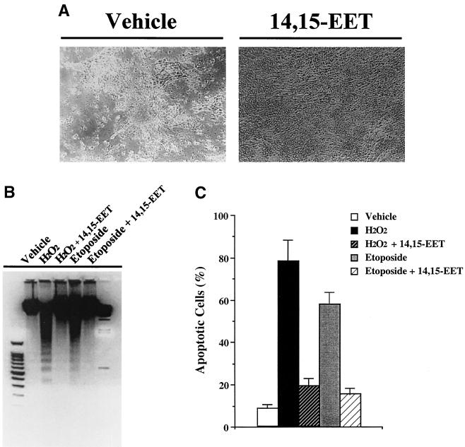
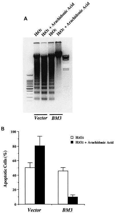
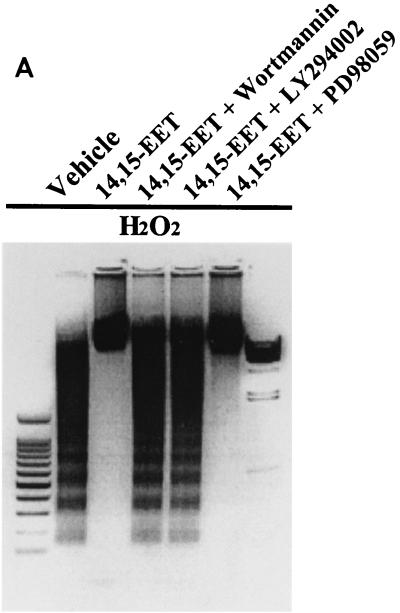
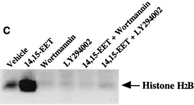
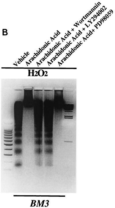
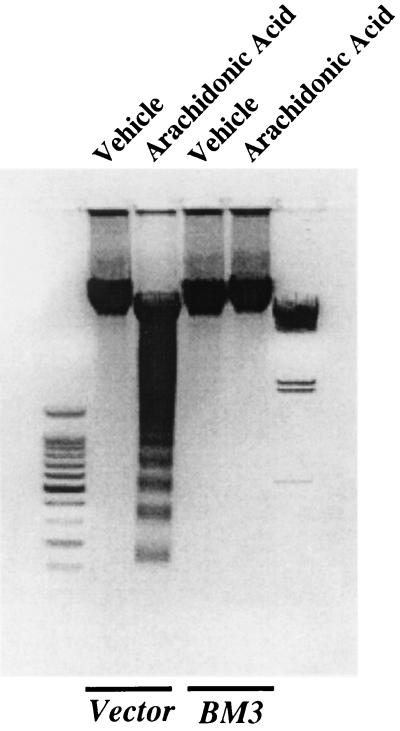
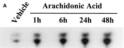
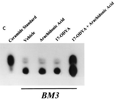
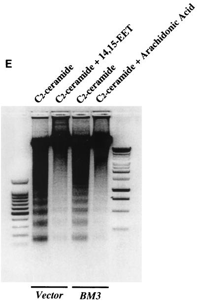
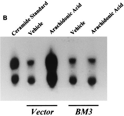
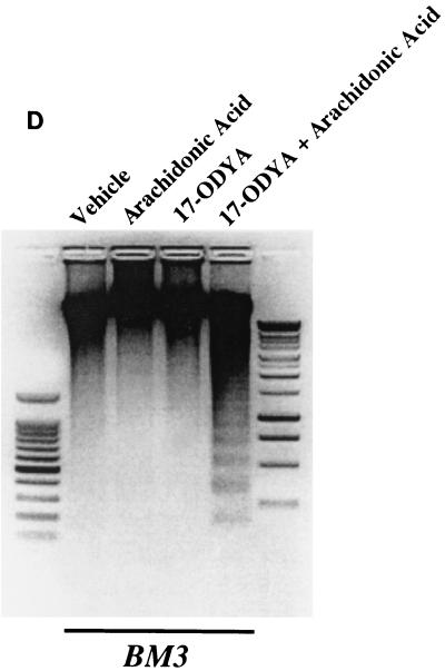
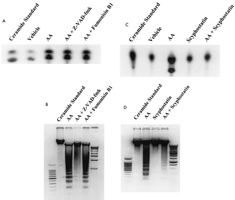
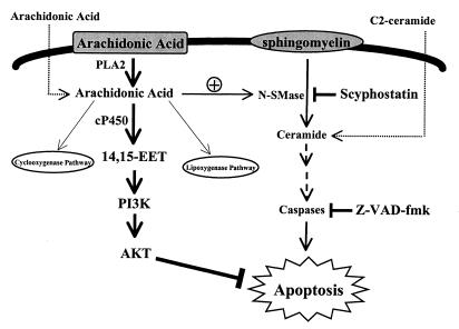
The ubiquitous cytochrome P450 hemoproteins play important functional roles in the metabolism and detoxification of foreign chemicals. However, other than established roles in cholesterol catabolism and steroid hormone biosynthesis, their cellular and/or organ physiological functions remain to be fully characterized. Here we show that the cytochrome P450 epoxygenase arachidonic acid metabolite 14,15-epoxyeicosatrienoic acid (14,15-EET) inhibits apoptosis induced by serum withdrawal, H(2)O(2), etoposide, or excess free arachidonic acid (AA), as determined by DNA laddering, Hoechst staining, and fluorescein isothiocyanate-labeled annexin V binding. In the stable transfectants (BM3 cells) expressing a mutant bacterial P450 AA epoxygenase, F87V BM3, which was genetically engineered to metabolize arachidonic acid only to 14,15-EET, AA did not induce apoptosis and protected against agonist-induced apoptosis. Ceramide assays demonstrated increased AA-induced ceramide production within 1 h and elevated ceramide levels for up to 48 h, the longest time tested, in empty-vector-transfected cells (Vector cells) but not in BM3 cells. Inhibition of cytochrome P450 activity by 17-octadecynoic acid restored AA-induced ceramide production in BM3 cells. Exogenous C2-ceramide markedly increased apoptosis in quiescent Vector cells as well as BM3 cells, and apoptosis was prevented by pretreatment of Vector cells with exogenous 14,15-EET and by pretreatment of BM3 cells with AA. The ceramide synthase inhibitor fumonisin B1 did not affect AA-induced ceramide production and apoptosis; in contrast, these effects of AA were blocked by the neutral sphingomyelinase inhibitor scyphostatin. The pan-caspase inhibitor Z-VAD-fmk had no effect on AA-induced ceramide generation but abolished AA-induced apoptosis. The antiapoptotic effects of 14,15-EET were blocked by two mechanistically and structurally distinct phosphatidylinositol-3 (PI-3) kinase inhibitors, wortmannin and LY294002, but not by the specific mitogen-activated protein kinase kinase inhibitor PD98059. Immunoprecipitation followed by an in vitro kinase assay revealed activation of Akt kinase within 10 min after 14,15-EET addition, which was completely abolished by either wortmannin or LY294002 pretreatment. In summary, the present studies demonstrated that 14,15-EET inhibits apoptosis by activation of a PI-3 kinase-Akt signaling pathway. Furthermore, cytochrome P450 epoxygenase promotes cell survival both by production of 14,15-EET and by metabolism of unesterified AA, thereby preventing activation of the neutral sphingomyelinase pathway and proapoptotic ceramide formation.


Figures

FIG. 1
FIG. 1
Antiapoptotic effects of exogenously administered 14,15-EET in LLCPKcl4 cells. (A) 14,15-EET prevented the renal epithelial cell line LLCPKcl4 from progressive loss of cell viability caused by serum deprivation. LLCPKcl4 cells grown to confluence were changed to serum-free medium. After 48 h, vehicle (Me2SO) or 14,15-EET (10 μM) was added every 3 days. After 10 days, the majority of the vehicle-treated cells had died while the 14,15-EET treated cells still maintained a confluent monolayer with normal morphology. (B) 14,15-EET inhibited DNA laddering induced by H2O2 or etoposide. After pretreatment with vehicle (Me2SO) or 14,15-EET (10 μM) for 1 h, quiescent LLCPKcl4 cells were exposed to H2O2 (500 μM) or etoposide (100 μM) for 24 h, and genomic DNA was extracted and electrophoresed on a 1.5% agarose gel containing ethidium bromide. Leftmost lane, 100-bp DNA ladder; rightmost lane, lambda DNA-HindIII DNA size marker (Promega). (C) 14,15-EET decreased H2O2- or etoposide-induced apoptotic-cell number; cells were scored for morphological evidence of apoptosis as described in Materials and Methods.
FIG. 2
FIG. 2
Effects of endogenously produced 14,15-EET in LLCPKcl4 cells. Arachidonic acid blocked H2O2-induced apoptosis in BM3 cells but not in Vector cells. Quiescent BM3 cells and Vector cells were pretreated with or without arachidonic acid (10 μM) for 1 h before exposure to H2O2 for 24 h, and DNA laddering (A) and apoptotic cell number (B) were assessed as described in Materials and Methods. (A) Leftmost lane, 100-bp DNA ladder; rightmost lane, lambda DNA-HindIII DNA size marker.
FIG. 3
FIG. 3
14,15-EET exerts its antiapoptotic effects in LLCPKcl4 cells by activation of a PI-3 kinase–Akt-signaling pathway. (A) 14,15-EET's antiapoptotic effects were abolished by inhibition of PI-3 kinase but not by inhibition of MAP kinase. After pretreatment with or without the specific PI-3 kinase inhibitor wortmannin (10 nM) or LY294002 (5 μM) or the MEK inhibitor PD98059 (10 μM) for 30 min, followed by 14,15-EET or vehicle for 1 h, LLCPKcl4 cells were exposed to H2O2 for 24 h, and DNA laddering was assessed. (B) Arachidonic acid's antiapoptotic effects in BM3 cells were abolished by inhibition of PI-3 kinase but not by inhibition of MAP kinase. Before induction of H2O2-mediated apoptosis, BM3 cells were pretreated with or without wortmannin, LY294002, or PD98059 and then treated with or without arachidonic acid. (A and B) Leftmost lane, 100-bp DNA ladder; rightmost lane, lambda DNA-HindIII size marker. (C) 14,15-EET increased Akt kinase activity, which was abolished by either wortmannin or LY294002. After treatment with or without the specific PI-3 kinase inhibitor wortmannin (10 nM) or LY294002 (5 μM), LLCPKcl4 cells were treated with or without 14,15-EET. The cells were then lysed and subjected to immunoprecipitation and Akt kinase activity assay with histone H2B as a substrate.
FIG. 3
FIG. 3
14,15-EET exerts its antiapoptotic effects in LLCPKcl4 cells by activation of a PI-3 kinase–Akt-signaling pathway. (A) 14,15-EET's antiapoptotic effects were abolished by inhibition of PI-3 kinase but not by inhibition of MAP kinase. After pretreatment with or without the specific PI-3 kinase inhibitor wortmannin (10 nM) or LY294002 (5 μM) or the MEK inhibitor PD98059 (10 μM) for 30 min, followed by 14,15-EET or vehicle for 1 h, LLCPKcl4 cells were exposed to H2O2 for 24 h, and DNA laddering was assessed. (B) Arachidonic acid's antiapoptotic effects in BM3 cells were abolished by inhibition of PI-3 kinase but not by inhibition of MAP kinase. Before induction of H2O2-mediated apoptosis, BM3 cells were pretreated with or without wortmannin, LY294002, or PD98059 and then treated with or without arachidonic acid. (A and B) Leftmost lane, 100-bp DNA ladder; rightmost lane, lambda DNA-HindIII size marker. (C) 14,15-EET increased Akt kinase activity, which was abolished by either wortmannin or LY294002. After treatment with or without the specific PI-3 kinase inhibitor wortmannin (10 nM) or LY294002 (5 μM), LLCPKcl4 cells were treated with or without 14,15-EET. The cells were then lysed and subjected to immunoprecipitation and Akt kinase activity assay with histone H2B as a substrate.
FIG. 4
FIG. 4
Arachidonic acid induced apoptosis in Vector cells but not in BM3 cells. Quiescent BM3 cells and Vector cells were treated with or without arachidonic acid (10 μM) for 48 h in the absence of other apoptosis-inducing stimuli and then subjected to assessment of DNA laddering. Leftmost lane, 100-bp DNA ladder; rightmost lane, lambda DNA-HindIII size marker.
FIG. 5
FIG. 5
Ceramide mediates arachidonic acid-induced apoptosis. (A) Time course of arachidonic acid induced-ceramide generation in LLCPKcl4 cells. Quiescent cells were treated with or without arachidonic acid (10 μM) for the indicated times, the cells were then harvested, and ceramide levels were quantitated as described in Materials and Methods. (B) Arachidonic acid increased ceramide generation in Vector cells but not in BM3 cells. Vector cells and BM3 cells were rendered quiescent and treated with or without arachidonic acid (10 μM) for 48 h. Ceramide levels were determined as described in Materials and Methods. (C and D) Arachidonic acid increased ceramide levels and induced apoptosis in BM3 cells pretreated with a P450 inhibitor. Quiescent BM3 cells were pretreated with or without the P450 inhibitor 17-ODYA (20 μM) and treated with or without arachidonic acid (10 μM). The cells were then harvested and ceramide levels were measured (C), and apoptosis was determined (D). (E) 14,15-EET and arachidonic acid inhibited apoptosis induced by administration of ceramide in Vector cells and BM3 cells, respectively. Vector cells and BM3 cells were made quiescent and pretreated with or without 14,15-EET (Vector cells) or arachidonic acid (BM3 cells); they were then exposed to the cell-permeable C2-ceramide (25 μM), and apoptosis was assessed. (D and E) Leftmost lane, 100-bp DNA ladder; rightmost lane, 1-kb DNA ladder.
FIG. 5
FIG. 5
Ceramide mediates arachidonic acid-induced apoptosis. (A) Time course of arachidonic acid induced-ceramide generation in LLCPKcl4 cells. Quiescent cells were treated with or without arachidonic acid (10 μM) for the indicated times, the cells were then harvested, and ceramide levels were quantitated as described in Materials and Methods. (B) Arachidonic acid increased ceramide generation in Vector cells but not in BM3 cells. Vector cells and BM3 cells were rendered quiescent and treated with or without arachidonic acid (10 μM) for 48 h. Ceramide levels were determined as described in Materials and Methods. (C and D) Arachidonic acid increased ceramide levels and induced apoptosis in BM3 cells pretreated with a P450 inhibitor. Quiescent BM3 cells were pretreated with or without the P450 inhibitor 17-ODYA (20 μM) and treated with or without arachidonic acid (10 μM). The cells were then harvested and ceramide levels were measured (C), and apoptosis was determined (D). (E) 14,15-EET and arachidonic acid inhibited apoptosis induced by administration of ceramide in Vector cells and BM3 cells, respectively. Vector cells and BM3 cells were made quiescent and pretreated with or without 14,15-EET (Vector cells) or arachidonic acid (BM3 cells); they were then exposed to the cell-permeable C2-ceramide (25 μM), and apoptosis was assessed. (D and E) Leftmost lane, 100-bp DNA ladder; rightmost lane, 1-kb DNA ladder.
FIG. 6
FIG. 6
Arachidonic acid induces apoptosis by activating neutral sphingomyelinase pathway. (A and B) Effects of ceramide synthase inhibitor and caspase inhibitor on arachidonic acid-induced ceramide production and apoptosis in LLCPKcl4 cells. Quiescent cells were pretreated with vehicle, fumonisin B1 (50 μM), or Z-VAD-fmk (50 μM) for 1 h, followed by treatment with or without arachidonic acid (10 μM). Cells were then harvested, ceramide levels were measured 1 h after administration of arachidonic acid (A), and apoptosis was determined 24 h after administration of arachidonic acid (B). (C and D) Effects of the neutral sphingomyelinase inhibitor scyphostatin on arachidonic acid-induced ceramide production and apoptosis in LLCPKcl4 cells. Quiescent cells were pretreated with or without scyphostatin (1 μM) for 1 h followed by treatment with or without arachidonic acid (10 μM). Cells were then harvested, ceramide levels were measured (C), and apoptosis was determined (D). (B and D) Leftmost lane, 100-bp DNA ladder; rightmost lane, 1-kb DNA ladder.
FIG. 7
FIG. 7
Arachidonic acid-ceramide-apoptosis-signaling pathway is regulated by cytochrome P450 in LLCPKcl4 cells. Arachidonic acid, an important constituent of cell membrane, is released by activation of specific phospholipases (PLA2) and further metabolized by cyclooxygenases, lipoxygenases, and cytochrome P450 pathways. If unesterified arachidonic acid is not metabolized, it activates neutral sphingomyelinase (N-SMase), which converts sphingomyelin to the second messenger, ceramide. Ceramide induces caspase activation, which leads to apoptosis. In cells such as the renal proximal tubule, in which cyclooxygenase and lipoxygenase are expressed at nearly undetectable levels, arachidonic acid metabolism is shunted to the cytochrome P450 pathway. This metabolism not only metabolizes and detoxifies excess unesterified arachidonic acid to prevent proapoptotic ceramide formation but also produces a metabolite, 14,15-EET, which activates a PI-3 kinase–Akt-signaling pathway. Thus, cytochrome P450 mediates cell survival by two complementary mechanisms.

References

    1. Amsler K, Cook J S. Development of Na+-dependent hexose transport in a cultured line of porcine kidney cells. Am J Physiol. 1982;242:C94–C101. - PubMed
    1. Ara G, Teicher B A. Cyclooxygenase and lipoxygenase inhibitors in cancer therapy. Prostaglandins Leukot Essent Fatty Acids. 1996;54:3–16. - PubMed
    1. Bonvalet J P, Pradelles P, Farman N. Segmental synthesis and actions of prostaglandins along the nephron. Am J Physiol. 1987;253:F377–F387. - PubMed
    1. Bortuzzo C, Hanif R, Kashfi K, Staiano-Coico L, Shiff S J, Rigas B. The effect of leukotrienes B and selected HETEs on the proliferation of colon cancer cells. Biochim Biophys Acta. 1996;1300:240–246. - PubMed
    1. Burgering B M, Coffer P J. Protein kinase B (c-Akt) in phosphatidylinositol-3-OH kinase signal transduction. Nature. 1995;376:599–602. - PubMed

Publication types