. 2001 Sep;21(18):6332-45.
doi: 10.1128/MCB.21.18.6332-6345.2001.

S phase-specific proteolytic cleavage is required to activate stable DNA binding by the CDP/Cut homeodomain protein

Affiliations

S phase-specific proteolytic cleavage is required to activate stable DNA binding by the CDP/Cut homeodomain protein

N S Moon et al. Mol Cell Biol. 2001 Sep.

Abstract

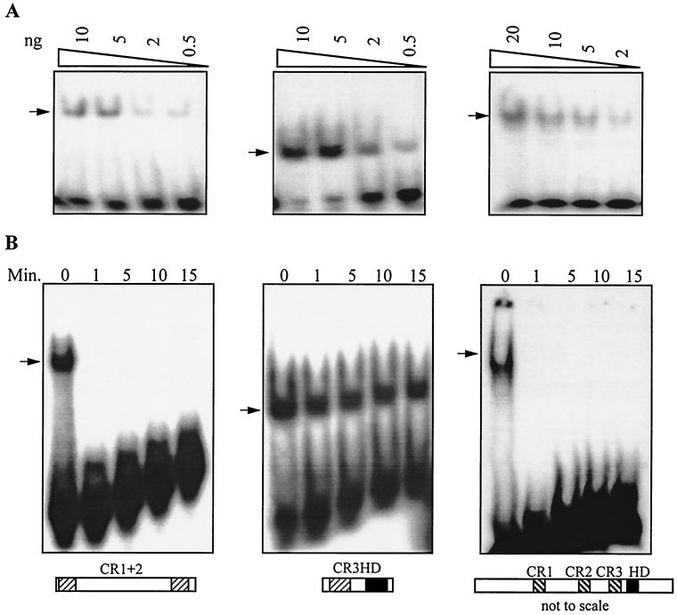
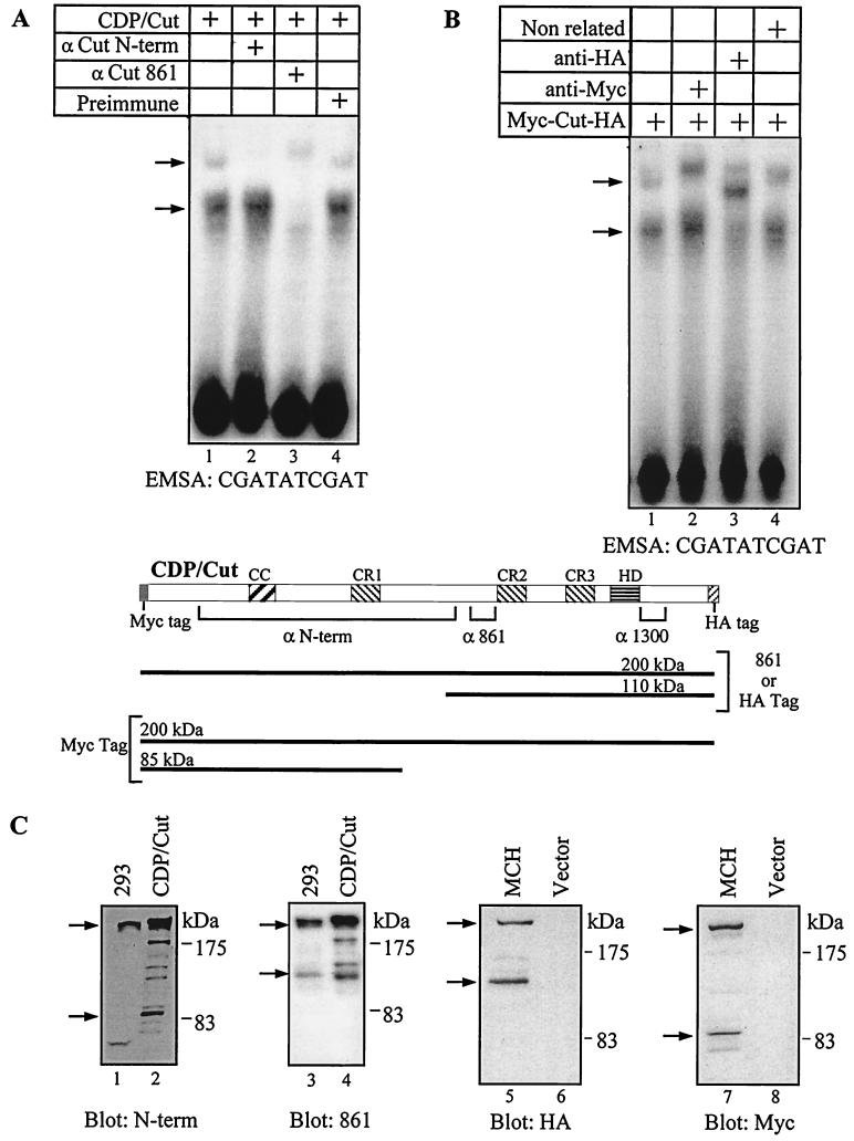
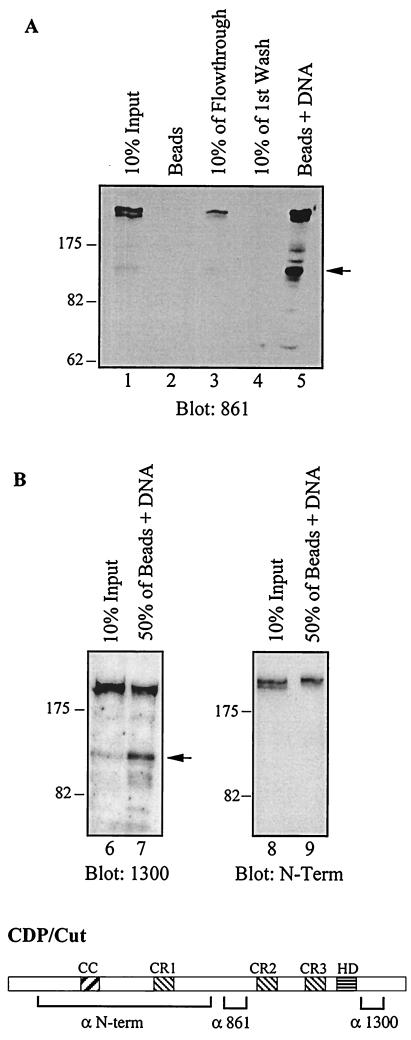
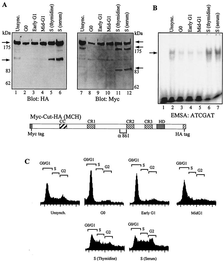
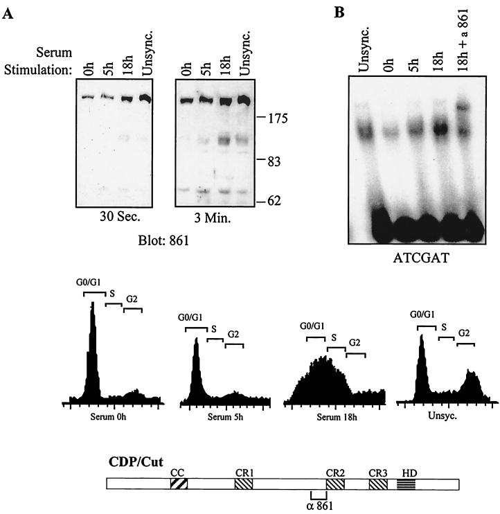
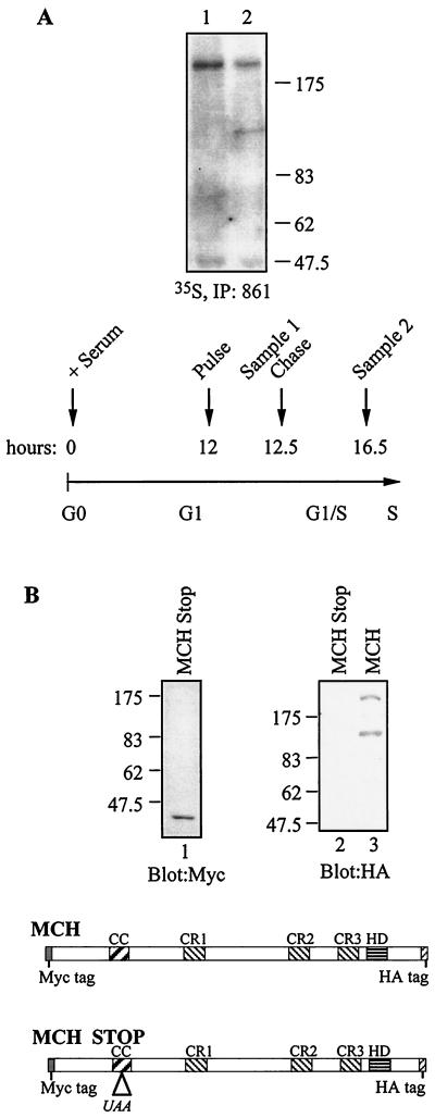
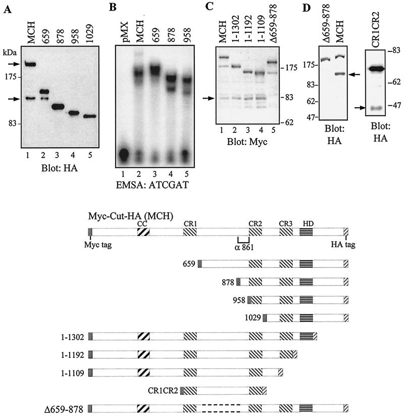
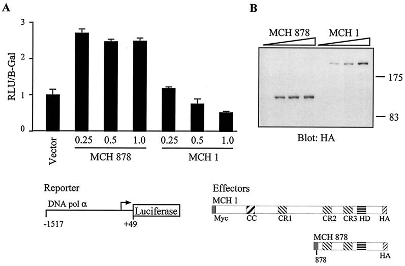
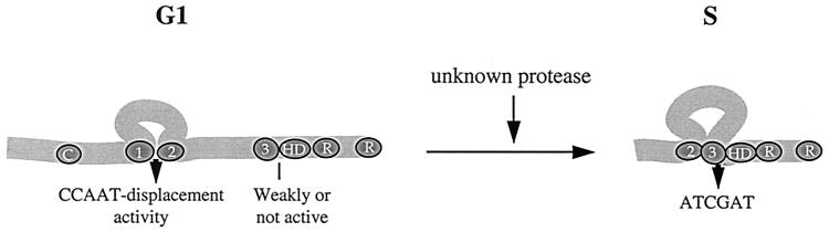
The CCAAT displacement protein (CDP), the homologue of the Drosophila melanogaster Cut protein, contains four DNA binding domains that function in pairs. Cooperation between Cut repeat 3 and the Cut homeodomain allows stable DNA binding to the ATCGAT motif, an activity previously shown to be upregulated in S phase. Here we showed that the full-length CDP/Cut protein is incapable of stable DNA binding and that the ATCGAT binding activity present in cells involves a 110-kDa carboxy-terminal peptide of CDP/Cut. A vector expressing CDP/Cut with Myc and hemagglutinin epitope tags at either end generated N- and C-terminal products of 90 and 110 kDa, suggesting that proteolytic cleavage was involved. In vivo pulse/chase labeling experiments confirmed that the 110-kDa protein was derived from the full-length CDP/Cut protein. Proteolytic processing was weak or not detectable in G(0) and G(1) but increased in populations of cells enriched in S phase, and the appearance of the 110-kDa protein coincided with the increase in ATCGAT DNA binding. Interestingly, the amino-truncated and the full-length CDP/Cut isoforms exhibited different transcriptional properties in a reporter assay. We conclude that proteolytic processing of CDP/Cut at the G(1)/S transition generates a CDP/Cut isoform with distinct DNA binding and transcriptional activities. These findings, together with the cleavage of the Scc1 protein at mitosis, suggest that site-specific proteolysis may play an important role in the regulation of cell cycle progression.


Figures

FIG. 1
FIG. 1
The full-length CDP/Cut protein exhibits DNA binding kinetics similar to that of CR1CR2. (A) DNA binding specificity of CR1CR2 (CR1+2), CR3HD, and CDP/Cut for CGATATCGAT. Decreasing amounts of the indicated fusion proteins were mixed with radiolabeled oligonucleotides containing the CGATATCGAT sequence at room temperature until equilibrium was reached. DNA-protein complexes were resolved on a nondenaturing polyacrylamide gel that was dried and autoradiographed. Note that oligonucleotides containing the CGATATCGAT sequence are recognized efficiently by CR1CR2, since two CGAT direct repeats are present (CGATATCGAT), and by CR3HD, since the ATCGAT sequence is present (CGATATCGAT). (B) DNA binding kinetics of CR1CR2, CR3HD, and CDP/Cut. One hundred nanograms of the indicated fusion protein was incubated with radiolabeled oligonucleotides containing the CGATATCGAT sequence at room temperature until the equilibrium was reached (15 min); 1,000-fold molar excess of unlabeled oligonucleotides was added, and at the indicated time points aliquots of the mixture were taken and analyzed by EMSA.
FIG. 2
FIG. 2
Two distinct retarded complexes are generated by CDP/Cut proteins, only one of which is supershifted by antibodies raised against the N-terminal region of the protein. (A and B) NIH 3T3 cells were transfected with a vector expressing the CDP/Cut protein (A) or Myc-Cut-HA, a CDP/Cut protein with Myc and HA epitope tags at its amino- and carboxy-termini, respectively (B). Nuclear extracts were prepared and analyzed in EMSA with oligonucleotides containing a universal CDP/Cut consensus-binding site (CGATATCGAT). The DNA and proteins were incubated either with no antibody (lane 1) or with the indicated antibodies (lanes 2 to 4). The preimmune serum is that for anti-861, the nonrelated antibody is a goat anti-rabbit immunoglobulin G. The arrows indicate the positions of the retarded complexes. Note that the lower retarded complex can be supershifted with anti-861 (A) and anti-HA (B) antibodies but not with anti-N-term (A) or anti-Myc (B) antibodies. A diagram of the protein is shown indicating the evolutionarily conserved domains and the regions recognized by the respective antibodies (CC, coiled-coil). (C) Nuclear extracts from untransfected 293 cells and transfected NIH 3T3 cells (CDP/Cut, MCH, Vector) were separated by electrophoresis on 8% polyacrylamide gels and analyzed in Western blots with CDP/Cut N-term, CDP/Cut 861, HA, and Myc antibodies. Note that the relative abundance of the full-length CDP/Cut protein appears less than that in subsequent blots using 6% polyacrylamide (Fig. 3, 4, and 5). This is because transfer of the 200-kDa protein to the membrane is less efficient in 8% polyacrylamide.
FIG. 3
FIG. 3
DNA affinity chromatography with the ATCGAT sequence leads to the specific enrichment of a 110-kDa CDP/Cut protein. (A) Nuclear extracts from HeLa cells were subjected to affinity chroma- tography using as bait biotinylated oligonucleotides containing the ATCGAT sequence. Samples were then separated by electrophoresis on 6% polyacrylamide gels and analyzed by Western blotting with the anti-CDP/Cut 861 (lanes 1 to 5), 1300 (lanes 6 to 7), and N-term (lanes 8 and 9) antibodies.
FIG. 4
FIG. 4
S phase-specific proteolytic cleavage of CDP/Cut. A cDNA encoding Myc-Cut-HA generates short proteins specifically in S phase. NIH 3T3 cells were transfected with a vector expressing Myc-Cut-HA, a CDP/Cut protein with Myc and HA epitope tags at its amino and carboxy termini, respectively. Cells were synchronized either by serum starvation and stimulation or by thymidine block, as described in Materials and Methods. Total extracts were prepared and analyzed in Western blots with anti-Myc and anti-HA antibodies (A) and in EMSA with an ATCGAT probe (B). Note that only a lower retarded complex is visible in EMSA with total extracts, whereas two complexes were observed with nuclear extracts (see Fig. 1). Cell cycle distribution was monitored by FACS analysis after staining of the DNA with propidium iodide (C). Unsync., unsynchronized.
FIG. 5
FIG. 5
CDP/Cut proteins (110 kDa) are expressed in S phase. NIH 3T3 cells were synchronized in G0 by serum starvation for 72 h and then stimulated with fresh DMEM plus 10% FBS. The indicated times correspond to the time elapsed since release from the G0 block. Nuclear extracts were prepared and analyzed in Western blots with anti-Cut 861 antibodies (A) and in EMSA with the ATCGAT probe (B). Cell cycle distribution was monitored by FACS analysis after staining of the DNA with propidium iodide. Unsync., unsynchronized.
FIG. 6
FIG. 6
35S-pulse-chase labeling demonstrates that the 110-kDa amino-truncated protein derives from the 200-kDa full-length CDP/Cut protein. (A) NIH 3T3 cells were transfected with cDNA expressing Myc-Cut-HA in two 150 mm-diameter plates and synchronized in G0 by serum starvation for 72 h and then stimulated with fresh DMEM plus 10% FBS. The indicated times correspond to the time elapsed since release from the G0 block. At 12 h, the medium was replaced with complete labeling medium containing 35S-labeled methionine and cysteine. At 12 h and 30 min, total extracts were prepared from one plate of cells, while the medium was replaced with complete medium with cold methionine and cysteine in the second plate. At 16 h and 30 min, total extracts were prepared from the second plate. Samples 1 and 2 were immunoprecipitated with 861 antibody, resolved by PAGE, and revealed by autoradiography. (B) A cDNA expressing Myc-Cut-HA and Myc-Cut-HA with a stop codon inserted in the coiled-coil region were transfected in NIH 3T3 cells. Nuclear extracts were prepared and analyzed by Western blot with Myc and HA antibody.
FIG. 7
FIG. 7
Proteolytic cleavage occurs downstream of CR1, between aa 659 and 878. NIH 3T3 cells were transfected with a series of vectors expressing amino- and carboxy-terminally truncated MCH proteins. Nuclear extracts were prepared and analyzed in Western blots with anti-HA or anti-Myc antibodies, as indicated, and in EMSA with the ATCGAT probe (B). A diagram of the proteins encoded by each construct is shown at the bottom. Note that only the full-length MCH and the 659 are processed into a shorter form. In EMSA, 659 generates two main retarded complexes, the higher of which migrates more slowly than that seen with MCH. This is because the nonprocessed 659 is more abundant than its processed derivative, and it is capable of stable binding to the ATCGAT probe.
FIG. 8
FIG. 8
An amino-terminally truncated CDP/Cut isoform is capable of transcriptional activation. HS578T cells were cotransfected with the DNA polymerase alpha luciferase reporter construct and a vector expressing either nothing, full-length Myc-Cut-HA (MCH 1), or a MCH protein starting at aa 878 (MCH 878). (A) Cytoplasmic extracts were prepared and processed to measure luciferase activity. Means of 6 transfections are shown and the results are expressed as relative light units (RLU) normalized to β-galactosidase activity from an internal control. (B) Total extracts were prepared in parallel and analyzed in Western blots with anti-HA antibodies.
FIG. 9
FIG. 9
Modulation of CDP/Cut at the G1/S transition. In early G1, CDP/Cut exists as a full-length protein that cannot stably bind to the ATCGAT sequence. As cells reach S phase, CDP/Cut is proteolytically processed into an amino-truncated 110-kDa protein in which the CR3HD bipartite DNA binding domain is fully active.

References

    1. Andres V, Chiara M D, Mahdavi V. A new bipartite DNA-binding domain: cooperative interaction between the cut repeat and homeo domain of the cut homeo proteins. Genes Dev. 1994;8:245–257. - PubMed
    1. Andres V, Nadal-Ginard B, Mahdavi V. Clox, a mammalian homeobox gene related to Drosophila cut, encodes DNA-binding regulatory proteins differentially expressed during development. Development. 1992;116:321–334. - PubMed
    1. Aufiero B, Neufeld E J, Orkin S H. Sequence-specific DNA binding of individual Cut repeats of the human CCAAT displacement/Cut homeodomain protein. Proc Natl Acad Sci USA. 1994;91:7757–7761. - PMC - PubMed
    1. Aza-Blanc P, Ramirez-Weber F A, Laget M P, Schwartz C, Kornberg T B. Proteolysis that is inhibited by hedgehog targets Cubitus interruptus protein to the nucleus and converts it to a repressor. Cell. 1997;89:1043–1053. - PubMed
    1. Aziz F, Vanwijnen A J, Vanghan P S, Wu S J, Shakoori A R, Lian J B, Soprano K J, Stein J L, Stein G S. The integrated activities of Irf-2 (Hinf-M), Cdp/Cut (Hinf-D) and H4tf-2 (Hinf-P) regulate transcription of a cell cycle controlled human histone H4 gene - mechanistic differences between distinct H4 genes. Mol Biol Rep. 1998;25:1–12. - PubMed

Publication types

LinkOut - more resources