. 2001 Aug 15;15(16):2054-9.
doi: 10.1101/gad.913601.

COUP-TFI: an intrinsic factor for early regionalization of the neocortex

Affiliations

COUP-TFI: an intrinsic factor for early regionalization of the neocortex

C Zhou et al. Genes Dev. .

Abstract

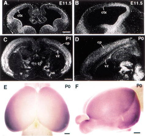
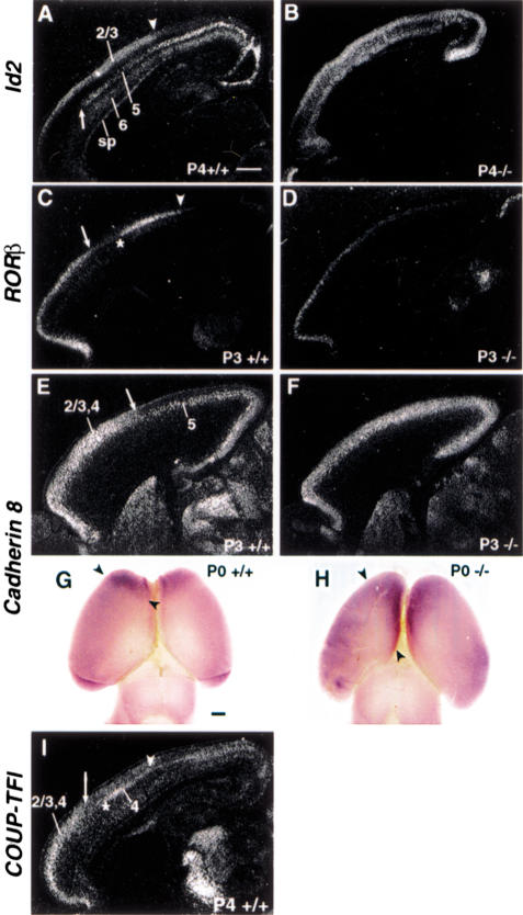
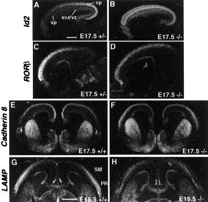
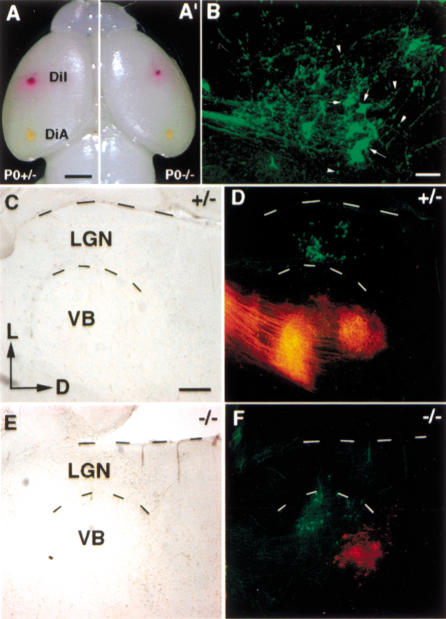
Regionalization of the cerebral cortex is thought to involve two phases: an early regionalization phase and a later refinement phase. It has been shown that early regionalization of the neocortex does not require thalamic inputs and is regulated by intrinsic factors. Recently, two such intrinsic factors, Pax6 and Emx2, have been identified. In this study, we identified COUP-TFI as a regulatory factor for early neocortical regionalization. The spatial and temporal expression pattern of COUP-TFI suggested a role in specification of the neocortex and in maintaining cortical identity. Altered region-specific expression of marker genes in the cortex as well as miswired area-specific connections between the cortex and the thalamus in COUP-TFI null mice indicate COUP-TFI plays a critical role in regulating early regionalization. Our results substantiate that COUP-TFI, an intrinsic factor, may work in concert with Pax6 and Emx2 to specify neocortical identity.


Figures

Figure 1
Figure 1
Graded expression of COUP-TFI during corticogenesis. In-situ hybridization using 35S-UTP-labeled COUP-TFI riboprobe on frontal (A) and sagittal (B) sections of E11.5, coronal (C) section of P1, and sagittal (D) section of P0 mouse brains. Whole-mount in situ hybridization using digoxigenin-labeled COUP-TFI riboprobe on P0 mouse brains (E,F). Because of limitation of probe penetration, only expression in superficial layers (layers 2/3) can be detected by whole-mount in situ hybridization. At E11.5, a high-lateral to low-medial and a high-caudal to low-rostral COUP-TFI gradient was observed in frontal (A) and sagittal (B) view. This high-caudolateral expression was detected in the cortical plate after birth as indicated by section (C,D) and whole-mount (E,F) in situ hybridization. cp, Cortical plate; ctx, cortex; vz, ventricular zone. Bar: A and B, 200 μm; C–F, 500 μm.
Figure 2
Figure 2
Changes in region-specific expression of Id2, RORβ, and Cadherin 8 in COUP-TFI mutants. Section in situ hybridization using Id2 (A,B), RORb (C,D), Cadherin 8 (E,F), and COUP-TFI (I) riboprobes on sagittal sections of P3 or P4 control (A,C,E,I) and COUP-TFI mutant (B,D,F) cortices. Whole-mount in situ hybridization using digoxigenin-labeled Cadherin 8 riboprobe on P0 control (G) and COUP-TFI mutant (H) brains. Id2 expression in the upper layer of layer 5 stopped abruptly at a rostral boundary (arrow in A) and in the rostral part of layers 2/3 with a boundary in the somatosensory area (arrowhead in A). However, Id2 expression in P4 COUP-TFI mutant cortex lost its lamina-specific pattern and was uniformly expressed in layers 5 and 2/3 (B). RORβ expression in layer-4 neurons showed a caudal boundary (arrowhead in C) and a region of much-reduced expression in a more rostral position (between arrow and asterisk in C) in P3 control cortex, while its expression in COUP-TFI mutant was more uniform and expanded caudally with a much lower expression level (D). Cadherin 8 was expressed in the rostral part of the neocortex in the layers 2/3 and 4 (arrow in E) in P3 control cortex (E). However, in P3 COUP-TFI mutant, Cadherin 8 expression in layers 2/3 and 4 lost its rostral restriction (F). This caudal expansion of Cadherin 8 expression in superficial layers also was observed by whole-mount in situ hybridization (arrowheads in G,H). In-situ hybridization with COUP-TFI riboprobe revealed that COUP-TFI was expressed throughout the neocortex (I). Specifically, a higher expression in layers 2/3 and 4 in the rostral part (arrow in I) and a high expression in layer 4 with a boundary in the somatosensory cortex (arrowhead in I) were observed. sp, Subplate. Bar, 500 μm.
Figure 3
Figure 3
Altered expression of Id2, RORβ, Cadherin 8, and LAMP during embryonic development. Section in situ hybridization using Id2 (A,B), RORβ (C,D), Cadherin 8 (E,F), and LAMP (G,H) riboprobes on sagittal (A–D) and coronal (E–H) sections of E17.5 or E15.5 control (A,C,E,G) and COUP-TFI mutant (B,D,F,H) cortices. Expression of Id2 exhibited a high-caudal to low-rostral gradient at E17.5 in control cortex (A), while it was uniformly expressed in COUP-TFI mutant (B). The high-rostral to low-caudal RORβ expression gradient in E17.5 control cortex (C) also became more uniform in COUP-TFI mutants (D). The high-medial to low-lateral Cadherin 8 expression (E) in control cortices was switched to high-lateral to low-medial in COUP-TFI mutants (F). LAMP transcripts were detected in the cortex of control E15.5 mice with a high-lateral to low-medial gradient (G). However, in COUP-TFI mutant mice, LAMP expression level in the cortex was greatly reduced with a rather uniform expression and an extended medial boundary (H). cp, Cortical plate; PR, the perirhinal cortex; SM, the sensorimotor cortex; sp, subplate; svz, subventricular zone; vz, ventricular zone. Bar, 500 μm.
Figure 4
Figure 4
Changes in region-specific connections between the cortex and the thalamus in COUP-TFI mutants. (A,A‘) Shows the positions of DiI and DiA crystals, which were placed in the somatosensory and the visual cortices, respectively. When visualized in the coronal sections of the thalamus using a laser-scanning confocal microscope (B), two types of labeling were observed: the large ends were retrogradely labeled thalamic neurons (arrows in B) and the small ends were boutons of anterogradely labeled axonal terminals of cortical neurons (arrowheads in B). C,D and E,F were conventional microscope images. C and E were bright fields. D and F were overlapping fluorescent images with Texas Red (for DiI labeling, red) and FITC (for DiA labeling, green) filters. Placing DiI and DiA crystals in control somatosensory or visual cortices resulted in labeling in the VB and the LGN, respectively (red and green in D). Consistent with our previous report of defects in thalamocortical projections, insertion of DiI in the somatosensory cortex of COUP-TFI mutants resulted in a much less labeling in the thalamus (red in F). Placing DiI in somatosensory cortex led to labeling in the VB as it did in the controls (red in F). However, when DiA crystals were placed in the presumptive visual cortex of COUP-TFI mutants, most of labeling was seen in the VB instead of the LGN (green in F). D, Dorsal; L, lateral; LGN, lateral geniculate nucleus; VB, ventrobasal thalamus. Bar: A, 1 mm ; B, 50 μm; C, 200 μm.
Figure 5
Figure 5
Normal-graded expression of Pax6 and Emx2 in the cortices of E13.5 COUP-TFI mutants. Section in situ hybridization using Pax6 (A–D) and Emx2 (E–H) riboprobes on sagittal (A,B,E,F) and frontal (C,D,G,H) E13.5 control (A,C,E,G) and COUP-TFI mutant (B,D,F,H) cortices. Pax6 was expressed in a high-rostral to low-caudal (A,B) and high-lateral to low-medial (C,D) pattern in control as well as in mutant cortices. Meanwhile, expression of Emx2 was in a high-caudal to low-rostral (E,F) and high-medial to low-lateral (G,H) pattern in control as well as in mutant cortices. Bar, 200 μm.

Similar articles

Cited by

References

    1. Bishop KM, Goudreau G, O’Leary DD. Regulation of area identity in the mammalian neocortex by Emx2 and Pax6. Science. 2000;288:344–349. - PubMed
    1. Bulfone A, Smiga SM, Shimamura K, Peterson A, Puelles L, Rubenstein JL. T-brain-1: A homolog of Brachyury whose expression defines molecularly distinct domains within the cerebral cortex. Neuron. 1995;15:63–78. - PubMed
    1. Caviness VS, Takahashi T, Nowakowski RS. Numbers, time and neocortical neuronogenesis: A general developmental and evolutionary model. Trends Neurosci. 1995;18:379–383. - PubMed
    1. Gulisano M, Broccoli V, Pardini C, Boncinelli E. Emx1 and Emx2 show different patterns of expression during proliferation and differentiation of the developing cerebral cortex in the mouse. Eur J Neurosci. 1996;8:1037–1050. - PubMed
    1. Horton HL, Levitt P. A unique membrane protein is expressed on early developing limbic system axons and cortical targets. J Neurosci. 1988;8:4653–4661. - PMC - PubMed

Publication types