. 2001 Aug 15;15(16):2083-93.
doi: 10.1101/gad.889201.

Suppression of cap-dependent translation in mitosis

Affiliations

Suppression of cap-dependent translation in mitosis

S Pyronnet et al. Genes Dev. .

Abstract

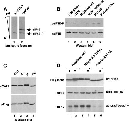
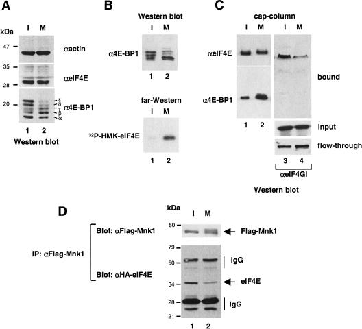
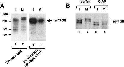
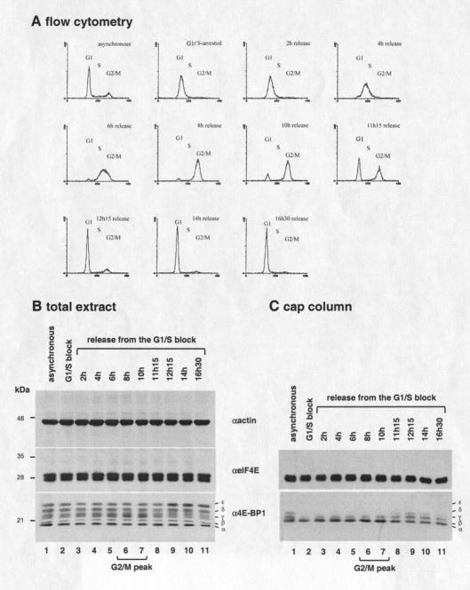
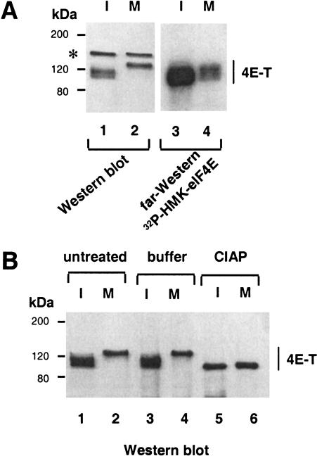
Cap-dependent translation is mediated by eIF4F, a protein complex composed of three subunits as follows: eIF4E, which recognizes the mRNA 5' cap structure; eIF4A, an RNA-helicase; and eIF4G, a scaffolding protein that binds eIF4E, eIF4A, and the eIF4E-kinase Mnk1 simultaneously. eIF4E is hypophosphorylated and cap-dependent translation is reduced at mitosis. Here, we show that 4E-BP1, a suppressor of eIF4E function, is also hypophosphorylated in mitosis, resulting in disruption of the eIF4F complex. Consequently, eIF4E is sequestered from the eIF4G/Mnk1 complex. These results explain the specific inhibition of cap-dependent translation in mitosis and also explain how eIF4E is rendered hypophosphorylated during mitosis. Furthermore, eIF4E interaction with eIF4GII is strongly decreased coincident with hyperphosphorylation of eIF4GII. Thus, inhibition of cap-dependent translation in mitosis results from a combination of phosphorylation modifications leading to eIF4F complex disruption.


Figures

Figure 1
Figure 1
Decreased eIF4E phosphorylation, but increased Mnk1 activity in mitotic cells. (A) Specificity of anti-phospho-specific eIF4E antibody. Total HeLa extract from asynchronous cells was resolved by isoelectric focusing (as described in Materials and Methods), and analyzed by Western blotting with anti-phospho-specific eIF4E (lane 1), or anti-eIF4E (lane 2) antibodies. (B) eIF4E phosphorylation at different phases of the cell cycle. Total HeLa extract from interphase, aphidicolin-treated (G1/S), mitotic (M shake-off), nocodazole-treated (M nocodazole), starved, or starved and TPA-treated cells was resolved by SDS-PAGE and analyzed by Western blotting with anti-phospho-specific eIF4E (top), or anti-eIF4E (bottom) antibody. (C) Mnk1 gel mobility. Total extract from cells transfected with Flag–Mnk1 and treated with aphidicolin (G1/S), thymidine (S), nocodazole (M), or extract from starved cells (G0) was resolved by SDS-PAGE, and analyzed by Western blotting with anti-Mnk1 (top) or anti-Flag (bottom) antibody. (D) Mnk1 activation during mitosis. Extract from interphase or mitotic (nocodazole-arrested) HeLa cells transfected with Flag–Mnk1 wild type or mutants was immunoprecipitated with anti-Flag antibody. Immunoprecipitates were used for immune complex kinase assay using [γ-32P]ATP and recombinant mouse eIF4E as a substrate (as described in Materials and Methods). Samples were resolved by SDS-PAGE, and revealed by autoradiography (bottom) or by Western blotting using the indicated antibodies (top, middle).
Figure 2
Figure 2
Dephosphorylation and increased binding of 4E-BP1 to eIF4E in mitosis. (A) Hypophosphorylation of 4E-BP1. HeLa cell extract from interphase (I) or nocodazole-arrested (M) cells was resolved by SDS-PAGE, and analyzed by Western blotting with anti-actin (top), anti-eIF4E (middle), or anti 4E-BP1 (bottom). (B) Increased binding of 4E-BP1 to eIF4E. HeLa cell extract from interphase (I) and nocodazole-treated (M) cells was resolved by SDS-PAGE and analyzed by Western blotting with anti-4E-BP1 (top) or by far-Western with 32P-HMK-eIF4E as a probe (bottom). (C) eIF4F complex disruption. Extract from interphase (I) or nocodazole-arrested (M) HeLa cells was incubated with an m7GDP resin. Input, flow-through, and bound material was resolved by SDS-PAGE and analyzed by Western blotting with the indicated antibodies. (D) Decreased eIF4E/Mnk1 interaction. Extracts from interphase (I) or nocodazole-arrested (M) HeLa cells transfected with Flag–Mnk1 and HA–eIF4E was immunoprecipitated with anti-Flag antibody. Immunoprecipitates were resolved by SDS-PAGE, and analyzed by Western blotting with anti-HA or anti-Flag antibody.
Figure 3
Figure 3
Hyperphosphorylation and decreased binding of eIF4GII to eIF4E in mitosis. (A) Slower electrophoretic mobility of eIF4GII and decreased binding to eIF4E. Total extract from interphase (I) or nocodazole-arrested (M) HeLa cells was resolved by SDS-PAGE and analyzed by Western blotting with anti-eIF4GII antibody, or by far-Western with an 32P-HMK–eIF4E probe. (B) Phosphatase treatment of eIF4GII. eIF4GII immunoprecipitated from interphase (I) or nocodazole-arrested (M) HeLa cells was untreated, treated with buffer alone, or with calf intestinal acid phosphatase (CIAP) as described in Materials and Methods. Immunoprecipitates resolved by SDS-PAGE were analyzed by Western blotting with anti-eIF4GII antibody.
Figure 4
Figure 4
eIF4E/4E-BP1 expression and interaction throughout the cell cycle. (A) Flow cytometry analysis of HeLa cells following release from a double thymidine block. (x axis) Fluorescence intensity; (y axis) cell number. (B) Expression of eIF4E and 4E-BP1 through the cell cycle. HeLa extract from interphase cells (lane 1), cells arrested at G1/S by double-thymidine block (lane 2), and from cells released from a double thymidine block (lanes 3–11) was resolved by SDS-PAGE and analyzed by Western blotting with the indicated antibodies. (C) eIF4E/4E-BP1 interaction through the cell cycle. The extract described in B was incubated with m7GDP resin. Bound proteins were resolved by SDS-PAGE and analyzed by Western blotting with the indicated antibodies. (D) eIF4F complex disruption in mitosis. HeLa extract from interphase cells (lane 1) or from cells collected by mitotic shake-off (lane 2) was incubated with m7GDP resin. Bound proteins were resolved by SDS-PAGE, and analyzed by Western blotting with the indicated antibodies. (E) Protein synthesis in mitotic versus interphase cells. [35S]methionine incorporated into TCA precipitable material was measured and expressed in arbitrary units. Values represent the average of two separate experiments.
Figure 5
Figure 5
Hyperphosphorylation and decreased binding of 4E-T to eIF4E in mitosis. (A) Slower electrophoretic mobility of 4E-T and decreased binding to eIF4E. Total extract from interphase (I) or nocodazole-arrested (M) HeLa cells was resolved by SDS-PAGE, and analyzed by Western blotting with anti-4E-T antibody (lanes 1,2) or by far-Western with an 32P-HMK—eIF4E probe (lanes 3,4). (*) Cross-reacting polypeptide detected in Western blotting, but not following immunoprecipitation. (B) Phosphatase treatment of 4E-T. 4E-T immunoprecipitated from interphase (I) or nocodazole-arrested (M) HeLa cells were untreated, treated with buffer alone, or with CIAP as described in Materials and Methods. Immunoprecipitates were resolved by SDS-PAGE, and analyzed by Western blotting with anti-4E-T antibody.

References

    1. Bonneau AM, Sonenberg N. Involvement of the 24-kDa cap-binding protein in the regulation of protein synthesis in mitosis. J Biol Chem. 1987;262:11134–11139. - PubMed
    1. Bu X, Haas DW, Hagedorn CH. Novel phosphorylation sites of eukaryotic initiation factor-4F and evidence that phosphorylation stabilizes interactions of the p25 and p220 subunits. J Biol Chem. 1993;268:4975–4978. - PubMed
    1. Buck CB, Shen X, Egan MA, Pierson TC, Walker CM, Siliciano RF. The human immunodeficiency virus type 1 gag gene encodes an internal ribosome entry site. J Virol. 2001;75:181–191. - PMC - PubMed
    1. Cornelis S, Bruynooghe Y, Denecker G, Van Huffel S, Tinton S, Beyaert R. Identification and characterization of a novel cell cycle-regulated internal ribosome entry site. Mol Cell. 2000;5:597–605. - PubMed
    1. Dostie J, Ferraiuolo M, Pause A, Adam SA, Sonenberg N. A novel shuttling protein, 4E-T, mediates the nuclear import of the mRNA 5′ cap-binding protein, eIF4E. EMBO J. 2000a;19:3142–3156. - PMC - PubMed

Publication types

MeSH terms

LinkOut - more resources