. 2001 Aug 15;15(16):2122-33.
doi: 10.1101/gad.204401.

A conserved MYB transcription factor involved in phosphate starvation signaling both in vascular plants and in unicellular algae

Affiliations

A conserved MYB transcription factor involved in phosphate starvation signaling both in vascular plants and in unicellular algae

V Rubio et al. Genes Dev. .

Abstract

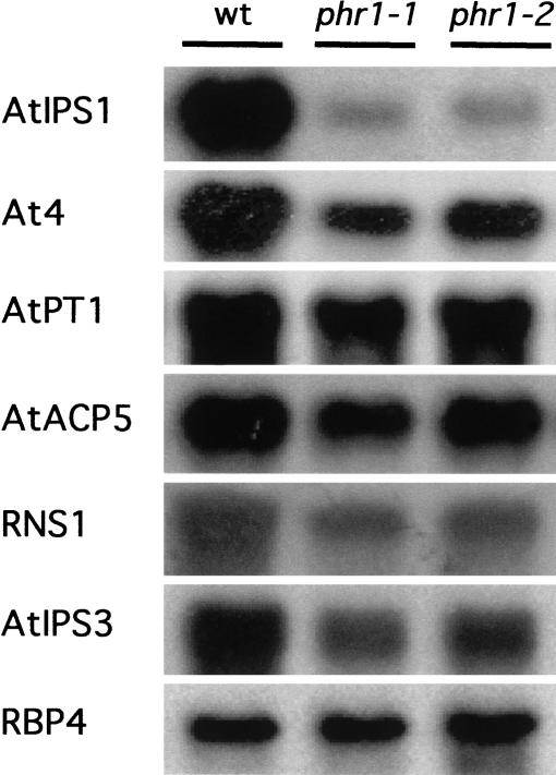
Plants have evolved a number of adaptive responses to cope with growth in conditions of limited phosphate (Pi) supply involving biochemical, metabolic, and developmental changes. We prepared an EMS-mutagenized M(2) population of an Arabidopsis thaliana transgenic line harboring a reporter gene specifically responsive to Pi starvation (AtIPS1::GUS), and screened for mutants altered in Pi starvation regulation. One of the mutants, phr1 (phosphate starvation response 1), displayed reduced response of AtIPS1::GUS to Pi starvation, and also had a broad range of Pi starvation responses impaired, including the responsiveness of various other Pi starvation-induced genes and metabolic responses, such as the increase in anthocyanin accumulation. PHR1 was positionally cloned and shown be related to the PHOSPHORUS STARVATION RESPONSE 1 (PSR1) gene from Chlamydomonas reinhardtii. A GFP::PHR1 protein fusion was localized in the nucleus independently of Pi status, as is the case for PSR1. PHR1 is expressed in Pi sufficient conditions and, in contrast to PSR1, is only weakly responsive to Pi starvation. PHR1, PSR1, and other members of the protein family share a MYB domain and a predicted coiled-coil (CC) domain, defining a subtype within the MYB superfamily, the MYB-CC family. Therefore, PHR1 was found to bind as a dimer to an imperfect palindromic sequence. PHR1-binding sequences are present in the promoter of Pi starvation-responsive structural genes, indicating that this protein acts downstream in the Pi starvation signaling pathway.


Figures

Figure 1
Figure 1
Characterization of the phr1 mutant alleles. (A) Histochemical analysis of GUS activity driven by the AtIPS1∷GUS reporter gene in response to phosphate starvation in wild type (wt; left) and in the phr1-1 mutant (right). Scale bar, 1 cm. (B) Histograms of metabolic (anthocyanin and Pi content) and developmental (root/shoot growth ratio and total weight) parameters of the wild-type (wt) and phr1-1 (1-1) and phr1-2 (1-2) mutant alleles, grown under different nutrient regimes; complete medium (+P), Pi starvation (−P), or nitrogen starvation (−N) regimes. (C) Plates containing the wild-type (bottom) and the phr1-1 (left) and phr1-2 (right) mutant alleles grown on different nutrient regimes. Scale bars, 1 cm. (D) Detail showing root hairs of wild type and phr1-1 grown under Pi starvation conditions. Scale bar, 0.5 mm. The analyses were conducted on plants grown in complete medium for 5 d, then transferred to complete medium or to medium lacking Pi or N for 7 d, except in the cases of the Pi and N starvation shown in C, in which the starvation lasted for 12 d. Data represent means of at least six independent measurements. Standard deviations are indicated by bars. Statistically significant differences using the Student's t test between wild-type and phr1 alleles were observed for anthocyanin accumulation, root-to-shoot growth ratio, and total weight for plants grown under Pi starvation conditions (P < 0.01), as well as for total Pi content for plants grown under Pi sufficient conditions (P < 0.02).
Figure 2
Figure 2
Northern analysis of the effect of phr1 mutations on the expression of Pi starvation-responsive genes. Wild-type and mutant phr1-1 and phr1-2 alleles were grown for 5 d in complete medium, transferred to medium lacking Pi, and collected at 7 d. Total RNA was isolated and RNA gel blots containing 10 μg of these samples were hybridized to the AtIPS1 probe and subsequently rehybridized to probes corresponding to the related gene as follows: At4 (Burleigh and Harrison 1999); Pi transporter AtPT1 (Muchhal et al. 1996); RNS1 gene (Bariola et al. 1994); type 5 acid phosphatase AtACP5 (del Pozo et al. 1999); AtIPS3 gene, encoding a protein of unknown function (J.C. del Pozo, J. Iglesias, V. Rubio, A. Leyva, and J. Paz-Ares, unpubl.); RBP4 gene (encoding the ribosome binding protein 4; C. Konnz, unpubl.) used as loading control.
Figure 3
Figure 3
Positional cloning and structure of the PHR1 gene. (A) PHR1 was first mapped between CAPS markers RPS2 and phra, and finally narrowed the physical localization between BACs F20O9 and F16A16. Within this region, a homolog to the PSR1 gene from C. reinhardtii (Wykoff et al. 1999) was identified (At4g28610). Sequencing of the region corresponding to the PSR1 homolog in the two alleles, phr1-1 and phr1-2 revealed that each had a mutation in this gene. The mutation in phr1-1 was a C-to-T transition, causing the introduction of a premature stop codon. The mutation in phr1-2 was also a G-to-A substitution, which impaired a GT splicing donor site. Nucleotides in the intron are shown in italics. The exon structure derived from comparison of the genomic and cDNA sequences is highlighted with boxes (empty, noncoding exons, or parts; full, coding exons, or parts). (B) Complementation of the phr1-1 mutant with plasmid pBIB∷PHR1, harboring the PHR1-coding region plus 2 kb upstream and 1.3 kb downstream sequences. T2 progeny of a transgenic plant harboring a copy of the pBIB∷PHR1 T-DNA (middle), displays a 3:1 segregation of the colored phenotype when germinated directly in Pi starvation medium. Control progeny from wild-type and phr1-1 homozygous plants are shown at left and right, respectively. Scale bar, 0.5 cm. (C) Nucleotide and deduced amino acid sequence from the PHR1 cDNA. The two regions conserved between PHR1 and the C. reinhardtii PSR1 protein, corresponding to the MYB domain and to a predicted coiled–coil domain, are highlighted in reverse contrast and gray, respectively. A putative nuclear localization signal is shown (underlined).
Figure 4
Figure 4
Sequence comparison among PHR1 and related proteins in databanks. (A) Alignment of the MYB (top) and predicted coiled–coil (bottom) conserved domains constructed by use of the CLUSTAL program (Higgins et al. 1996). The protein accession number given in the SPTREMBL or EMBL databanks is preceded by a species identifier as follows: A. thaliana (At), Nicotiana tabacum (Nt), Mesembryanthemum crystallinum (Mc), and C. reinhardtii (Cr). Arrowhead indicates the position of an insertion of 8 and 16 amino acid residues in the Q9SVP8 and Q9LRN5 sequences, respectively. Alignment was colored according to the average BLOSUM62 score (0.5–1.49, light gray; 1.5–2.9, gray; ≥3.0, black). (B) Phylogram of proteins described in A sharing the MYB and predicted coiled–coil conserved domains, constructed by use of the CLUSTAL (Higgins et al. 1996) program and the neighbor-joining method (Saitou and Nei 1987). The bootstrap (Felsenstein 1992) value of each node is indicated (of 1000 samples). Scale bar, 0.05 substitutions/site. To construct the alignment and the tree, only the two conserved regions were considered.
Figure 4
Figure 4
Sequence comparison among PHR1 and related proteins in databanks. (A) Alignment of the MYB (top) and predicted coiled–coil (bottom) conserved domains constructed by use of the CLUSTAL program (Higgins et al. 1996). The protein accession number given in the SPTREMBL or EMBL databanks is preceded by a species identifier as follows: A. thaliana (At), Nicotiana tabacum (Nt), Mesembryanthemum crystallinum (Mc), and C. reinhardtii (Cr). Arrowhead indicates the position of an insertion of 8 and 16 amino acid residues in the Q9SVP8 and Q9LRN5 sequences, respectively. Alignment was colored according to the average BLOSUM62 score (0.5–1.49, light gray; 1.5–2.9, gray; ≥3.0, black). (B) Phylogram of proteins described in A sharing the MYB and predicted coiled–coil conserved domains, constructed by use of the CLUSTAL (Higgins et al. 1996) program and the neighbor-joining method (Saitou and Nei 1987). The bootstrap (Felsenstein 1992) value of each node is indicated (of 1000 samples). Scale bar, 0.05 substitutions/site. To construct the alignment and the tree, only the two conserved regions were considered.
Figure 5
Figure 5
Sequence-specific DNA-binding properties of PHR1. (A) EMSA of in vitro translated PHR1 protein binding to overlapping DNA fragments from the 5′ region of AtIPS1. Mock-translated reticulocyte lysate was used in the indicated lanes. The DNA fragments used in the experiment are represented diagrammatically at top, and span 707 bp upstream of the first ATG of AtIPS1 (a, from −726 to −568; b, from −612 to −440; c, from −484 to −343; d, from −384 to −245; e, from −285 to −130; f, from −152 to −19). Arrows show PHR1-bound or free DNA (B or F, respectively). Other bands detected in the autoradiograph are shared for each fragment between the lanes corresponding to the mock-translated and to the in vitro-translated PHR1 protein, and thus represent interactions between the fragment and proteins of the reticulocyte lysate. (B) EMSA of amino-terminal and carboxy-terminal deletion derivatives binding to the overlapping region between fragments a and b (from −612 to −568). A diagrammatic representation of the PHR1 protein is shown at top. The black and gray boxes represent the MYB and the predicted coiled–coil domains, respectively. Arrows show the amino terminus and the carboxyl terminus of the amino- and carboxy-terminally truncated derivatives, respectively. (C) Scan mutagenesis of the 10 bp sequence containing the PHR1-binding site (P1BS). Wild-type P1BS is shown with the imperfect palindromic repeats indicated by arrows. Base changes in the mutants tested are indicated in the lines below. Broken lines indicate positions with the same base as in the wild-type P1BS. (D) EMSA of PHR1 to the P1BS-related sequence present in the AtIPS3 promoter (see Table 1). (E) PHR1 homodimerization. EMSA was conducted with the full-size PHR1 protein and with the Δ207Nt deletion derivative binding to the sequence used in B. Proteins were translated in vitro, alone, or in combination.
Figure 6
Figure 6
Northern analysis of PHR1 gene expression. A. thaliana plants were grown in complete medium for 5 d, then transferred to medium containing Pi (+P) for 7 d or lacking Pi (−P) for 2 or 7 d. Poly A+-enriched RNA was isolated from these samples and RNA gel blots containing 0.5 μg of these samples were hybridized to the PHR1 probe and subsequently rehybridized to a probe corresponding to the RBP4 gene used as loading control.
Figure 7
Figure 7
Subcellular localization of a GFP∷PHR1 fusion protein in wild-type plants grown under Pi sufficient and Pi starvation conditions. Microscopic images of root cells from transgenic A. thaliana Col-0 plants harboring a control gene 35S∷GFP (top) or a 35S∷GFP∷PHR1 fusion gene (bottom). Plants were grown in complete medium for 5 d, then transferred to medium containing Pi (+P, left) or lacking Pi (−P, right ) for 7 d before analysis. Scale bar, 20 μm.

References

    1. Altschul SF, Gish W, Miller W, Myers EW, Lipman DJ. Basic local alignment search tool. J Mol Biol. 1990;215:403–410. - PubMed
    1. Ames BN. Assay of inorganic phosphate, total phosphate and phosphatases. Methods Enzymol. 1966;8:115–118.
    1. Bajwa W, Meyhack B, Rudolph H, Schweingruber AM, Hinnen A. Structural analysis of the two tandemly repeated acid phosphatase genes in yeast. Nucleic Acids Res. 1984;12:7721–7739. - PMC - PubMed
    1. Bariola PA, Howard CJ, Taylor CB, Verburg MT, Jaglan VD, Green PJ. The Arabidopsis ribonuclease gene RSN1 is tightly controlled in response to phosphate limitation. Plant J. 1994;6:673–685. - PubMed
    1. Bates TR, Lynch JP. Stimulation of root hair elongation in Arabidopsis thaliana by low phosphorus availability. Plant Cell Environ. 1996;19:529–538.

Publication types

MeSH terms

Associated data