. 2001 Aug 15;15(16):2134-45.
doi: 10.1101/gad.894801.

The loop domain of heat shock transcription factor 1 dictates DNA-binding specificity and responses to heat stress

Affiliations

The loop domain of heat shock transcription factor 1 dictates DNA-binding specificity and responses to heat stress

S G Ahn et al. Genes Dev. .

Abstract

Eukaryotic heat shock transcription factors (HSF) regulate an evolutionarily conserved stress-response pathway essential for survival against a variety of environmental and developmental stresses. Although the highly similar HSF family members have distinct roles in responding to stress and activating target gene expression, the mechanisms that govern these roles are unknown. Here we identify a loop within the HSF1 DNA-binding domain that dictates HSF isoform specific DNA binding in vitro and preferential target gene activation by HSF family members in both a yeast transcription assay and in mammalian cells. These characteristics of the HSF1 loop region are transposable to HSF2 and sufficient to confer DNA-binding specificity, heat shock inducible HSP gene expression and protection from heat-induced apoptosis in vivo. In addition, the loop suppresses formation of the HSF1 trimer under basal conditions and is required for heat-inducible trimerization in a purified system in vitro, suggesting that this domain is a critical part of the HSF1 heat-stress-sensing mechanism. We propose that this domain defines a signature for HSF1 that constitutes an important determinant for how cells utilize a family of transcription factors to respond to distinct stresses.


Figures
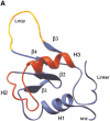
Figure 1
Figure 1
Structure of the heat shock transcription factor (HSF) DNA-binding domain. (A) The central helix-turn-helix motif (red) is composed of α-helix2 and α-helix3, where the latter is the DNA recognition helix. The structure was modeled using the Ribbons program from the coordinates of NMR structure of the Drosophila DBD (Vuister et al. 1994a). The loop (yellow) was determined to be solvent exposed. (B) Alignment of the amino acid sequences for the mouse HSF1, HSF2, Drosophila HSF1, and Kluyveromyces lactis HSF DBDs. Predicted secondary structure for the mouse HSFs is based on homology to the solved K. lactis and Drosophila HSF DBD structures (Harrison et al. 1994; Vuister et al. 1994a), where cylinders represent α-helicies and arrows represent β-sheets. Residues conserved between HSF1 and HSF2 are boxed; residues identical among all four sequences are indicated by a solid oval; those positions that have conserved substitutions are indicated by an open oval. (C) Diagram of specific chimeras used in this study. HSF1 sequences (white); HSF2 sequences (black). (L) Linker; (HR A) trimerization domain.
Figure 1
Figure 1
Structure of the heat shock transcription factor (HSF) DNA-binding domain. (A) The central helix-turn-helix motif (red) is composed of α-helix2 and α-helix3, where the latter is the DNA recognition helix. The structure was modeled using the Ribbons program from the coordinates of NMR structure of the Drosophila DBD (Vuister et al. 1994a). The loop (yellow) was determined to be solvent exposed. (B) Alignment of the amino acid sequences for the mouse HSF1, HSF2, Drosophila HSF1, and Kluyveromyces lactis HSF DBDs. Predicted secondary structure for the mouse HSFs is based on homology to the solved K. lactis and Drosophila HSF DBD structures (Harrison et al. 1994; Vuister et al. 1994a), where cylinders represent α-helicies and arrows represent β-sheets. Residues conserved between HSF1 and HSF2 are boxed; residues identical among all four sequences are indicated by a solid oval; those positions that have conserved substitutions are indicated by an open oval. (C) Diagram of specific chimeras used in this study. HSF1 sequences (white); HSF2 sequences (black). (L) Linker; (HR A) trimerization domain.
Figure 2
Figure 2
Differential binding to the hsp70 heat shock element (HSE) maps to the heat shock transcription factor (HSF) DNA-binding domain. (A) DNase I footprinting by mHSF1 and mHSF2 to the hsp70 HSE. The hsp70 HSE probe was labeled at the 5′ end of the coding strand, and the concentration of the probe in all reaction mixtures was 0.1 nM. The amounts of bacterial lysates in lanes A–E were 0.5, 1, 2, 4, and 8 μL, respectively. Control reaction shows DNase I footprinting reaction in the absence of protein. The extent of HSF1 and HSF2 protection are indicated at the left with brackets. The positions of HSEs 1 to 5 are marked by arrows. (B) Specificity in DNA-binding maps to the DNA-binding domain. Chimeras in which the DBD was interchanged were expressed and purified as in Materials and Methods and used for DNase I footprinting. Lane assignments are as described for panel A. The extent of protein binding is delimited by the brackets.
Figure 3
Figure 3
Extended occupancy of the hsp70 heat shock element (HSE) requires the HSF1 loop. (A) DNase I footprinting by chimeric heat shock transcription factor (HSF) proteins to the hsp70 HSE. HSF2Helix1 (mHSF2 in which aa 18–43 were substituted with aa 26– 51 from mHSF1) and HSF2Loop1 (mHSF2 aa 73–91 were substituted with aa 81–99 from mHSF1) were purified and used for DNase I footprint experiments, as described in Figure 2. The positions of the HSEs are shown by arrows. (B) Sequence of the hsp70 HSE with the five inverted HSE repeats are indicated with arrows. The boundaries of protection by HSF1 and HSF2 and the chimeras are indicated by the brackets.
Figure 4
Figure 4
The HSF1 loop dictates preferential activation from an extended (heat shock element) HSE in yeast. (A) Cartoon illustration of the arrangement of HSE repeats in the SSA3 and CUP1 gene promoters. (B) RNA levels for lacZ or ACT1 were measured by RNase protection assays using RNA isolated from control (C) or heat-shocked (HS; 40°C for 15 min) yeast expressing the indicated mouse heat shock transcription factor (HSF). The abbreviations for the HSF chimeras are identical to those described in the Figure 1 legend.
Figure 5
Figure 5
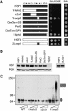
The HSF1 loop contributes to suppression of HSF1 activity in yeast. (A) Complementation assay for the ability of mouse HSF1, HSF2, and chimeras to substitute for yeast (heat-shock transcription factor) HSF. Cells harboring a chromosomal disruption of yeast hsf1 and bearing a plasmid with (heat stress transcription factor) HSF under the control of the GAL1 promoter were transformed with another plasmid expressing the indicated mouse HSF from a constitutive promoter. Transformants were diluted serially (from left to right, OD650, 0.1, 0.01, 0.001) and spotted onto glucose- or galactose-containing medium, incubated for 3 d at 30°C and photographed. (B) Western analysis for expression of mouse HSF isoforms in yeast using antibodies specific to HSF1 or HSF2. Levels of PGK are shown for normalization of loading. The 1Sph2 isoform is not detected with the HSF1 antibody because the primary epitopes recognized by this antibody map to the carboxy-terminal regions of the protein, which have been replaced by HSF2 in the chimera. (C) Crosslinking of HSF reveals that the HSF1 loop suppresses trimerization. Whole-cell extracts were prepared from yeast cells expressing the indicated forms of mouse HSF and subjected to in vitro cross-linking with 0, 0.5, or 2 mM EGS (indicated by wedge). HSF proteins were detected by immunoblotting with HSF1-specific antibody. The monomeric and trimeric HSF species are depicted with a single or triple oval, respectively.
Figure 6
Figure 6
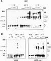
The HSF1 loop confers heat shock inducible multimerization in vitro. (A) Temperature-dependent trimerization of HSF1. Purified HSF1 (4 μM) was incubated at 15°C, 32°C, or 42°C for 30 min and then subjected to EGS cross-linking (EGS concentration indicated by the wedge 0, 0.1, 0.2, 0.5, 1.0, 2.5 mM from left to right) and detection by Western blotting using a HSF1-specific antibody. The predicted monomer, dimer, trimer, and putative hexamers are indicated by the ovals. (B) The HSF1 loop confers heat inducible trimerization to HSF2. Purified HSF2 and HSF2loop1 proteins, incubated at 15°C, 32°C, or 42°C, were subjected to EGS cross-linking (0, 0.5, 2.5 mM left to right) and analyzed as in A.
Figure 7
Figure 7
The HSF1 loop confers heat-inducible activation of Hsp gene expression in hsf1−/− mouse embryonic fibroblast (MEF) cells. Immunoblot analyses for levels of HSP70, HSP27, HSC70, and actin were performed on extracts prepared from control (C) or heat-shocked (HS; 42°C for 1 h, then at 37°C for 3 h ) hsf1−/− MEF cells transfected with pcDNA3.1 (V) or vector containing the indicated mouse HSF isoform. Extracts from isogenic wild-type (WT) MEFs are shown for comparison.
Figure 8
Figure 8
Expression of HSF2Loop1 in hsf1−/− cells prevents heat-induced apoptosis. hsf1−/− mouse embryonic fibroblasts (MEFs) were transfected with pcDNA3 alone (Vector) or harboring the indicated HSF sequence. Cells were exposed for 1 h to 44°C or kept at 37°C (control) and then cultured for another 24 h at 37°C prior to detection of apoptosis by flow cytometry of annexin V-FITC and propidium iodide (PI)-stained cells. The percentage of cells for each population of viable, apoptotic or apoptotic and necrotic, falling within each quadrant is indicated. Viable cells sort to the lower left quadrant.

Similar articles

Cited by

References

    1. Abravaya K, Myers MP, Murphy SP, Morimoto RI. The human heat shock protein hsp70 interacts with HSF, the transcription factor that regulates heat shock gene expression. Genes & Dev. 1992;6:1153–1164. - PubMed
    1. Ali A, Bharadwaj S, O'Carroll R, Ovsenek N. HSP90 interacts with and regulates the activity of heat shock factor 1 in Xenopus oocytes. Mol Cell Biol. 1998;18:4949–4960. - PMC - PubMed
    1. Bharadwaj S, Ali A, Ovsenek N. Multiple components of the HSP90 chaperone complex function in regulation of heat shock factor 1 in vivo. Mol Cell Biol. 1999;19:8033–8041. - PMC - PubMed
    1. Bonner JJ, Ballou C, Fackenthal DL. Interactions between DNA-bound trimers of the yeast heat shock factor. Mol Cell Biol. 1994;14:501–508. - PMC - PubMed
    1. Christians E, Davis AA, Thomas SD, Benjamin IJ. Maternal effect of Hsf1 on reproductive success. Nature. 2000;407:693–694. - PubMed

Publication types