Comparative Study
. 2001 Aug 15;15(16):2161-72.
doi: 10.1101/gad.200601.

Functional comparison of the nematode Hox gene lin-39 in C. elegans and P. pacificus reveals evolutionary conservation of protein function despite divergence of primary sequences

Affiliations
Comparative Study

Functional comparison of the nematode Hox gene lin-39 in C. elegans and P. pacificus reveals evolutionary conservation of protein function despite divergence of primary sequences

K Grandien et al. Genes Dev. .

Abstract

Hox transcription factors have been implicated in playing a central role in the evolution of animal morphology. Many studies indicate the evolutionary importance of regulatory changes in Hox genes, but little is known about the role of functional changes in Hox proteins. In the nematodes Pristionchus pacificus and Caenorhabditis elegans, developmental processes can be compared at the cellular, genetic, and molecular levels and differences in gene function can be identified. The Hox gene lin-39 is involved in the regulation of nematode vulva development. Comparison of known lin-39 mutations in P. pacificus and C. elegans revealed both conservation and changes of gene function. Here, we study evolutionary changes of lin-39 function using hybrid transgenes and site-directed mutagenesis in an in vivo assay using C. elegans lin-39 mutants. Our data show that despite the functional differences of LIN-39 between the two species, Ppa-LIN-39, when driven by Cel-lin-39 regulatory elements, can functionally replace Cel-lin-39. Furthermore, we show that the MAPK docking and phosphorylation motifs unique for Cel-LIN-39 are dispensable for Cel-lin-39 function. Therefore, the evolution of lin-39 function is driven by changes in regulatory elements rather than changes in the protein itself.


Figures

Figure 1
Figure 1
Summary of ventral epidermal cell fate specification in C. elegans (Cel) and P. pacificus (Ppa) and sequence comparison of Cel–LIN-39 and Ppa–LIN-39. (A) Schematic representation of the 12 ventral epidermal Pn.p cells in a C. elegans L1 stage larva. (B) In C. elegans (left panel), lin-39 activity defines the P(3–8).p cells as the vulval equivalence group. P6.p (black oval) has the 1° fate and forms the inner part of the vulva, whereas P(5,7).p (hatched ovals) have the 2° fate and form the outer part of the vulva. P(3,4,8).p (diagonally striped ovals) remain epidermal and have the 3° fate. P(5–7).p are induced by the anchor cell (AC) to undergo vulval differentiation. The inductive signal from the AC is encoded by an EGF-like molecule and is transmitted through an EGFR/RAS/MAPK signaling pathway, which modulates the activity of several transcription factors, including LIN-39. In the absence of lin-39 activity (Cel–lin-39), the P(3–8).p cells, like the other Pn.p cells, fuse with the surrounding hypodermis (white ovals). If lin-39 activity is provided during the early step but not during AC induction (Cel hs::lin39 early), the P(3–8).p cells take on the 3° fate. In P. pacificus (right panel), lin-39 defines P(5–8).p as the vulval equivalence group. P(5–7).p form the vulva with a 2°-1°-2° fate pattern. P8.p has a special fate designated as 4° (filled striped oval). Cells not receiving lin-39 activity (Ppa–lin-39) undergo programmed cell death (X). If programmed cell death is inhibited, a normal vulva is formed, indicating that lin-39 is dispensable for vulval formation (Ppa–lin-39; ced-3). It is not known if the EGF/RAS/MAPK pathway is involved in vulval induction by the gonad in P. pacificus. (C) Sequence alignment of Cel–LIN-39 and Ppa–LIN-39. The highly conserved homeodomain and hexapeptide motifs are overlined by a solid and a hatched bar, respectively. The putative phosphorylation site in Cel–LIN-39 is indicated by S/TP, and the motif similar to a MAPK docking site (Jacobs et al. 1999) is indicated by FXFP. Note that these motifs are absent in Ppa–LIN-39. (D) Schematic representation of the LIN-39 primary structure. (Hex) Hexapeptide motif, (HD) homeodomain. Solid and open arrowheads above the bar indicate the positions of all published nonsense and missense mutations, respectively, found in C. elegans lin-39 alleles (Clark et al. 1993; Wang et al. 1993). Arrows indicate nonsense mutations introduced in C. elegans rescue constructs. Solid arrowheads below the bar indicate all nonsense mutations found in P. pacificus lin-39 alleles (Eizinger and Sommer 1997).
Figure 2
Figure 2
Characterization of regulatory regions in the C. elegans lin-39 gene by the use of LacZ reporter constructs. (A) Schematic description of the LacZ reporter constructs analyzed and the obtained expression patterns. Rectangles indicate lin-39 exons; (red) homeodomain-encoding exons; (filled and hatched) other coding exons; (open) 5′ and 3′ untranslated exons. The green bar indicates the unc-54 3′ UTR in pKG5. The LacZ reporter gene is indicated by a blue, hatched rectangle. Bent arrows represent two putative transcription start sites. Note that pKG7 is based on the P. pacificus lin-39 gene. For each construct at least three transgenic lines were investigated. (++) Apparently normal expression; (+) weaker than normal expression; (e) ectopic expression; (−) no expression. (B) Representative examples of expression patterns obtained for the constructs pKG1, pKG2, pKG5, and KG6. See text for detailed description.
Figure 3
Figure 3
Summary of egg-laying rescue experiments. All transgenic constructs were analyzed in the strain MT4498 carrying the null allele Cel–lin-39(n1880), which is completely egg-laying defective. At least three transgenic lines were tested for each construct. (Filled rectangles) Coding exons; (open rectangles) 5′ and 3′ noncoding exons; (bent arrows) transcription start sites; (S) the unique restriction site in pKG11–pKG14; (diagonal bars) introduced frame-shift mutations. Inserted cDNAs in pKG12–pKG14 are indicated.
Figure 4
Figure 4
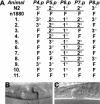
Vulval cell fate in Cel–lin-39(n1880) animals rescued by Ppa–lin-39 expression. (A) Vulval cell fate patterns obtained by Nomarski microscopy for 11 worms transgenic for the construct pKG13. 1°, 2°, 3°, and F fates are defined in Figure 1. Underlined cells form one continuous vulval invagination. Vulval cell fates for wild-type (N2) and lin-39(n1880) animals are shown as reference. (B) Example of completely rescued vulval cell fates in the L4 larval stage. (C) Example of partially rescued vulval cell fates with one 1° and one 2° cell in the L4 larval stage.
Figure 5
Figure 5
Rescue of the fate of the VC neurons in Cel–lin-39(n1880) by Ppa–lin-39 expression. Schematic representation of the P cell lineage with n denoting cell number. In wild-type C. elegans, the P(3–8).aap cells form VC neurons, whereas P(1,2,9–12).aap cells undergo programmed cell death (X). (B) Schematic representation of the positions of the six VC neurons. Note especially the positions of VC4 and VC5 in close proximity to the vulva (data not shown). (CF) Expression of unc-4::GFP in VC4 and VC5 neurons in lin-39(n1880) animals rescued by P. pacificus lin-39 expression. No VC neurons are formed in lin-39(n1880) mutant animals. (C,E) GFP expression. Open arrowheads indicate GFP expressing cells. (D,F) The same pictures as in C and E, respectively, overlaid with the corresponding Nomarski pictures. Filled arrowheads indicate the vulva.
Figure 6
Figure 6
In vitro MAPK phosphorylation. Wild-type or phosphorylation-defective (T230V) Cel–LIN-39 as well as human Elk-1 were generated by in vitro translation in the presence of 35S-methionine (lanes 1,4,5,8,9,12) or unlabeled methionine (lanes 2,3,6,7,10,11) and were subsequently used for in vitro phosphorylation by p42 MAPK. Unlabeled proteins were phosphorylated with [γ-32P]ATP, leading to MAPK-dependent and MAPK-independent labeling of many proteins in the in vitro translation reaction (lanes 2,3,6,7,10,11). Proteins labeled with 35S were phosphorylated with nonradioactive ATP. Phosphorylation of hElk-1 was readily observed; note the difference in migration (lanes 9,12) for 35S-labeled protein and the increase in [γ-32P]ATP-dependent phosphorylation (lanes 10,11). In contrast, no sign of phosphorylation of Cel–LIN-39 could be observed (cf. lanes 1 and 4, 2 and 3). Note that a protein of slightly faster mobility than Cel–LIN-39 shows a partially MAPK-dependent phosphorylation (cf. lanes 2 and 3, 6 and 7, 11 and 12). An arrow indicates the size of Cel–LIN-39, a filled arrowhead phosphorylated hElk-1, and an open arrowhead unphosphorylated hElk-1, respectively.

Similar articles

Cited by

References

    1. Anderson P, Kimble J. mRNA and translation. In: Riddle DL, et al., editors. C. elegans II. Cold Spring Harbor, NY: Cold Spring Harbor Laboratory Press; 1997. pp. 185–208. - PubMed
    1. Beitel GJ, Tuck S, Greenwald I, Horvitz HR. The Caenorhabditis elegans gene lin-1 encodes an ETS-domain protein and defines a branch of the vulval induction pathway. Genes & Dev. 1995;9:3149–3162. - PubMed
    1. Brenner S. The genetics of Caenorhabditis elegans. Genetics. 1974;77:71–94. - PMC - PubMed
    1. The C. elegans Sequencing Consortium. Genome sequence of the nematode C. elegans: A platform for investigating biology. Science. 1998;282:2012–2018. - PubMed
    1. Clandinin TR, Katz WS, Sternberg PW. Caenorhabditis elegans hom-C genes regulate the response of vulval precursor cells to inductive signal. Dev Biol. 1997;182:150–161. - PubMed

Publication types

MeSH terms

Substances