. 2001 Sep;183(18):5334-42.
doi: 10.1128/JB.183.18.5334-5342.2001.

Mutational analysis of the Rhizobium lupini H13-3 and Sinorhizobium meliloti flagellin genes: importance of flagellin A for flagellar filament structure and transcriptional regulation

Affiliations

Mutational analysis of the Rhizobium lupini H13-3 and Sinorhizobium meliloti flagellin genes: importance of flagellin A for flagellar filament structure and transcriptional regulation

B Scharf et al. J Bacteriol. 2001 Sep.

Abstract

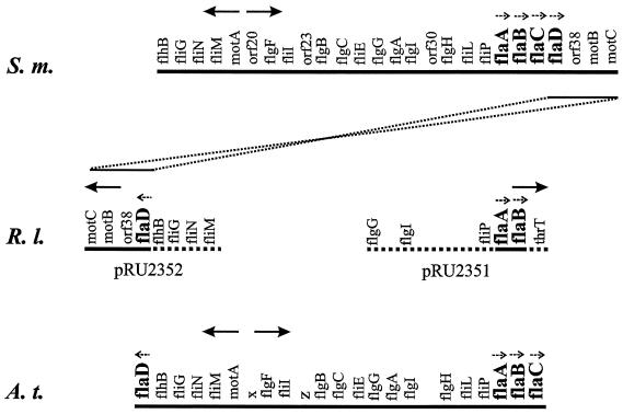
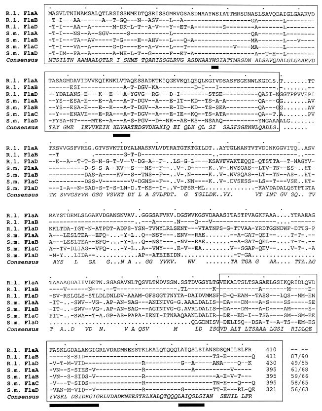
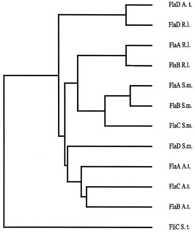
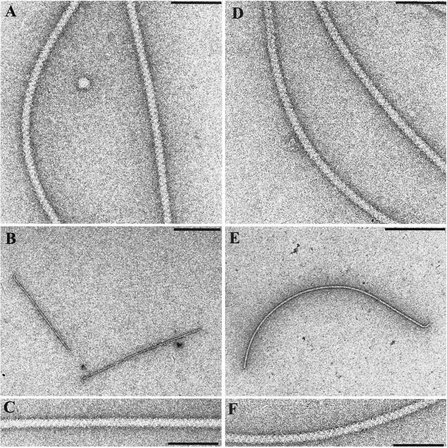
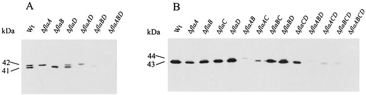
Complex flagellar filaments are unusual in their fine structure composed of flagellin dimers, in their right-handed helicity, and in their rigidity, which prevents a switch of handedness. The complex filaments of Rhizobium lupini H13-3 and those of Sinorhizobium meliloti are composed of three and four flagellin (Fla) subunits, respectively. The Fla-encoding genes, named flaA through flaD, are separately transcribed from sigma(28)-specific promoters. Mutational analysis of the fla genes revealed that, in both species, FlaA is the principal flagellin and that FlaB, FlaC, and FlaD are secondary. FlaA and at least one secondary Fla protein are required for assembling a functional flagellar filament. Western analysis revealed a ratio close to 1 of FlaA to the secondary Fla proteins (= FlaX) present in wild-type extracts, suggesting that the complex filament is assembled from FlaA-FlaX heterodimers. Whenever a given mutant combination of Fla prevented the assemblage of an intact filament, the biosynthesis of flagellin decreased dramatically. As shown in S. meliloti by reporter gene analysis, it is the transcription of flaA, but not of flaB, flaC, or flaD, that was down-regulated by such abortive combinations of Fla proteins. This autoregulation of flaA is unusual. We propose that any combination of Fla subunits incapable of assembling an intact filament jams the flagellar export channel and thus prevents the escape of an (as yet unidentified) anti-sigma(28) factor that antagonizes the sigma(28)-dependent transcription of flaA.


Figures

FIG. 1
FIG. 1
Aligned partial maps of the S. meliloti (S.m.) (36), R. lupini (R.l.) (this work) and A. tumefaciens (A.t.) (5) flagellar gene clusters. Solid lines signify fully sequenced genomic regions, and dashed lines signify partially sequenced genomic regions. Gene loci are not drawn to scale; relevant transcription units and their polarities are marked by small dashed arrows, and general transcription polarity is marked by large arrows. The nomenclature of the S. meliloti flaC and flaD genes (36) has been changed in accordance with their map order, and the R. lupini flaD gene has been named by analogy to its A. tumefaciens paralogue (5). An R. lupini flaC paralogue was not detected. R. lupini (and A. tumefaciens) genes translocated and inverted with respect to their positions on the S. meliloti map are connected by dotted lines.
FIG. 2
FIG. 2
Comparison of the three R. lupini (R.l.) and four S. meliloti (S.m.) deduced flagellin polypeptide sequences. Numbering refers to amino acid residues in each line. Dashes signify identical residues with respect to the R. lupini FlaA sequence, and dots signify gaps. The consensus sequence includes all residues that form a homology group with a weighted relative frequency of 0.5 or greater. Conserved N- and C-terminal domains are boxed, and black bars denote amino acid residuces unique to right-handed helical flagellar filaments (41). Identity/similarity values (percentages) relative to R. lupini FlaA are listed at the end of each sequence.
FIG. 3
FIG. 3
Dendrogram based on full-length flagellin sequences from A. tumefaciens (Fla A. t.), R. lupini (Fla R. l.), and S. meliloti (Fla S. m.) and constructed by using the progressive, pairwise alignment method of PileUp from the Genetics Computer Group package (6). The Salmonella enterica serovar Typhimurium flagellin (FliC S. t.) sequence was used as an outgroup marker.
FIG. 4
FIG. 4
Electron micrographs of R. lupini and S. meliloti wild-type and mutant flagellar filaments negatively stained with uranyl acetate. Wild-type R. lupini (A) and S. meliloti (D) complex flagellar filaments are dominated by a prominent three-start helical band pattern (zigzag pattern). Mutant filaments lacking the FlaA subunit from either R. lupini (B) or S. meliloti (E) are shown. Their complex surface structures appear to be normal (C and F). Bars: 100 nm (A, C, D, and F) and 500 nm (B and E).
FIG. 5
FIG. 5
Immunoblot analysis of flagellin subunit proteins present in wild-type and mutant strains of R. lupini (A) and S. meliloti (B). Equal amounts of total cell protein (of ca. 107 cells contained in 20 μl at an OD600 of 0.3) of each strain listed were separated by denaturing gel electrophoresis, blotted on a nitrocellulose membrane, and detected with polyclonal antibody raised against purified flagella from R. lupini (A) and S. meliloti (B), respectively. Approximate band intensities are listed in Table 3.
FIG. 6
FIG. 6
DNA sequence alignment of putative promoter sequences upstream of the R. lupini (Rl) and S. meliloti (Sm) fla genes. flaA∗ denotes a secondary promoter sequence further upstream from the flaA gene (28). Capital letters indicate homology with consensus residues at a given alignment position. The R. lupini and S. meliloti (Rl/Sm) flagellin promoter consensus sequence was derived from a minimum plurality of four matches (except for position 3 of the −10 box, which was adapted to the highly active flaA promoter sequences). The consensus sequences of V. parahaemolyticus polar flagellar (VPA) (23) and E. coli ς28 (11) promoters are shown for comparison.

References

    1. Aizawa S-I. Encyclopedia of microbiology. 2nd ed. Vol. 2. New York, N.Y: Academic Press, Inc.; 2000. Flagella; pp. 380–389.
    1. Bachman B J. Linkage map of Escherichia coli K-12, 8th ed. Microbiol Rev. 1990;54:130–197. - PMC - PubMed
    1. Chesnokova O, Coutinho J B, Khan I H, Mikhail M S, Kado C I. Characterization of flagella genes of Agrobacterium tumefaciens, and the effect of a bald strain on virulence. Mol Microbiol. 1997;23:579–590. - PubMed
    1. Cohen-Krausz S, Trachtenberg S. Helical perturbations of the flagellar filament: Rhizobium lupini H13–3 at 13 Å resolution. J Struct Biol. 1998;122:267–282. - PubMed
    1. Deakin W J, Parker V E, Wright E L, Ashcroft K J, Loake G J, Shaw C H. Agrobacterium tumefaciens possesses a fourth flagellin gene located in a large gene cluster concerned with flagellar structure, assembly and motility. Microbiology. 1999;145:1397–1407. - PubMed

Publication types

Associated data