. 2001 Aug 20;194(4):427-38.
doi: 10.1084/jem.194.4.427.

Major histocompatibility complex class II-positive cortical epithelium mediates the selection of CD4(+)25(+) immunoregulatory T cells

Affiliations

Major histocompatibility complex class II-positive cortical epithelium mediates the selection of CD4(+)25(+) immunoregulatory T cells

S J Bensinger et al. J Exp Med. .

Abstract

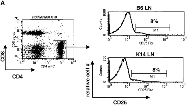
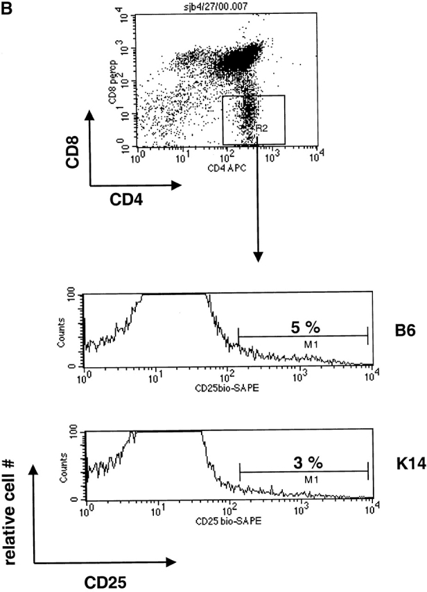
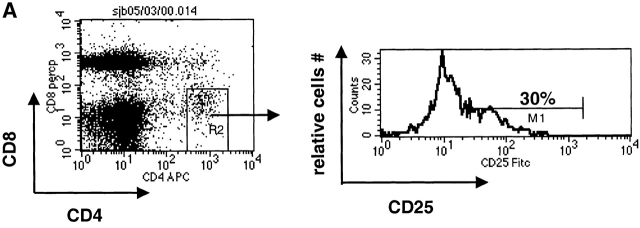
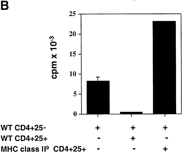
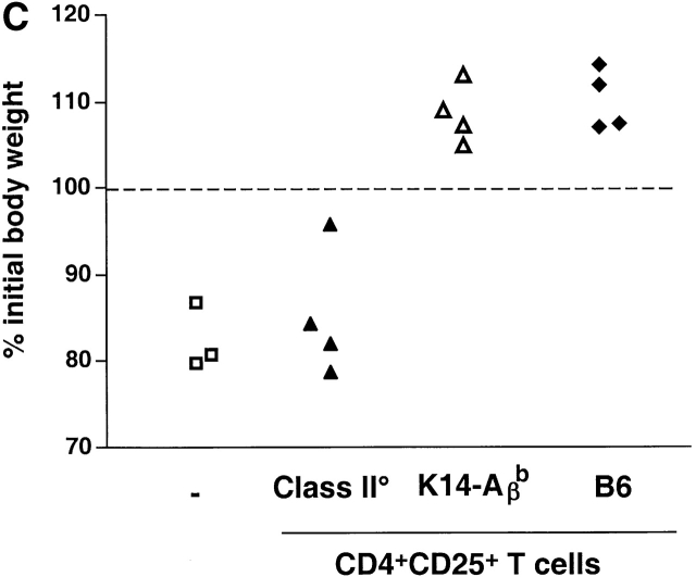
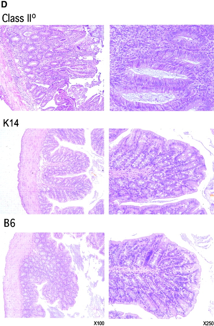
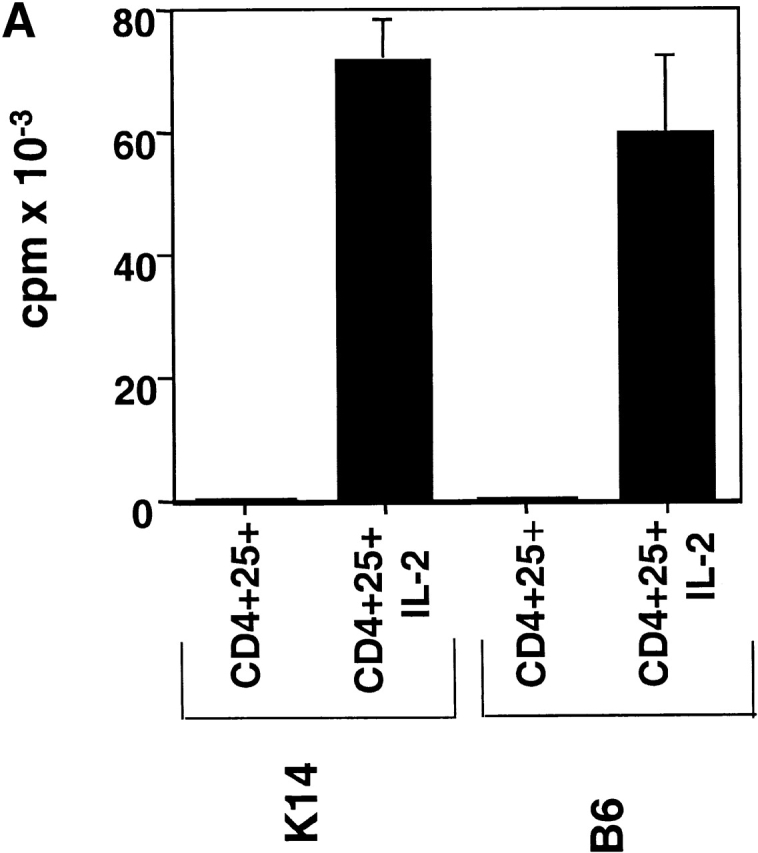
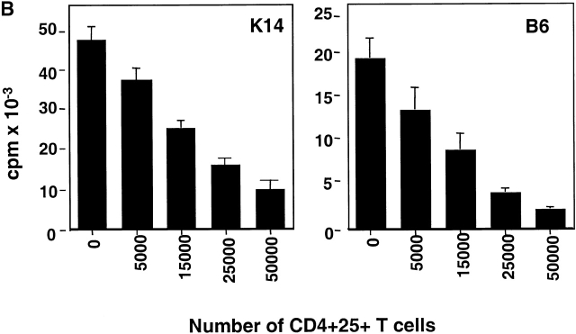
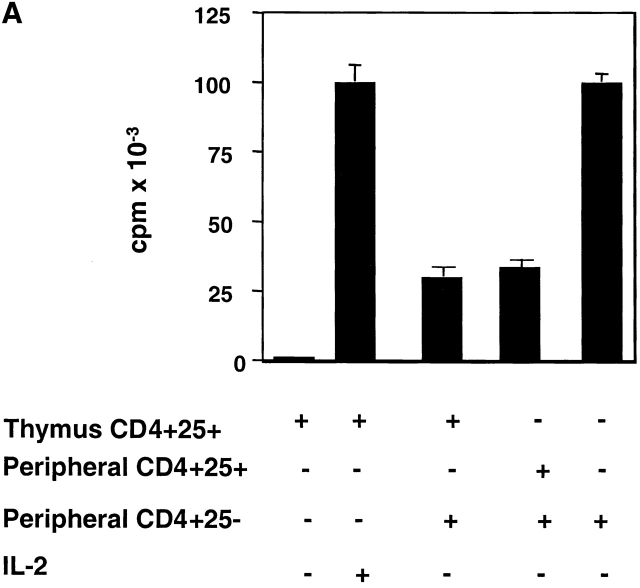
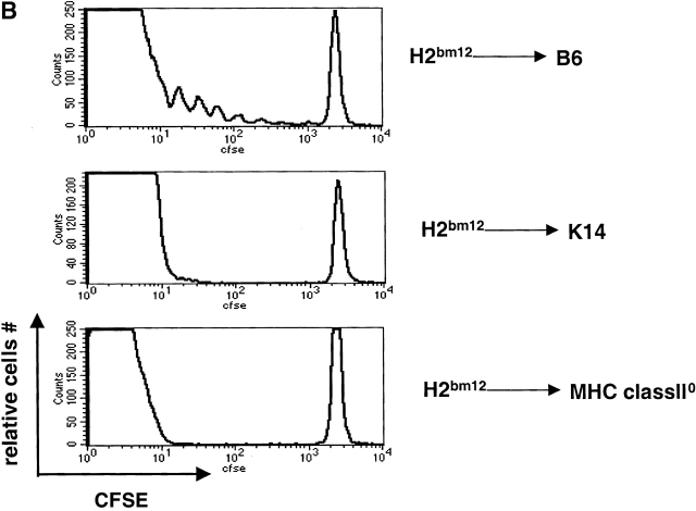
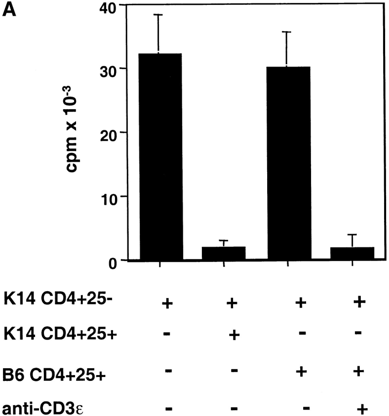
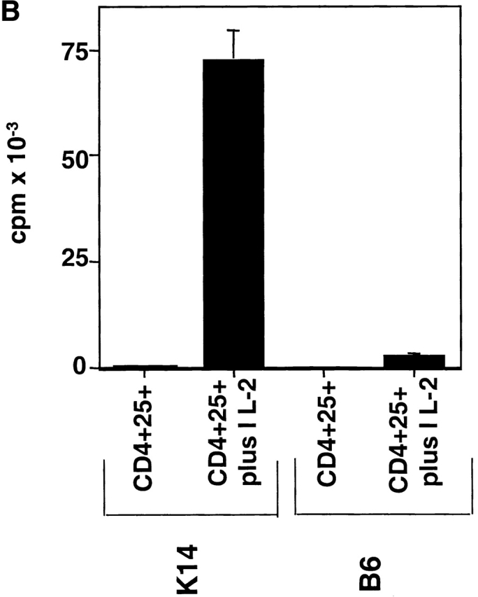
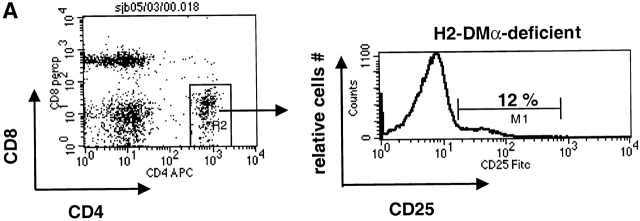
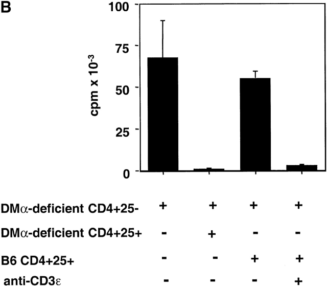
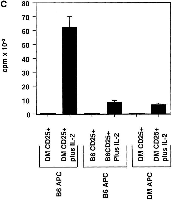
CD4(+)25(+) T cells are a unique population of immunoregulatory T cells which are critical for the prevention of autoimmunity. To address the thymic selection of these cells we have used two models of attenuated thymic deletion. In K14-A(beta)(b) mice, major histocompatibility complex (MHC) class II I-A(b) expression is limited to thymic cortical epithelium and deletion by hematopoietic antigen-presenting cells does not occur. In H2-DMalpha-deficient mice, MHC class II molecules contain a limited array of self-peptides resulting in inefficient clonal deletion. We find that CD4(+)25(+) T cells are present in the thymus and periphery of K14-A(beta)(b) and H2-DMalpha-deficient mice and, like their wild-type counterparts, suppress the proliferation of cocultured CD4(+)25(-) effector T cells. In contrast, CD4(+)25(+) T cells from MHC class II-deficient mice do not suppress responder CD4(+) T cells in vitro or in vivo. Thus, development of regulatory CD4(+)25(+) T cells is dependent on MHC class II-positive thymic cortical epithelium. Furthermore, analysis of the specificities of CD4(+)25(+) T cells in K14-A(beta)(b) and H2-DMalpha-deficient mice suggests that a subset of CD4(+)25(+) T cells is subject to negative selection on hematopoietic antigen-presenting cells.


Figures

Figure 1
Figure 1
K14-Aβ b mice have CD4+25+ T cells. (A) Total LN cells from K14-Aβ b or B6 (WT) were stained with APC-conjugated anti-CD4, Percp-conjugated anti-CD8, and FITC-conjugated anti-CD25 as in Materials and Methods. Percentages indicated in histograms are of live CD4+ lymphocytes. (B) Total thymocytes from K14-Aβ b or B6 (WT) mice were stained with APC-conjugated anti-CD4, Percp-conjugated anti-CD8, and biotin-conjugated anti-CD25 followed by PE-conjugated streptavidin. Histograms are gated through live CD4-single positive thymocytes. Percentages indicated are of live CD4-single positive thymocytes.
Figure 1
Figure 1
K14-Aβ b mice have CD4+25+ T cells. (A) Total LN cells from K14-Aβ b or B6 (WT) were stained with APC-conjugated anti-CD4, Percp-conjugated anti-CD8, and FITC-conjugated anti-CD25 as in Materials and Methods. Percentages indicated in histograms are of live CD4+ lymphocytes. (B) Total thymocytes from K14-Aβ b or B6 (WT) mice were stained with APC-conjugated anti-CD4, Percp-conjugated anti-CD8, and biotin-conjugated anti-CD25 followed by PE-conjugated streptavidin. Histograms are gated through live CD4-single positive thymocytes. Percentages indicated are of live CD4-single positive thymocytes.
Figure 3
Figure 3
MHC class II–deficient CD4+25+ T cells are not functional suppressors. (A) Total LN cells from MHC class II–deficient mice were stained with APC-conjugated anti-CD4, Percp-conjugated anti-CD8, and FITC-conjugated anti-CD25 as in Materials and Methods. Percentages indicated in histograms are of live CD4+ lymphocytes. (B) CD4+25 cells (5 × 10⁴) from B6 mice were incubated with B6 APCs (105), soluble anti-CD3ε (1 μg/ml), and the indicated number of CD4+25+ cells from MHC class II–deficient or B6 mice for 72 h, pulsed with [3H]thymidine for the final 16 h and harvested. Results are representative of two experiments. (C and D) Class II–deficient CD4+25+ cells can not prevent wasting and inflammatory bowel disease. B6-Rag-2° recipients received 3 × 10⁵ B6 CD4+45RBhigh cells either alone or with 3 × 10⁵ CD4+25+ cells from K14-Aβ b, MHC class II–deficient, or B6 CD4+25+ cells. Results are representative of two experiments. (C) Shown are the percentages of initial body weight for individual mice on the day of killing. (D) H&E histology of colonic sections from individual animals which received effector cells and either MHC class II–deficient, K14-Aβ b, or B6 CD4+25+ cells. Note the extensive mononuclear cell infiltrates, mucosal hyperplasia, and crypt abscesses in the recipients of MHC class II–deficient CD4+25+ cells.
Figure 3
Figure 3
MHC class II–deficient CD4+25+ T cells are not functional suppressors. (A) Total LN cells from MHC class II–deficient mice were stained with APC-conjugated anti-CD4, Percp-conjugated anti-CD8, and FITC-conjugated anti-CD25 as in Materials and Methods. Percentages indicated in histograms are of live CD4+ lymphocytes. (B) CD4+25 cells (5 × 10⁴) from B6 mice were incubated with B6 APCs (105), soluble anti-CD3ε (1 μg/ml), and the indicated number of CD4+25+ cells from MHC class II–deficient or B6 mice for 72 h, pulsed with [3H]thymidine for the final 16 h and harvested. Results are representative of two experiments. (C and D) Class II–deficient CD4+25+ cells can not prevent wasting and inflammatory bowel disease. B6-Rag-2° recipients received 3 × 10⁵ B6 CD4+45RBhigh cells either alone or with 3 × 10⁵ CD4+25+ cells from K14-Aβ b, MHC class II–deficient, or B6 CD4+25+ cells. Results are representative of two experiments. (C) Shown are the percentages of initial body weight for individual mice on the day of killing. (D) H&E histology of colonic sections from individual animals which received effector cells and either MHC class II–deficient, K14-Aβ b, or B6 CD4+25+ cells. Note the extensive mononuclear cell infiltrates, mucosal hyperplasia, and crypt abscesses in the recipients of MHC class II–deficient CD4+25+ cells.
Figure 2
Figure 2
K14-Aβ b CD4+25+ cells are anergic and suppressive. (A) CD4+25+ cells (5 × 10⁴) from K14-Aβ b or B6 (WT) were cultured with MHC class II-negative APCs (105), soluble anti-CD3ε (1 μg/ml), and rIL-2 (10 U/ml) for 72 h, pulsed with [3H]thymidine for 16 h and harvested. (B) CD4+25 cells (5 × 10⁴) from K14-Aβ b or B6 mice were incubated with MHC class II-negative APCs (105), soluble anti-CD3ε (1 μg/ml), and the indicated number of CD4+25+ cells from K14-Aβ b or B6 mice for 72 h, pulsed with [3H]thymidine for the final 16 h and harvested. Results are representative of three experiments.
Figure 2
Figure 2
K14-Aβ b CD4+25+ cells are anergic and suppressive. (A) CD4+25+ cells (5 × 10⁴) from K14-Aβ b or B6 (WT) were cultured with MHC class II-negative APCs (105), soluble anti-CD3ε (1 μg/ml), and rIL-2 (10 U/ml) for 72 h, pulsed with [3H]thymidine for 16 h and harvested. (B) CD4+25 cells (5 × 10⁴) from K14-Aβ b or B6 mice were incubated with MHC class II-negative APCs (105), soluble anti-CD3ε (1 μg/ml), and the indicated number of CD4+25+ cells from K14-Aβ b or B6 mice for 72 h, pulsed with [3H]thymidine for the final 16 h and harvested. Results are representative of three experiments.
Figure 4
Figure 4
K14-Aβ b CD4+25+ T cells are generated in the thymus. (A) CD4+25 cells (5 × 10⁴) from K14-Aβ b mice were incubated with MHC class II-negative APCs (105), soluble anti-CD3ε (1 μg/ml), and the indicated number of K14-Aβ b CD4+25+ thymocytes or peripheral K14-Aβ b CD4+25+ cells for 72 h, pulsed with [3H]thymidine for 16 h, and harvested. Results are representative of three experiments. (B) CFSE-labeled, alloreactive H-2bm12CD4+ cells (107) were intravenously injected into indicated hosts. After 72 h animals were killed. LN and spleen cells were stained with APC-conjugated anti-CD4, and analyzed by FACS®. Histograms are gated on live CD4+ splenocytes. This figure is representative of four independent experiments.
Figure 4
Figure 4
K14-Aβ b CD4+25+ T cells are generated in the thymus. (A) CD4+25 cells (5 × 10⁴) from K14-Aβ b mice were incubated with MHC class II-negative APCs (105), soluble anti-CD3ε (1 μg/ml), and the indicated number of K14-Aβ b CD4+25+ thymocytes or peripheral K14-Aβ b CD4+25+ cells for 72 h, pulsed with [3H]thymidine for 16 h, and harvested. Results are representative of three experiments. (B) CFSE-labeled, alloreactive H-2bm12CD4+ cells (107) were intravenously injected into indicated hosts. After 72 h animals were killed. LN and spleen cells were stained with APC-conjugated anti-CD4, and analyzed by FACS®. Histograms are gated on live CD4+ splenocytes. This figure is representative of four independent experiments.
Figure 5
Figure 5
K14-Aβ b CD4+25+ cells are activated by I-Ab-positive APCs. (A) CD4+25 T cells (5 × 10⁴) from K14-Aβ b mice were incubated with T cell–depleted B6 splenocytes. The indicated wells also received either K14-Aβ b CD4+25+ cells (5 × 10⁴), B6 CD4+25+ cells (5 × 10⁴), or WT CD4+25+ cells (5 × 10⁴), and anti-CD3ε (1 μg/ml). Wells were cultured for 96 h and pulsed with [3H]thymidine for 16 h and harvested. (B) CD4+25+ cells (5 × 10⁴) from K14-Aβ b or B6 mice were cultured with B6 splenocytes (105) and rIL-2 (10 U/ml) in the indicated wells for 96 h, pulsed with [3H]thymidine for the final 16 h, and harvested. Results are representative of three experiments.
Figure 5
Figure 5
K14-Aβ b CD4+25+ cells are activated by I-Ab-positive APCs. (A) CD4+25 T cells (5 × 10⁴) from K14-Aβ b mice were incubated with T cell–depleted B6 splenocytes. The indicated wells also received either K14-Aβ b CD4+25+ cells (5 × 10⁴), B6 CD4+25+ cells (5 × 10⁴), or WT CD4+25+ cells (5 × 10⁴), and anti-CD3ε (1 μg/ml). Wells were cultured for 96 h and pulsed with [3H]thymidine for 16 h and harvested. (B) CD4+25+ cells (5 × 10⁴) from K14-Aβ b or B6 mice were cultured with B6 splenocytes (105) and rIL-2 (10 U/ml) in the indicated wells for 96 h, pulsed with [3H]thymidine for the final 16 h, and harvested. Results are representative of three experiments.
Figure 6
Figure 6
DMα-deficient CD4+25+ T cells also contain I-Ab reactive specificities. (A) Total LN cells from DMα-deficient or B6 mice were stained with APC-conjugated anti-CD4, Percp-conjugated anti-CD8, and FITC-conjugated anti-CD25. Percentages in histogram are live CD4+ lymphocytes. (B) CD4+25 cells (5 × 10⁴) from DMα-deficient or B6 mice were incubated with T cell–depleted B6 splenocytes (2 × 10⁵). Additionally, indicated wells received either DMα-deficient CD4+25+ cells (5 × 10⁴), B6 CD4+25+ cells (5 × 10⁴), or B6 CD4+25+ cells (5 × 10⁴) and anti-CD3ε (1 μg/ml). Wells were cultured for 96 h and pulsed with [3H]thymidine for the final 16 h and harvested. (C) CD4+25+ cells (5 × 10⁴) from DMα-deficient or B6 mice were cultured with either DMα-deficient or B6, T cell–depleted splenocytes (2 × 10⁵), and rIL-2 (10 U/ml) as indicated, for 96 h. Wells were pulsed with [3H]thymidine for the final 16 h and harvested. Results are representative of three experiments.
Figure 6
Figure 6
DMα-deficient CD4+25+ T cells also contain I-Ab reactive specificities. (A) Total LN cells from DMα-deficient or B6 mice were stained with APC-conjugated anti-CD4, Percp-conjugated anti-CD8, and FITC-conjugated anti-CD25. Percentages in histogram are live CD4+ lymphocytes. (B) CD4+25 cells (5 × 10⁴) from DMα-deficient or B6 mice were incubated with T cell–depleted B6 splenocytes (2 × 10⁵). Additionally, indicated wells received either DMα-deficient CD4+25+ cells (5 × 10⁴), B6 CD4+25+ cells (5 × 10⁴), or B6 CD4+25+ cells (5 × 10⁴) and anti-CD3ε (1 μg/ml). Wells were cultured for 96 h and pulsed with [3H]thymidine for the final 16 h and harvested. (C) CD4+25+ cells (5 × 10⁴) from DMα-deficient or B6 mice were cultured with either DMα-deficient or B6, T cell–depleted splenocytes (2 × 10⁵), and rIL-2 (10 U/ml) as indicated, for 96 h. Wells were pulsed with [3H]thymidine for the final 16 h and harvested. Results are representative of three experiments.

References

    1. Benoist C., Mathis D. Positive selection of the T cell repertoirewhere and when does it occur? Cell. 1989;58:1027–1033. - PubMed
    1. Berg L.J., Pullen A.M., Fazekas de St B., Groth D., Mathis C., Benoist, Davis M.M. Antigen/MHC-specific T cells are preferentially exported from the thymus in the presence of their MHC ligand. Cell. 1989;58:1035–1046. - PubMed
    1. Kisielow P., Bluthmann H., Staerz U.D., Steinmetz M., von Boehmer H. Tolerance in T-cell-receptor transgenic mice involves deletion of nonmature CD4+8+ thymocytes. Nature. 1988;333:742–746. - PubMed
    1. Lo D., Sprent J. Identity of cells that imprint H-2-restricted T-cell specificity in the thymus. Nature. 1986;319:672–675. - PubMed
    1. Marrack P., Lo D., Brinster R., Palmiter R., Burkly L., Flavell R.H., Kappler J. The effect of thymus environment on T cell development and tolerance. Cell. 1988;53:627–634. - PubMed

Publication types

Substances