. 2001 Aug;12(8):2308-27.
doi: 10.1091/mbc.12.8.2308.

Biogenesis of Golgi stacks in imaginal discs of Drosophila melanogaster

Affiliations
Free PMC article

Biogenesis of Golgi stacks in imaginal discs of Drosophila melanogaster

V Kondylis et al. Mol Biol Cell. 2001 Aug.
Free PMC article

Abstract

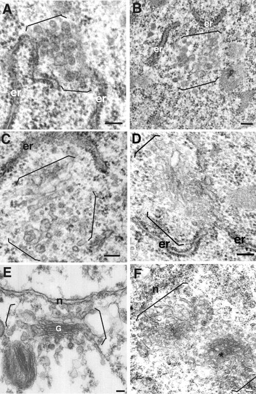
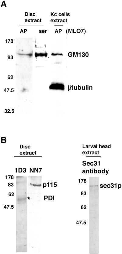
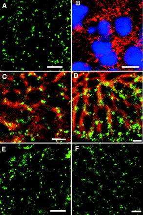
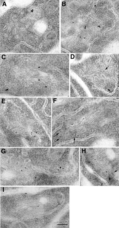
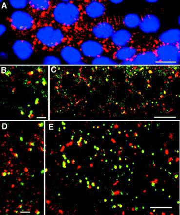
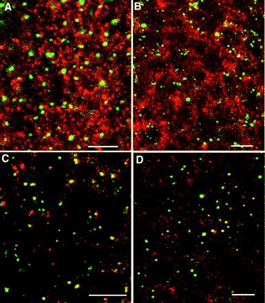
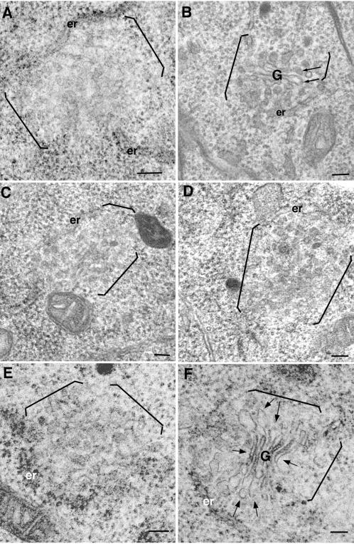
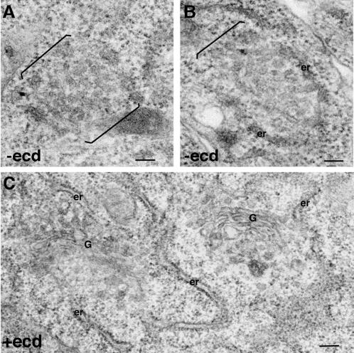
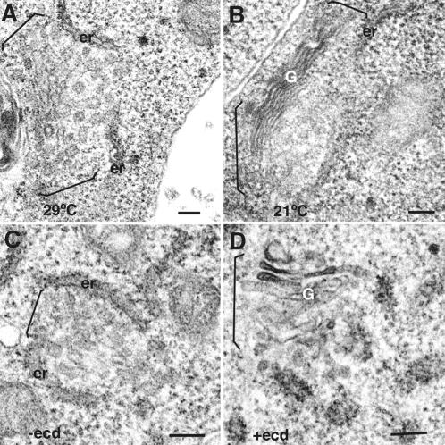
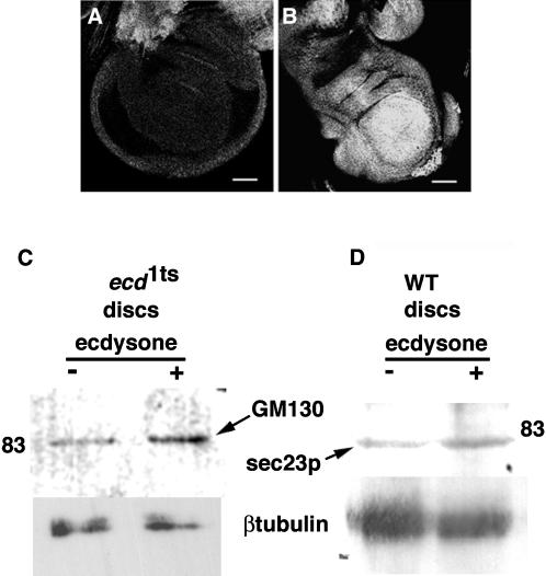
We provide a detailed description of Golgi stack biogenesis that takes place in vivo during one of the morphogenetic events in the lifespan of Drosophila melanogaster. In early third-instar larvae, small clusters consisting mostly of vesicles and tubules were present in epithelial imaginal disk cells. As larvae progressed through mid- and late-third instar, these larval clusters became larger but also increasingly formed cisternae, some of which were stacked. In white pupae, the typical Golgi stack was observed. We show that larval clusters are Golgi stack precursors by 1) localizing various Golgi-specific markers to the larval clusters by electron and immunofluorescence confocal microscopy, 2) driving this conversion in wild-type larvae incubated at 37 degrees C for 2 h, and 3) showing that this conversion does not take place in an NSF1 mutant (comt 17). The biological significance of this conversion became clear when we found that the steroid hormone 20-hydroxyecdysone (ecdysone) is critically involved in this conversion. In its absence, Golgi stack biogenesis did not occur and the larval clusters remained unaltered. We showed that dGM130 and sec23p expression increases approximately three- and fivefold, respectively, when discs are exposed to ecdysone in vivo and in vitro. Taken together, these results suggest that we have developed an in vivo system to study the ecdysone-triggered Golgi stack biogenesis.


Figures

Figure 1
Figure 1
Larval clusters and Golgi areas in discs from third-instar larvae and white pupae. Larval clusters and Golgi area were visualized in leg and wing discs of early third-instar larvae (A and B), mid third-instar larvae (C), late third-instar larvae (D), and white pupae (E and F). Brackets mark the borders of the larval clusters and Golgi areas not bounded by ER cisternae. Stacks are marked with a G. In F, the asterisks indicate an en face view of a fenestrated cisternae. Note the proximity of the larval clusters to ER cisternae (er) in A–D. n, nuclear envelope. Bars, 100 nm.
Figure 2
Figure 2
Western blot of Drosophila tissues and Kc cells. (A) Imaginal disc and Kc cell extracts were blotted with either the MLO7 serum (ser) or the MLO7 affinity-purified antibody (AP) raised against the 73 N-terminal amino acids of human GM130. One strong band just above the 83-kDa molecular weight marker is visible in all lanes. The Kc cell extract was also blotted with the anti-β-tubulin mAb and a band at 50 kDa is visible. Disc extract protein (45 μg, equivalent of 8–10 discs) and 100 μg of Kc cell extract protein were loaded on the 8% acrylamide gel. (B) Disc extract was blotted with 1D3. One band at 50 kDa (asterisk) was revealed in addition to two fainter bands just below and one above the 83-kDa marker. Disc extracts (45 μg) were also blotted with NN7 and one strong band was revealed at ∼83 kDa. Disc extract protein (equivalent of 8–10 discs) was loaded on the 10% acrylamide gel. Larval head extract was blotted with an anti-Sec31p antibody. One band was revealed at ∼130 kDa. Protein (40 μg) was loaded (equivalent to 5 heads) on a 6% acrylamide gel. The numbers on the left of the blots indicate the molecular weight of the prestained markers.
Figure 3
Figure 3
Immunofluorescence imaging of the larval clusters in imaginal disc cells. dGM130 immunofluorescence pattern was visualized in mid-third-instar larval leg and wing discs labeled with rabbit polyclonal MLO7 serum followed by an anti-rabbit IgG conjugated with FITC (green) (A). Note the dots surrounding a black space (nucleus). Discs were also labeled with mouse monoclonal 1D3 followed by an anti-mouse IgG conjugated with Texas Red (red), and DAPI (blue) (B). Dots are numerous and also surround the nucleus. Discs were double immunolabeled with the use of MLO7 and 1D3 followed by an anti-rabbit IgG conjugated with FITC (green) and an anti-mouse IgG conjugated with Texas Red (red) (C). Note that the green dots overlap partially with the red staining representing the ER. Discs were double immunolabeled with MLO7 and the mouse monoclonal anti-β-tubulin antibody followed by an anti-rabbit IgG conjugated with FITC (green) and an anti-mouse IgG conjugated with Texas Red (red) (D). Discs were also labeled with NN7 (the anti-p115 antibody) (E) and the anti-δ-AP3 antibody (F) followed by a anti-rabbit IgG conjugated with FITC. Note that the pattern in A, E, and F are similar. Bars, 5 μm.
Figure 4
Figure 4
Immuno-EM localization of Golgi proteins on larval clusters and Golgi stacks. Unicryl-embedded mid-third-instar discs expressing low level of Fringe-DXD-myc were sectioned and labeled with 9E10 (anti-myc antibody) followed by anti-mouse IgG conjugated to 15-nm gold (A–C) MLO7 (anti-GM130 antibody) followed by anti-rabbit IgG conjugated to 10-nm gold (D–F), and NN7 (an anti-p115 antibody) followed by anti-rabbit IgG conjugated to 10-nm gold (G–I). Larval clusters (A, B, D, E, G, and H) and Golgi areas comprising a stack (C, F, and I) were labeled by the three antibodies. The arrows in D, F, and H indicate gold particles that may be difficult to see. Bar, 100 nm.
Figure 5
Figure 5
dGM130, dp115, and Fringe in mid-third-instar larval discs cells. UAS-Fringe-DXD-myc (stock 919)/HsGAL4 mid-third-instar larval discs were immunolabeled after heat shocking the larvae to induce the synthesis of myc-tagged Fringe-DXD. These discs were immunolabeled with the monoclonal anti-myc antibody 9E10 followed by an anti-mouse IgG conjugated with Texas Red (red), and DAPI (blue) (A). Note the dots around the nucleus. These discs were also double labeled with 9E10 and either MLO7 (B and C) or NN7 (D and E) followed by an anti-mouse IgG conjugated with Texas Red (red) and an anti-rabbit IgG conjugated with FITC (green). Note the colocalization of the two proteins (yellow) and also the structures that display all three colors (yellow, green, and red). Bars, 5 μm.
Figure 6
Figure 6
Immunofluorescence labeling of the larval clusters with components of the COPII machinery. UAS-Fringe-DXD-myc/HsGAL4 mid-third-instar larval discs (Fig .5) were immunolabeled with the anti-Sec23p antibody (A and C) or Sec31p antibody (B and D) together with 1D3 (A and B) and 9E10 (C and D) followed by an anti-rabbit IgG conjugated with FITC and an anti-mouse IgG conjugated to Texas Red. Note in C and D structures in which dSec23p, dSec31p, and Fringe overlap completely or partially together with structures that contain only dSec23p or dSec31p or only Fringe. Bars, 5 μm.
Figure 7
Figure 7
Effect of the comt 17 mutation on the biogenesis of Golgi stacks. Mid-third-instar WT larvae were incubated at 22°C (A) or at 37°C (B) for 2 h. followed by dissection of their discs. Comt17 homozygote larvae were incubated at 37°C for 2 h followed by disc dissection (C) or semidissected and incubated at 37°C for 2 h in the presence of ecdysone (D). comt 17/Y; TM6/+ (E) and comt 17/Y; TM3 HSN, Sb/+ (F) larvae were semidissected and incubated at 37°C for 2 h in the presence of ecdysone. The discs were fixed and processed for conventional EM. The larval clusters and the Golgi areas are marked between brackets when ER cisternae are not marking the natural boundaries, and the stack marked with a G. Note the cisternae in B and F (arrows). er, endoplasmic reticulum. Bars, 100 nm.
Figure 8
Figure 8
In vitro recapitulation of Golgi stack formation in the presence of ecdysone. WT mid-third-instar larvae were semidissected, incubated in M3 or Schneider medium either supplemented (C) with ecdysone and fly extract or without (A and B). Their discs were fixed and processed for conventional EM. The larval clusters and the Golgi areas are marked between brackets when ER cisternae are not marking the natural boundaries, and the stack marked with a G. er, endoplasmic reticulum. Bars, 100 nm.
Figure 9
Figure 9
Effect of ecdysone on the Golgi stacks biogenesis. ecd1ts mutant larvae were maintained at the restrictive temperature of 29°C and either kept at 29°C (A) or released at 22°C for 18 h (B). Their disks were processed for EM and Golgi areas (either Golgi stacks or larval clusters) examined. Alternatively, ecd1ts mutant larvae maintained at 29°C were semidissected and incubated in the absence (C) or the presence (D) of 3 μM ecdysone. The larval clusters and the Golgi areas are marked between brackets when ER cisternae are not marking the natural boundaries, and the stack marked with a G. er, endoplasmic reticulum. Bars, 100 nm.
Figure 10
Figure 10
Effect of ecdysone on dGM130 and dSec23 protein expression. Mid- (A) and late (B) third-instar larval discs were immunolabeled with the use of MLO7 as described in Figures 3 and 5. (C) ecd1ts mutants were maintained at 29°C, their discs dissected and incubated at 22°C in the absence (−) or the presence (+) of 3 μM ecdysone for 18 h. Thirty leg and 10 wing discs were dissected from each incubation. Disc extract protein (30 μg) was loaded on each lane of a 10% acrylamide gel. Immunoblotting was performed with the use of the affinity-purified MLO7 and the mAb to β-tubulin. (D) Mid-third-instar WT larvae were semidissected and incubated in the absence (−) or in the presence (+) of 3 μM ecdysone. Sixty leg and 20 wing discs were dissected from each incubation. 60 μg of disc extract protein was loaded on each lane of a 10% acrylamide gel. Immunoblotting was performed with the use of the rabbit polyclonal antibody to Sec23p and the mAb to β-tubulin. Bar, 30 μm (A) and 40 μm (B).

Similar articles

Cited by

References

    1. Acharya U, Jacobs R, Peters JM, Watson N, Farquhar MG, Malhotra V. The formation of Golgi stacks from vesiculated Golgi membranes requires two distinct fusion events. Cell. 1995;82:895–904. - PubMed
    1. Acharya U, Mallabiabarrena A, Acharya JK, Malhotra V. Signaling via mitogen-activated protein kinase kinase (MEK1) is required for Golgi fragmentation during mitosis. Cell. 1998;92:183–192. - PubMed
    1. Adams MD, et al. The genome sequence of Drosophila melanogaster. Science. 2000;287:2185–2195. - PubMed
    1. Antony C, Huchet M, Changeux JP, Cartaud J. Developmental regulation of membrane traffic organization during synaptogenesis in mouse diaphragm muscle. J Cell Biol. 1995;130:959–968. - PMC - PubMed
    1. Ashburner M, et al. An exploration of the sequence of a 2.9-Mb region of the genome of Drosophila melanogaster: the Adh region. Genetics. 1999;153:179–219. - PMC - PubMed

Publication types

MeSH terms