. 2001 Aug;12(8):2453-68.
doi: 10.1091/mbc.12.8.2453.

Targeting of Shiga toxin B-subunit to retrograde transport route in association with detergent-resistant membranes

Affiliations

Targeting of Shiga toxin B-subunit to retrograde transport route in association with detergent-resistant membranes

T Falguières et al. Mol Biol Cell. 2001 Aug.

Abstract

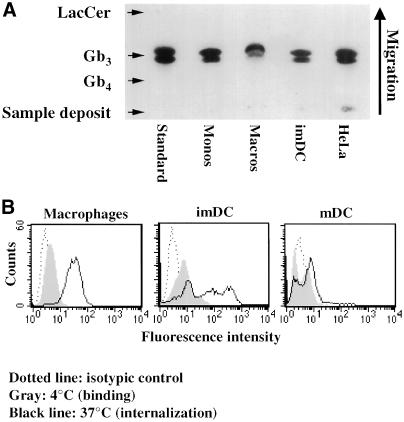
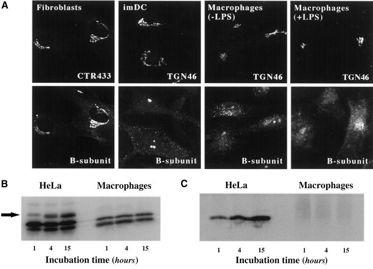
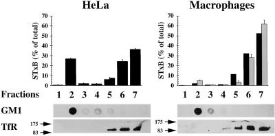
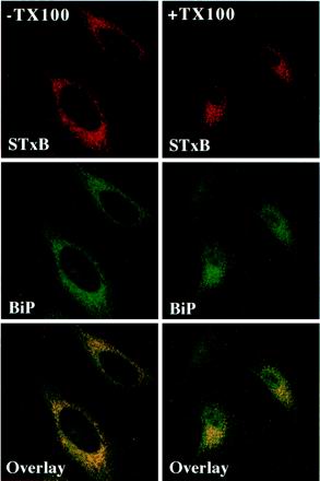
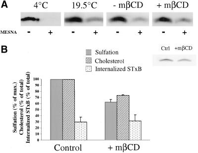
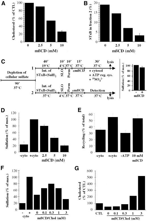
In HeLa cells, Shiga toxin B-subunit is transported from the plasma membrane to the endoplasmic reticulum, via early endosomes and the Golgi apparatus, circumventing the late endocytic pathway. We describe here that in cells derived from human monocytes, i.e., macrophages and dendritic cells, the B-subunit was internalized in a receptor-dependent manner, but retrograde transport to the biosynthetic/secretory pathway did not occur and part of the internalized protein was degraded in lysosomes. These differences correlated with the observation that the B-subunit associated with Triton X-100-resistant membranes in HeLa cells, but not in monocyte-derived cells, suggesting that retrograde targeting to the biosynthetic/secretory pathway required association with specialized microdomains of biological membranes. In agreement with this hypothesis we found that in HeLa cells, the B-subunit resisted extraction by Triton X-100 until its arrival in the target compartments of the retrograde pathway, i.e., the Golgi apparatus and the endoplasmic reticulum. Furthermore, destabilization of Triton X-100-resistant membranes by cholesterol extraction potently inhibited B-subunit transport from early endosomes to the trans-Golgi network, whereas under the same conditions, recycling of transferrin was not affected. Our data thus provide first evidence for a role of lipid asymmetry in membrane sorting at the interface between early endosomes and the trans-Golgi network.


Figures

Figure 1
Figure 1
(A) TLC analysis of Gb3 expression in various cells. Neutral glycolipids were extracted from 1 × 10⁷ monocytes, 2 × 10⁷ macrophages, 2 × 10⁷ imDCs, and 1 × 10⁶ HeLa cells, separated by TLC, and Gb3 was detected and quantified (Table 1) by overlay with STxB. (B) FACS analysis of STxB binding to and internalization into monocyte-derived cells. Fluorescein-labeled STxB (0.1 μM; 5 μg/ml) was incubated on ice (gray profiles) or for 30 min at 37°C (black lines) with the indicated cells that were then analyzed by FACS. Background signal of nonlabeled cells is also shown (dotted lines). Note the weak but detectable binding after incubation on ice and strong signal after incubation at 37°C.
Figure 2
Figure 2
Receptor-dependent STxB internalization into macrophages. (A) Fab-fragment of 13C4 mAb inhibits STxB binding to Gb3. Gb3 was spotted on TLC plates that were then incubated with STxB prebound (+Fab) or not (−Fab) to the Fab-fragment of 13C4, followed by detection with the use of the STxB overlay technique. Note that Fab-bound STxB did not interact with Gb3. (B) FACS analysis of the effect on internalization of STxB prebinding to the Fab-fragment of 13C4. STxB (0.1 μM; 5 μg/ml) was prebound to increasing doses of the Fab-fragment, as indicated. The complex was then incubated with macrophages for 30 min at 37°C. Cells were put on ice, fixed, permeabilized, and incubated with Fab-fragment (to reveal STxB that had not been prebound to the Fab-fragment) and secondary fluorescein-coupled anti-mouse antibody, followed by FACS analysis. Means of three experiments (± SEM) are shown. (C) Immunofluorescence analysis of the effect on internalization of STxB prebinding to the Fab-fragment of 13C4. Experiments as described in B, except that the cells wereviewed by indirect immunofluorescence. STxB (0.1 μM; 5 μg/ml) was prebound (+Fab) or not (−Fab) to the Fab-fragment at a molar ration of 1:1 and then cointernalized with fluorescein-coupled Dex3 for 30 min at 37°C into macrophages. Insets show high-magnification views. Note that some Dex3-containing structures contained only low amounts of STxB.
Figure 3
Figure 3
STxB is internalized into macrophages by micropinocytosis. (A–C) STxB (0.1 μM; 5 μg/ml) was cointernalized for 30 min into macrophages with Dex2000 at 37°C (A), with Dex3 at 18°C (B), or with Dex2000 at 19.5°C (C). Cells were then fixed and stained for STxB. Note that STxB extensively colocalized with Dex3, but not with Dex2000.
Figure 4
Figure 4
Analysis of STxB transport to the late endocytic pathway in macrophages and DC. (A) Iodinated STxB-Glyc-KDEL (50 nM; 2.5 μg/ml) was incubated for 30 min at 37°C with macrophages (speckled columns) or bound to HeLa cells (▪) on ice (A). The cells were then washed on ice, chased at 37°C for the indicated times, and TCA-soluble counts (percentage of the total cell associated radioactivity) were determined. Means (±SEM) of three independent experiments are shown. The appearance of TCA-soluble counts in macrophages indicated that in these cells, STxB was transported to late endosomes/lysosomes. (B) Experiment on macrophages as described in A, in which during the chase period of 30 min, 1 μM Bafi and 50 mM ammonium chloride (NH4Cl) were added. (C) STxB internalized at 19.5°C colocalized extensively with the early endosomal protein EEA1. STxB was continuously internalized for 1 h. The cells were then fixed and stained with the indicated antibodies. (D) After a 30-min shift to 37°C after internalization at 19.5°C, STxB appeared to be lost from the cells. (Insets) Enhancement of the remaining signal showed a limited codistribution of the remaining STxB labeling with the lysosomal glycoprotein Lamp2, suggesting that STxB might be targeted to lysosomes for degradation. (E) A 30-min shift to 37°C in the presence of Bafi allowed detecting a significant degree of codistribution between STxB and Lamp2. (F) As in E, macrophages were shifted to 37°C in the presence of Bafi and then stained for the indicated proteins. Note that even when STxB degradation was prevented, no significant accumulation in the Golgi region marked by Rab6 could be detected.
Figure 5
Figure 5
Analysis of STxB transport to the biosynthetic/secretory pathway in macrophages and DC. (A) Immunofluorescence analysis of STxB transport in different primary human cells. Wild-type STxB was internalized constitutively for 45 min at 37°C into macrophages and imDCs, and after binding into fibroblast and LPS-treated macrophages. The cells were then fixed and stained for STxB (bottom) and the Golgi marker CTR433 or the TGN marker TGN46 (top). In macrophages and imDCs, STxB was detected in vesicular cytoplasmic structures, but not in the TGN, whereas in fibroblasts, STxB readily entered the TGN. In LPS-treated macrophages, STxB accumulated in a number of cytoplasmic locations that only partly covered the Golgi region. (B) Glycosylation analysis. Iodinated STxB-Glyc-KDEL (50 nM; 2.5 μg/ml) was internalized after prebinding into HeLa cells or continuously into macrophages for the indicated times. At the end of each incubation period, the cells were lysed and lysates were analyzed by gel electrophoresis. The same numbers of counts were loaded on gels. The arrow indicates the glycosylation product, i.e., ER-localized STxB, which was observed in HeLa cells, but not in macrophages. The lowest bands are proteolytical cleavage products. (C) Sulfation analysis. 0.2 μM of the fusion protein STxB-Sulf2 (10 μg/ml) was internalized after prebinding into HeLa cells or continuously into macrophages for the indicated times in the presence of radioactive sulfate. The cells were then lysed, STxB was immunoprecipitated, and immunoprecipitates were analyzed by gel electrophoresis. Sulfated STxB-Sulf2, i.e., protein that had passed through the TGN, was detected in HeLa cells, but not in macrophages.
Figure 6
Figure 6
Analysis of STxB association with DRMs. Iodinated STxB (0.2 μM; 10 μg/ml) was bound to control macrophages (▪), LPS-treated macrophages (░⃞), or HeLa cells on ice. The cells were washed, lysed in 1% Triton X-100 buffer, and DRMs were floated in Optiprep step gradients. The gradients were fractionated and the fractions were counted in a gamma counter to determine the distribution of STxB. Furthermore, the fractions were analyzed by dot-blot for GM1 and by gel electrophoresis for the TfR. Note that in HeLa cells, STxB was readily detected in the DRM fraction 2, whereas association with DRMs was at background levels in non-LPS treated macrophages and only slightly above background in LPS-treated cells. Means of two to four experiments (±SEM) are shown.
Figure 7
Figure 7
Analysis of STxB association with DRMs along the retrograde pathway. (A–C) Cy3-labeled STxB was internalized for 45 min at 19.5°C (A) or 37°C (B), or Cy3-STxB-Glyc-KDEL (C) for 4 h at 37°C into HeLa cells. The cells were then either fixed directly (−Triton X-100), or preextracted with 1% Triton X-100 solution (+Triton X-100) before fixation. Permeabilized cells were then stained for GM1 and the TfR. Under these conditions, GM1 and the EE- (A), Golgi- (B), or ER (C)-associated STxB resisted extraction, whereas the TfR was readily extracted. (D–E) STxB-Sulf2 was internalized in the presence of radioactive sulfate for 45 min at 37°C into HeLa cells (D), and iodinated STxB-Glyc-KDEL was internalized for 15 h (E). The cells were then washed, lysed in 1% Triton X-100 buffer, and DRMs were floated in Optiprep step gradients. The gradients were fractionated, STxB in the fractions was immunoprecipitated with 13C4 antibody, and immunoprecipitates were analyzed by gel electrophoresis. DRM fraction 2 contained a significant amount of the sulfated (D) or glycosylated (E; uppermost bands) STxB.
Figure 8
Figure 8
STxB and BiP colocalize even after extraction with Triton X-100. Cy3-coupled STxB-Glyc-KDEL was bound to the plasma membrane of HeLa cells on ice. The cells were washed, shifted to 37°C for 4 h, placed on ice, incubated (+TX100) or not (−TX100) for 1 min in 1% Triton X-100-containing buffer, and then fixed at room temperature for 15 min. The fixed and permeabilized (saponin) cells were incubated with the indicated antibodies, mounted in Mowiol, and viewed by confocal microscopy. Note that even after extraction with Triton X-100, BiP labeling colocalized with STxB-Glyc-KDEL-specific labeling.
Figure 9
Figure 9
EE-to-TGN transport is inhibited under cholesterol extraction conditions. (A) Biotinylated STxB mutant was bound to HeLa cells on ice (condition 4°C) or internalized at 19.5°C into EE (conditions 19.5°C, −mβCD, +mβCD). The cells were then either directly placed on ice (4 and 19.5°C), or incubated with (+mβCD) or without (−mβCD) 10 mM mβCD for 60 min at 19.5°C before passage on ice. After MESNA (+) or mock (−) treatment on ice, the cells were lysed and biotinylated STxB was detected by Western. (B) STxB-Sulf2 was internalized and cholesterol was extracted as described in A. Cells were then used to determine the amount of free cellular cholesterol (control cells contained 7.93 ± 1.88 μg of cholesterol/106 cellules) or were shifted to 37°C for 30 min in the presence of radioactive sulfate. Note that transport to the TGN (sulfation) was significantly inhibited by cholesterol extraction. The inset shows a typical result of the sulfation reactions in the −mβCD and the +mβCD conditions.
Figure 10
Figure 10
Establishment of a cause-and-effect relationship between STxB association with DRMs and its transport from EE to the TGN. (A) A 15-min treatment at 37°C of SLO-permeabilized HeLa cells with the indicated doses of mβCD led to a dose-dependent extraction of free cellular cholesterol. The means of two to three experiments are shown. Nontreated SLO-permeabilized cells had 5 μg of cholesterol/106 cells. (B) Cholesterol extraction displaces STxB from DRM fraction 2. Note that STxB association with fraction 2 is lower than in Figure 6 due to an additional incubation at 37°C for cholesterol extraction (see MATERIALS AND METHODS). The means of two to three experiments are shown. (C) Generic protocols used to reconstitute EE-to-TGN transport of STxB. Perm., permeabilization in intracellular transport buffer/dithiothreitol at 37°C (see MATERIALS AND METHODS). Detection: incubation of permeabilized cells with radioactive sulfate in the absence of cytosol. Protocol 1: standard protocol (see below, D). Protocol 2: Comparison of sulfation efficiencies on TGN-localized STxB (40 min transport at 37°C in intact cells) in SLO-permeabilized and mβCD- or mock-treated cells (for results, see insert; means of three experiments). (D) STxB transport from EE to the TGN on SLO-permeabilized cells is inhibited when DRMs are destabilized by 15-min extraction of cellular cholesterol with the indicated doses of mβCD. Transport efficiency in the presence of cytosol is put to 100%. The means (± SEM) of three to six experiments are shown. (E) ATP- and cytosol-dependent Tf recycling to the plasma membrane is not affected by cellular cholesterol extraction. The percentage of released Tf over total cell associated Tf was determined, and the means (± SEM) of 6–10 experiments are shown. (F) Cholesterol back-addition. In a variation of protocol 1 (C), the cells were extracted for 10 min at 37°C with 10 mM mβCD from permeabilization on, incubated for 10 min at 37°C with the indicated concentrations of cholesterol-saturated mβCD, and then shifted for 30 min to 37°C in the presence of cytosol and radioactive sulfate. Note that cholesterol back addition partially rescued transport to the TGN. (G) Quantification of cholesterol back-addition. The quantities of total free cellular cholesterol under the indicated conditions were determined. In F and G, a representative of two experiments is shown.

References

    1. Abe A, Inokuchi J, Jimbo M, Shimeno H, Nagamatsu A, Shayman JA, Shukla GS, Radin NS. Improved inhibitors of glucosylceramide synthase. J Biochem. 1992;111:191–196. - PubMed
    1. Advani RJ, Yang B, Prekeris R, Lee KC, Klumperman J, Scheller RH. VAMP-7 mediates vesicular transport from endosomes to lysosomes. J Cell Biol. 1999;146:765–776. - PMC - PubMed
    1. Arab S, Lingwood CA. Intracellular targeting of the endoplasmic reticulum/nuclear envelope by retrograde transport may determine cell hypersensitivity to verotoxin via globotriaosyl ceramide fatty acid isoform traffic. J Cell Physiol. 1998;177:646–660. - PubMed
    1. Bagnat M, Keranen S, Shevchenko A, Simons K. Lipid rafts function in biosynthetic delivery of proteins to the cell surface in yeast. Proc Natl Acad Sci USA. 2000;97:3254–3259. - PMC - PubMed
    1. Banchereau J, Steinman RM. Dendritic cells and the control of immunity. Nature. 1998;392:245–252. - PubMed

Publication types

LinkOut - more resources