. 2001 Aug;12(8):2482-96.
doi: 10.1091/mbc.12.8.2482.

Targeting of a tail-anchored protein to endoplasmic reticulum and mitochondrial outer membrane by independent but competing pathways

Affiliations
Free PMC article

Targeting of a tail-anchored protein to endoplasmic reticulum and mitochondrial outer membrane by independent but competing pathways

N Borgese et al. Mol Biol Cell. 2001 Aug.
Free PMC article

Abstract

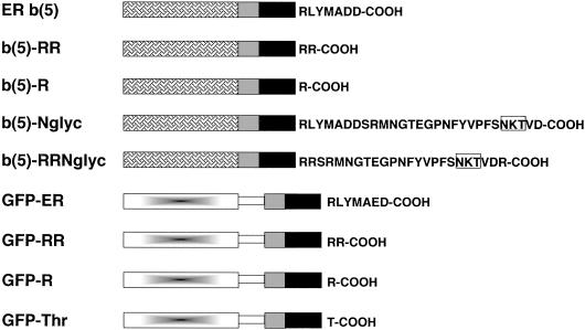
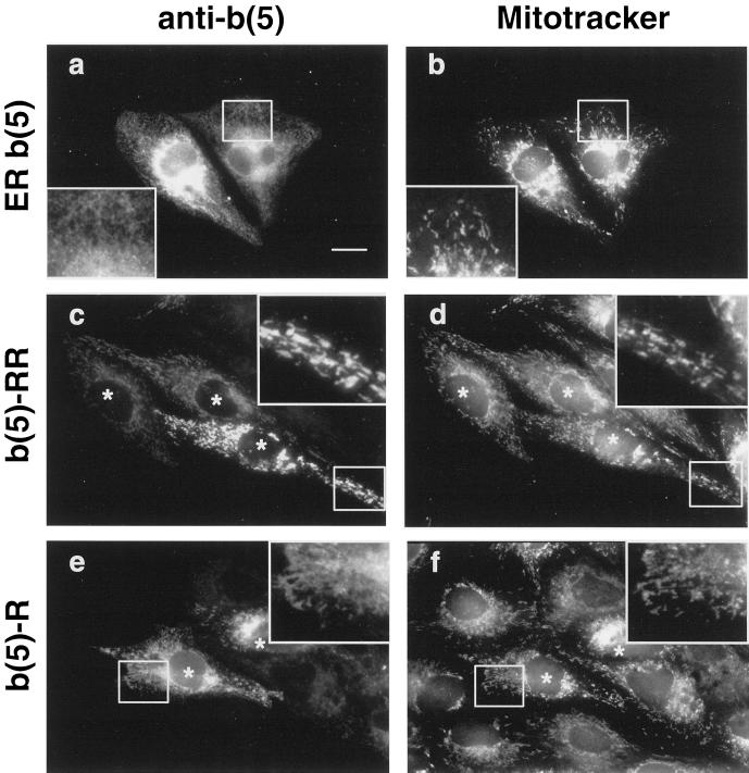
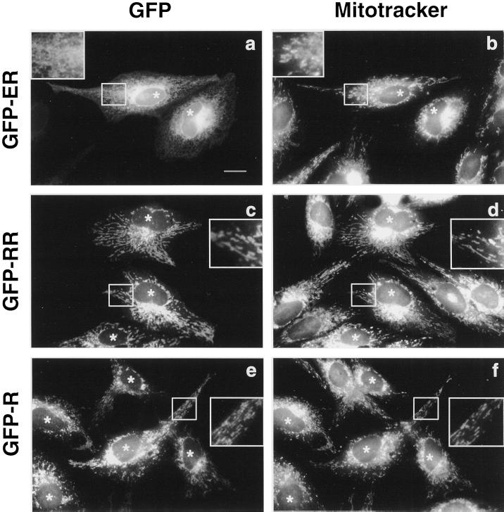
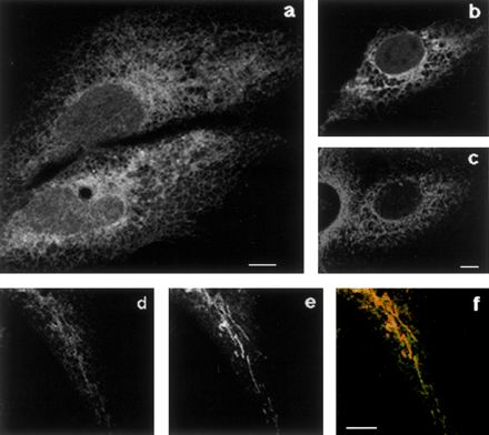
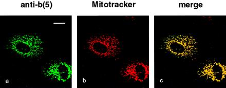
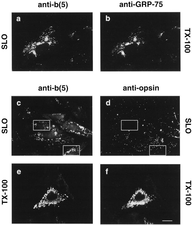
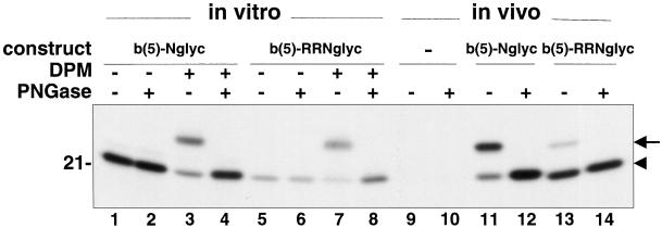
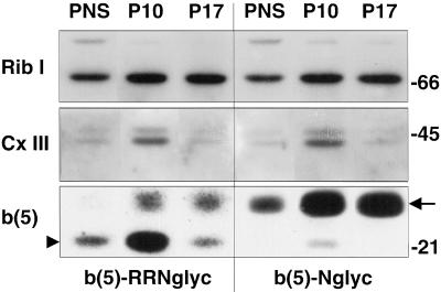
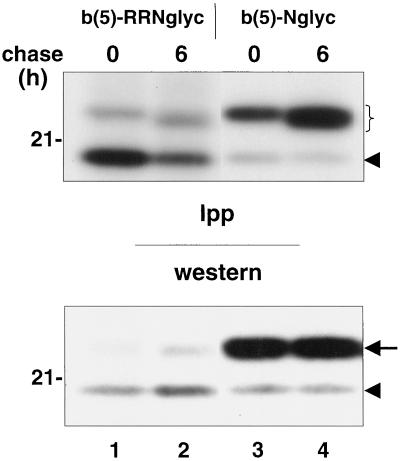
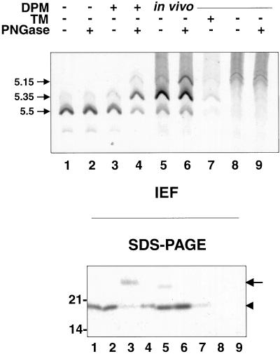
Many mitochondrial outer membrane (MOM) proteins have a transmembrane domain near the C terminus and an N-terminal cytosolic moiety. It is not clear how these tail-anchored (TA) proteins posttranslationally select their target, but C-terminal charged residues play an important role. To investigate how discrimination between MOM and endoplasmic reticulum (ER) occurs, we used mammalian cytochrome b(5), a TA protein existing in two, MOM or ER localized, versions. Substitution of the seven C-terminal residues of the ER isoform or of green fluorescent protein reporter constructs with one or two arginines resulted in MOM-targeted proteins, whereas a single C-terminal threonine caused promiscuous localization. To investigate whether targeting to MOM occurs from the cytosol or after transit through the ER, we tagged a MOM-directed construct with a C-terminal N-glycosylation sequence. Although in vitro this construct was efficiently glycosylated by microsomes, the protein expressed in vivo localized almost exclusively to MOM, and was nearly completely unglycosylated. The small fraction of glycosylated protein was in the ER and was not a precursor to the unglycosylated form. Thus, targeting occurs directly from the cytosol. Moreover, ER and MOM compete for the same polypeptide, explaining the dual localization of some TA proteins.


Figures

Figure 1
Figure 1
Schematic representation of the constructs used in this study. Grilled box, heme-binding, cytosolic domain of ER b(5); gray box, polar flanking region of ER b(5) upstream to the TMD; black box, transmembrane domain of ER b(5); gradient-filled box, GFP; open rectangle, artificial linker (myc epitope followed by [Gly4-Ser]3. Residues downstream to the TMD are indicated with the single letter code. The N-glycosylation consensus site used in the b(5)-Nglyc and b(5)-RRNglyc constructs is boxed.
Figure 2
Figure 2
Replacement of the C-terminal polar sequence of ER b(5) with a single or double arginine relocates the protein to mitochondria. CV-1 cells transfected with ER b(5) (a and b), b(5)-RR (c and d), or b(5)-R (e and f) were stained with Mitotracker CMX rose then fixed, permeabilized, and incubated with polyclonal anti-b(5) antibodies followed by FITC-conjugated secondary antibodies. The panels on the left and right side of the figure represent the same field of cells viewed by conventional epifluorescence under the fluorescein [cyt b(5)] or rhodamine (Mitotracker) filter, respectively. ER b(5) yields a classical ER pattern, and colocalization with mitochondria is not detectable (compare a and b). b(5)-RR and b(5)-R staining are mainly or partially superimposed with Mitotracker (compare c and d, or e and f, respectively). In cells transfected with b(5)-R, a residue of reticular staining is also evident (compare e and f, insets), that is not visible in b(5)-RR-transfected cells (compare c and e, insets). Bar, 20 μm. Insets, 2.3× magnification. Areas magnified in the insets are boxed. Asterisks in c–f are placed over the nuclei of transfected cells.
Figure 3
Figure 3
The heme-binding domain of cyt b(5) is not required for targeting. Mitotracker-loaded CV-1 cells expressing GFP-ER, GFP-RR, or GFP-R were fixed and observed by conventional epifluorescence. GFP fluorescence (a, c, and e) and Mitotracker labeling (b, d, and f) are compared in the same fields. GFP-ER retains the ER localization and colocalization with mitochondria is not detectable (compare a and b). GFP-RR and GFP-R are relocalized to mitochondria (compare c and d, or e and f, respectively). Bar, 20 μm; insets. Areas enlarged in the insets (2.3×) are boxed. Asterisks are placed over the nuclei of transfected cells.
Figure 4
Figure 4
GFP-Thr localizes both to the ER and to mitochondria. CV-1 cells transfected with GFP-Thr were allowed to express the cDNA for 18 h, after which cells were further incubated in the presence of 20 μg/ml cycloheximide for 7 h. At this time the cells had nearly undetectable GFP fluorescence, as assessed by observation of the living cells. Cycloheximide was removed, and 45 min later cells were fixed (a–c) or loaded with Mitotracker and then fixed (d–f). The sample shown in b and c was stained with anti-calnexin antibodies followed by incubation with Texas Red-conjugated secondary antibodies. Single confocal sections are shown. (a) GFP fluorescence shows clear reticular staining. (b and c) GFP (b) and Texas Red (c, calnexin) fluorescence were acquired separately from the same field. Comparison of the two images shows a partial colocalization between GFP-Thr and the ER marker. (d–f) Comparison between GFP fluorescence (d) and Mitotracker (e) shows localization of GFP-Thr also to mitochondria (f, merge). Bars, 10 μm.
Figure 5
Figure 5
b(5)-RRNglyc is targeted to mitochondria. Mitotracker-loaded CV-1 cells expressing b(5)-RRNglyc were fixed, permeabilized with Triton X-100, processed for immunofluorescence with anti-b(5) antibodies followed by FITC-conjugated secondary antibodies, and observed by confocal microscopy. Images of the same field were acquired separately with fluorescein [a, b(5)] and Texas Red (b, Mitotracker) filters. A single confocal section is shown. (c) Merged image of the two acquisitions. Bar, 20 μm.
Figure 6
Figure 6
b(5)-RRNglyc C-terminal polar residues are sequestered from antibodies restricted to the cytosol. CV-1 cells expressing b(5)-RRNglyc were permeabilized with 40 U/ml SLO (see MATERIALS AND METHODS) in the presence of anti-b(5) antibodies alone (a and b), or anti-b(5) antibodies and anti-opsin mAbs together (c and d), before fixation. Another sample was fixed and permeabilized with Triton X-100 (TX-100) without prior incubation with SLO, and doubly stained with anti-b(5) antibodies (e) and anti-opsin mAbs (f). Cells were visualized by conventional epifluorescence microscopy. (a and b) SLO-treated cells incubated with anti-b(5) antibodies were counterstained with anti-GRP-75 mAbs after fixation and permeabilization. Stained cells were examined under the fluorescein filter to visualize b(5) (a) or under the rhodamine filter to visualize anti-GRP-75-stained mitochondria (b). Anti-b(5) antibodies added to unfixed cells recognize b(5)-RRNglyc on the cytosolic side of the mitochondria as shown by colocalization with mitochondrial GRP-75. (c and d) In SLO-permeabilized cells, incubated with both anti-b(5) antibodies and anti-opsin mAbs together, the anti-opsin mAbs, revealed by rhodamine-conjugated secondary antibodies (d), shows only nonspecific staining that does not correspond to the anti-b(5) staining, shown in c. Boxes highlight areas in which the difference between staining with the two antibodies is clearly visible. After Triton X-100 permeabilization (e and f), anti-b(5) (e) and anti-opsin (f) antibodies stain the same structures. Bar, 20 μm. c–f were photographed with the same exposure. Each pair of images (c and d) and (e and f) was acquired from negatives and processed with Adobe Photoshop software under identical conditions.
Figure 7
Figure 7
Different degree of glycosylation of b(5)-RRNglyc obtained in vitro and in vivo. Lanes 1–8, b(5)-Nglyc (lanes 1–4) or b(5)-RRNglyc (lanes 5–8) mRNAs were translated in vitro in the reticulocyte lysate system in the presence of [35S]methionine. After blocking translation with cycloheximide, half of each sample was further incubated for another hour in the presence of DPMs, as indicated. The translation products were immunoselected with anti-opsin mAbs, treated (lanes 2, 4, 6, and 8) or mock treated (lanes 1, 3, 5, and 7) with PNGase F, and analyzed by SDS-PAGE/fluorography. The arrowhead indicates the position of the unglycosylated product. In the presence of DPMs, both constructs show an additional major band with lower electrophoretic mobility (lanes 3 and 7, arrow), which is converted to the more rapidly migrating polypeptide by PNGase F (lanes 4 and 8), demonstrating that it is glycosylated. Lanes 10–14, CV-1 cells were transfected with b(5)-Nglyc (lanes 11 and 12), b(5)-RRNglyc (lanes 13 and 14), or mock transfected (lanes 9 and 10) and labeled for 2 h with [35S]Met/Cys. Cleared cell lysates were subjected to anti-opsin mAb immunoselection. Immunocomplexes were digested with PNGase F (lanes 10, 12, and 14) or mock digested (lanes 9, 11, and 13). Note the lower proportion of glycosylated b(5)-RRNglyc in vivo (lane 13, arrow), compared with that obtained in vitro (lane 7). The numbers on the left of the panel indicates the migration of soybean trypsin inhibitor (21 kDa).
Figure 8
Figure 8
Subcellular distribution of glycosylated and unglycosylated b(5)-RRNglyc investigated by cell fractionation. HeLa cells, transfected with b(5)-RRNglyc or b(5)-Nglyc, were subjected to subcellular fractionation by differential centrifugation, to isolate P10 and P17 fractions as described in MATERIALS AND METHODS. The distribution of glycosylated (arrow) and unglycosylated (arrowhead) expression products was analyzed by Western blotting [b(5), bottom] and compared with that of a marker for the ER (ribophorin I, top) and for mitochondria (∼45-kDa complex III polypeptide, middle). The glycosylated forms of the constructs codistribute with ribophorin I, whereas the unglycosylated polypeptides have a distribution similar to the mitochondrial marker. Numbers on the right indicate the position and size (kDa) of markers (Bio-Rad, Richmond, CA).
Figure 9
Figure 9
Glycosylated b(5)-RRNglyc is stable on the ER membrane. CV-1 cells transfected with b(5)-RRNglyc or b(5)-Nglyc cDNAs were labeled for 2 h with [35S]Met/Cys. Expression products were immunoselected with anti-opsin mAbs from cleared cell lysates prepared at the end of pulse or after 6 h of chase and subjected to SDS-PAGE/fluorography (top, Ipp). An aliquot of each lysate was also analyzed directly by Western blot with the use of enhanced chemiluminescence to reveal bound antibodies (bottom, Western). The bracket in the top panel and the arrow in the bottom panel indicate the position of the glycosylated form of both constructs. The arrowhead in both panels indicates the unglycosylated form. For both constructs, the glycosylated form is stable, although a slight downward mobility shift is observed at 6 h of chase, probably due to mannose trimming. The position of the 21-kDa size marker is indicated on the left.
Figure 10
Figure 10
b(5)-RRNglyc does not undergo intracellular deglycosylation. Lanes 1–4, b(5)-RRNglyc was translated in vitro in the presence of [35S]methionine. After blocking translation with cycloheximide, half of each sample was further incubated for another hour in the presence of DPMs, as indicated. After immunoprecipitation with anti-opsin mAbs, the immunoselected products were digested with PNGase F (lanes 2 and 4) or mock digested (lanes 1 and 3) then analyzed by isoelectrofocusing (top) or SDS-PAGE followed by autoradiography (bottom). Numbers on the left of the top panel indicate the pIs of the observed species. The pI 5.5 and pI 5.35 forms produced in the presence of DPMs (lane 3) are shifted to pI 5.35 and 5.15 by PNGase F treatment (lane 4), whereas those produced in the absence of DPMs are unaffected by PNGase digestion (lanes 1 and 2). Lanes 5–9, CV-1 cells were transfected with b(5)-RRNglyc cDNA (lanes 5–7) or mock transfected (lanes 8 and 9) and labeled for 2 h with [35S]Met/Cys in the presence (lane 7) or absence (lanes 5, 6, 8, and 9) of TM. Immunoprecipitates obtained with anti-opsin mAbs were treated with PNGase F (lanes 6 and 9) or mock digested (lanes 5, 7, and 8). Samples were analyzed by IEF or SDS-PAGE. Both TM-treated and untreated cells yield a major band focused at pH 5.35, indicating that b(5)-RRNglyc has not undergone deglycosylation in vivo. Arrow and arrowhead in the bottom panel indicate the positions of the glycosylated and unglycosylated polypeptide, respectively. Molecular weight standards are indicated as in Figures 7–9. See text for further explanation.

References

    1. Adamus G, Arendt A, Hargrave PA. Genetic control of antibody response to bovine rhodopsin in mice: epitope mapping of rhodopsin structure. J Neuroimmunol. 1991;34:89–97. - PubMed
    1. Akao Y, Otsuki YS, Kataoka S, Ito Y, Tsujimoto Y. Multiple subcellular localization of bcl-2: detection in nuclear outer membrane, endoplasmic reticulum, and mitochondrial membrane. Cancer Res. 1994;54:2468–2471. - PubMed
    1. Ausubel FM, Brent R, Kingston RE, Moore DD, Seidman JG, Smith JA, Struhl K. Current Protocols in Molecular Biology. New York: John Wiley & Sons; 1987.
    1. Bhakdi S, Weller U, Walev I, Martin E, Jonas D, Palmer M. A guide to the use of pore-forming toxins for controlled permeabilization of cell membranes. Med Microbiol Immunol. 1993;182:167–175. - PubMed
    1. Borgese N, Aggujaro D, Carrera P, Pietrini G, Bassetti M. A role for N-myristoylation in protein targeting: NADH-cytochrome b5 reductase requires myristic acid for association with outer mitochondrial but not endoplasmic reticulum membranes. J Cell Biol. 1996;135:1501–1513. - PMC - PubMed

Publication types

MeSH terms

Grants and funding

LinkOut - more resources