. 2001 Sep 1;21(17):6480-91.
doi: 10.1523/JNEUROSCI.21-17-06480.2001.

Inflammatory neurodegeneration mediated by nitric oxide from activated glia-inhibiting neuronal respiration, causing glutamate release and excitotoxicity

Affiliations

Inflammatory neurodegeneration mediated by nitric oxide from activated glia-inhibiting neuronal respiration, causing glutamate release and excitotoxicity

A Bal-Price et al. J Neurosci. .

Abstract

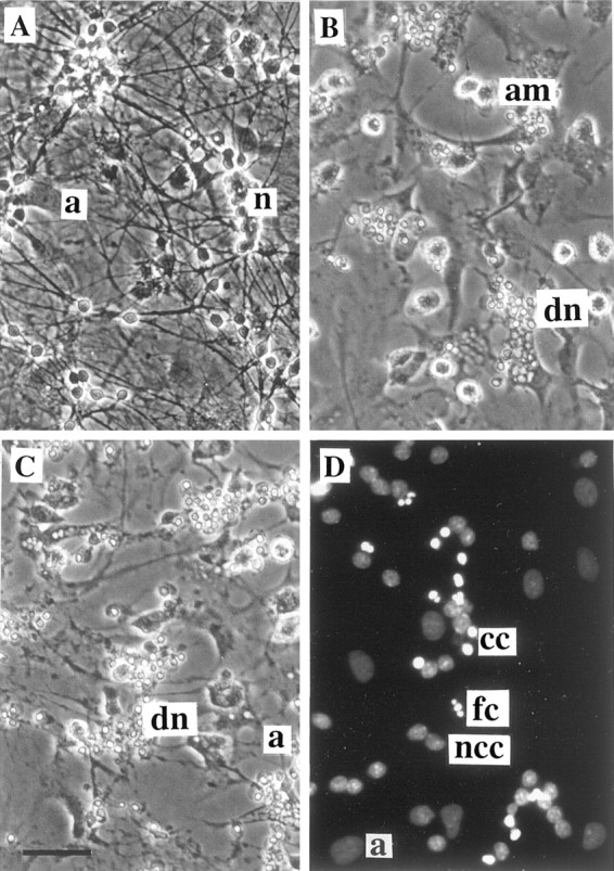
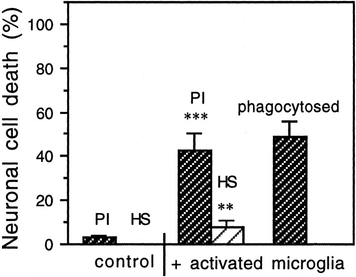
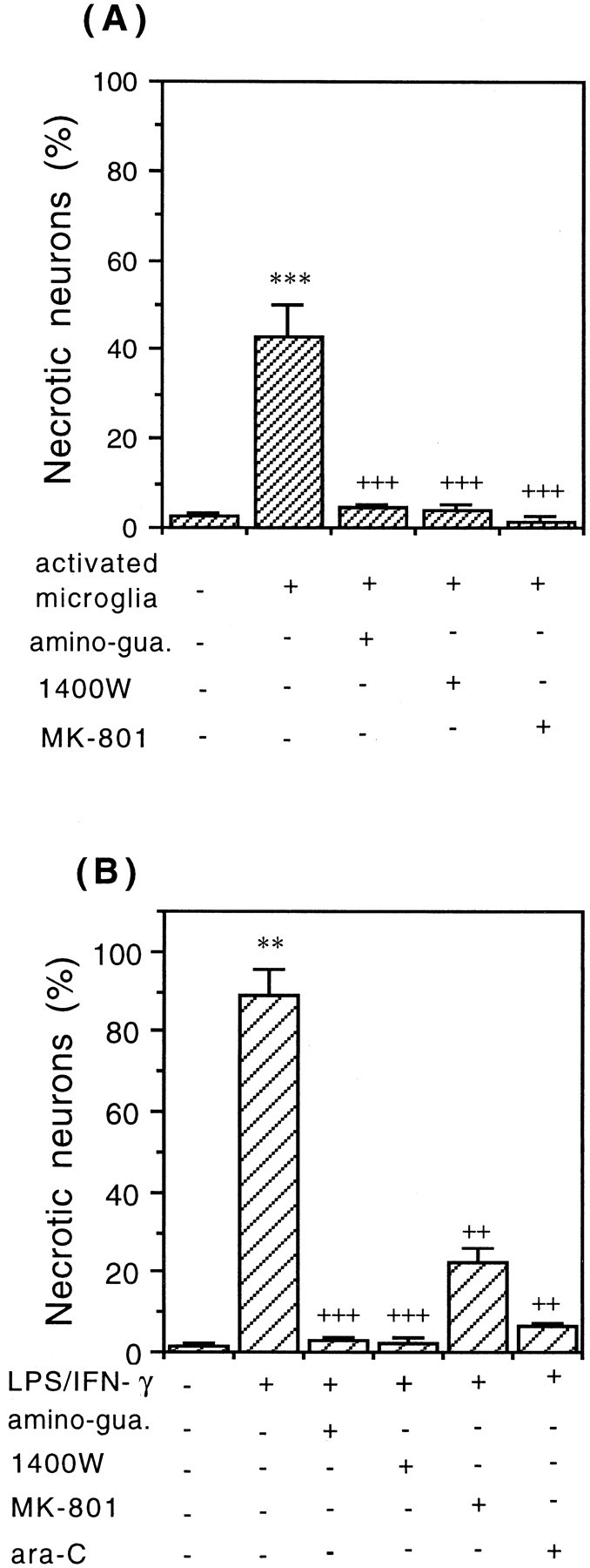
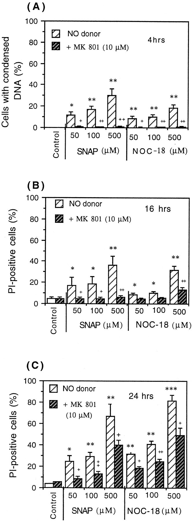
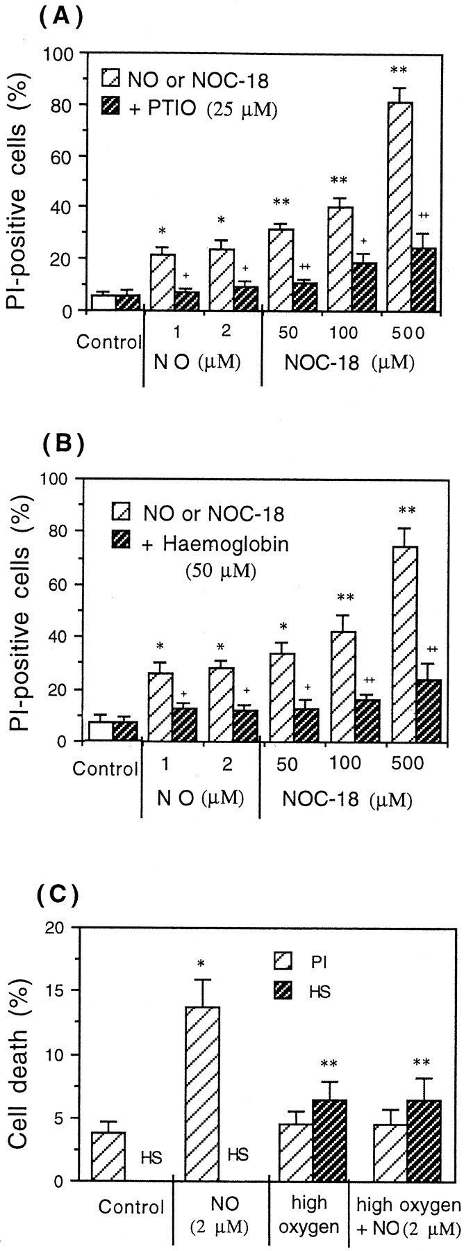
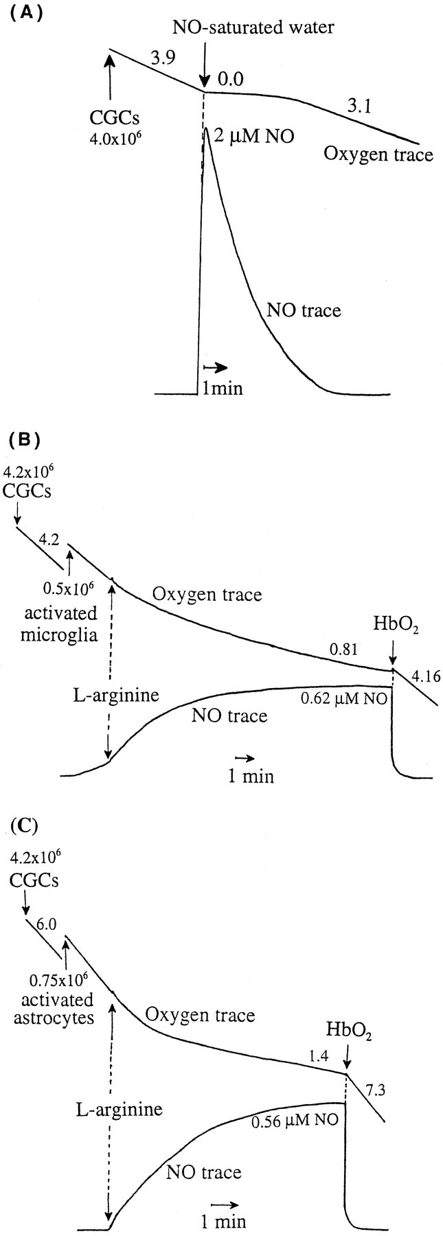
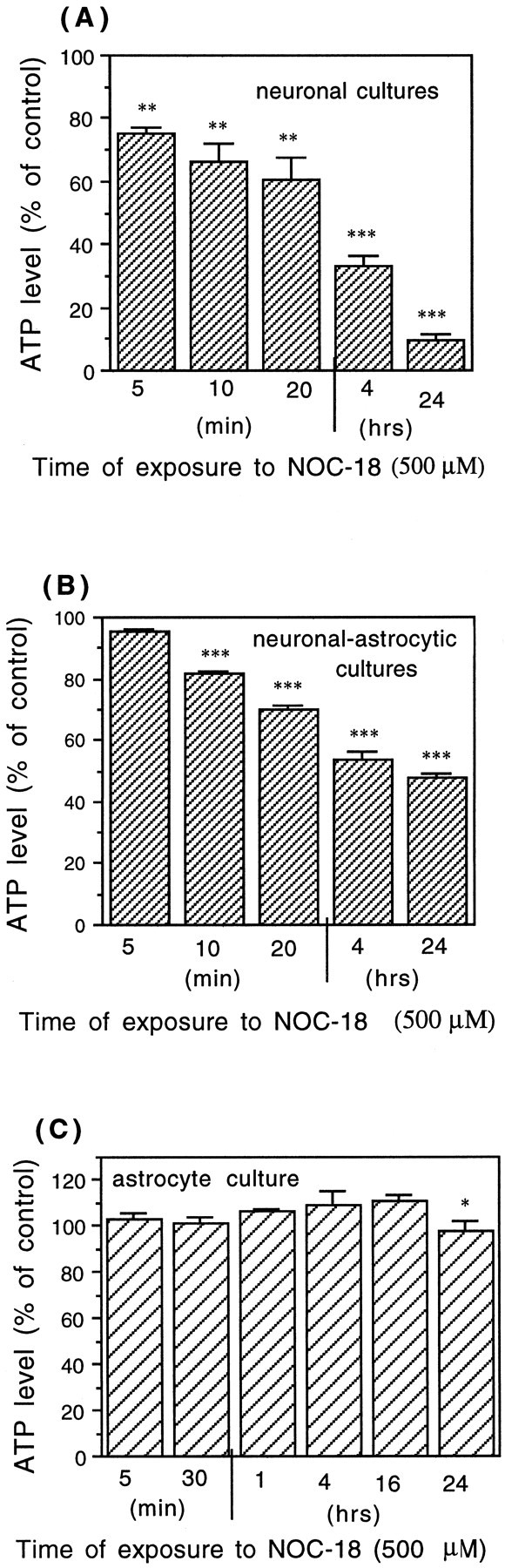
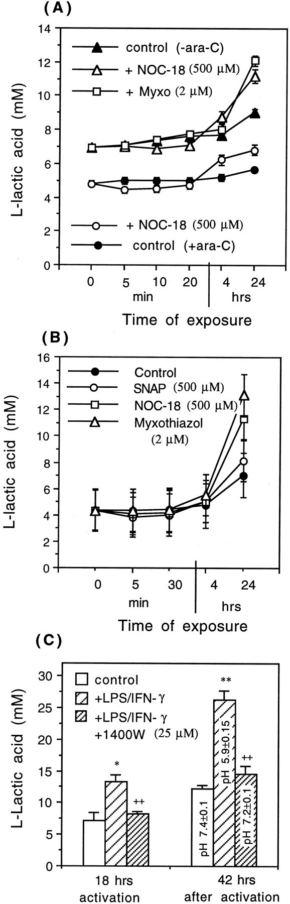
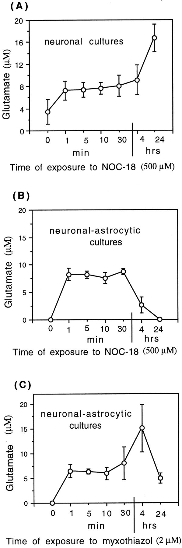
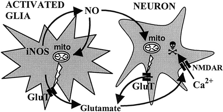
Glia undergo inflammatory activation in most CNS pathologies and are capable of killing cocultured neurons. We investigated the mechanisms of this inflammatory neurodegeneration using a mixed culture of neurons, microglia, and astrocytes, either when the astrocytes were activated directly with lipopolysaccharide (LPS) and interferon-gamma (IFN-gamma) or LPS/IFN-gamma-activated microglia were added to mixed neuronal cultures. In either case, activated glia caused 75-100% necrotic cell death within 48 hr, which was completely prevented by inhibitors of inducible nitric oxide synthase (iNOS) (aminoguanidine or 1400W). Activated astrocytes or microglia produced nitric oxide (NO) (steady-state level approximately 0.5 microm), which immediately inhibited the cellular respiration of cocultured neurons, as did authentic NO. NO donors also decreased ATP levels and stimulated lactate production by neurons, consistent with NO-induced respiratory inhibition. NO donors or a specific respiratory inhibitor caused rapid (<1 min) release of glutamate from neuronal and neuronal-astrocytic cultures and subsequent neuronal death that was blocked by an antagonist of NMDA receptor (MK-801). MK-801 also blocked neuronal death induced by activated glia. High oxygen also prevented NO-induced neuronal death, consistent with death being induced by NO inhibition of cytochrome c oxidation in competition with oxygen. Thus activated glia kill neurons via NO from iNOS, which inhibits neuronal respiration resulting in glutamate release and subsequent excitotoxicity. This may contribute to neuronal cell death in inflammatory, infectious, ischemic, and neurodegenerative diseases.


Figures

Fig. 1.
Fig. 1.
Cell death of CGCs (neuronal–astrocytic cultures, 9 DIV) induced by coculture with activated microglia or by direct exposure to proinflammatory cytokines or NOC-18 (NO donor).A, In the control culture of CGCs, note the phase-bright normal cell bodies and dense neuritic network. B, Coculture of CGCs (9 DIV, 0.25 × 10⁶cells/cm2) with LPS/IFN-γ-activated microglia (0.2 × 10⁶cells/cm2) for 24 hr induced cell death of all neurons. Note shrunken cell bodies and nuclei and loss of all neurites (phase-contrast photographs). C, Addition of LPS (4 ng/ml) and IFN-γ (100 U/ml) simultaneously for 48 hr to the CGCs, cultured in the presence of glial cells (9 DIV, nontreated with ara-C), caused severe neuronal cell death, but dead cells were not phagocytosed. D, Morphological analysis of nuclear chromatin in CGC culture (neuronal–astrocytic, 9 DIV) exposed to 500 μm NOC-18 for 4 hr using DNA-binding fluorochrome Hoechst 33342 (fluorescence microscope). Viable cells showed round nuclei with weak fluorescence, but some nuclei had strongly condensed chromatin (bright fluorescence), predominantly without fragmentation. However, a few nuclei with fragmented chromatin were also present. Particular cell or nuclear types are indicated by the following abbreviations (placed to the right of the cell): n, healthy neurons; a, astrocyte; am, activated microglia; dn, dead neurons; cc, condensed chromatin; fn, fragmented chromatin;ncc, non-condensed chromatin. Scale bar (shown inC): AC, 40 μm;D, 20 μm.
Fig. 2.
Fig. 2.
Cell death of CGCs (neuronal–astrocytic cultures, 9 DIV) induced by addition of LPS/IFN-γ-activated microglia (0.2 × 10⁶ cells/cm2) for 24 hr. The presence of activated microglia caused predominantly necrotic cell death (PI-positive cells); however, Hoechst 33342-stained (HS) neurons (with condensed chromatin, occasionally with fragmentation) were also present. Also, some CGCs disappeared completely (presumably because of phagocytosis by activated microglia). Values represent the means ± SD of three or more separate cultures. In each experiment, three wells per treatment were analyzed, and the cells in three fields per well were counted (∼80 ± 25 cells per field). **p < 0.01, ***p < 0.001 from control groups.
Fig. 3.
Fig. 3.
Necrotic cell death of CGCs (0.25 × 10⁶cells/cm2) induced by activated microglia (A) or LPS/IFN-γ (B). Addition of 0.2 – 106cells/cm2 of LPS/IFN-γ-activated microglia for 24 hr (A) or LPS/IFN-γ for 48 hr (B) to the culture of CGCs (neuronal–astrocytic) caused necrosis (PI-positive cells). The necrotic neuronal cell death was completely prevented by iNOS inhibitors aminoguanidine (200 μm) or 1400W (25 μm), was wholly or partially blocked by an NMDA receptor antagonist (10 μm MK-801), and in the case of (B) was completely prevented by pretreatment with ara-C (to eliminate glial cells). Percentages of necrotic neurons (PI-positive cells) were calculated, and the values represent the means ± SD of at least three independent experiments. ***p < 0.001 from control groups and+++p < 0.001 from activated microglia (A) or ++p < 0.01, +++p < 0.001 from LPS/IFN-γ treatment (B).
Fig. 4.
Fig. 4.
Induction of neuronal death in CGC cultures (neuronal–astrocytic, 9 DIV) by NO donors (NOC-18 or SNAP) after 4 hr (A), 16 hr (B), and 24 hr (C). After 4 hr of incubation with either NO donor (SNAP or NOC-18), Hoechst-positive neurons were observed (condensed chromatin, rarely with fragmentation) (Fig.1D), but no propidium iodide-staining cells were seen. After 16 or 24 hr of incubation, neurons died mainly by necrosis (PI-positive cells). Neuronal cell death induced by both NO donors (SNAP or NOC-18) was prevented by MK-801 (NMDA receptor antagonist) after 4 and 16 hr (but not 24 hr) of incubation. Values represent means ± SD (bars) of determinations made in three separate cultures. *p < 0.05, **p < 0.01, ***p < 0.001 from control groups and +p < 0.05,++p < 0.01,+++p < 0.001 from SNAP or NOC-18 treatment.
Fig. 5.
Fig. 5.
Necrotic cell death of CGCs (PI-positive cells) induced by NO-saturated water or NO donor (NOC-18) after 24 hr of exposure was prevented by NO scavengers PTIO (A) or hemoglobin (B) or by high oxygen (95% O2/5% CO2) (C). The data present the mean ± SD of three independent experiments. *p < 0.05, **p < 0.01 from control groups and+p < 0.05,++p < 0.01 from NO or NOC-18 treatment.
Fig. 6.
Fig. 6.
Rapid inhibition of mitochondrial respiration of CGCs by addition of 2 μm NO-saturated water (A) or NO produced by activated microglia (B) or activated astrocytes (C). A, Oxygen consumption (top trace) of CGCs (∼4.0 × 10⁶ of cells) before addition of NO-saturated water was 3.9 natom of O/min/106 cells; after addition of NO-saturated water (2.0 μm NO), oxygen consumption was completely and immediately inhibited for the first few minutes, but the rate recovered as the NO level declined (3.1 natom of O/min/106 cells). B, Addition of activated microglia (∼0.5 × 10⁶cells; NO level 0.62 μm) and arginine (200 μm; NOS substrate) caused rapid and strong inhibition of neuronal respiration (∼4.2 × 10⁶cells; oxygen consumption, 4.2 natom of O/min/106 cells and 0.81 natom of O/min/106 cells before and after addition of activated microglia, respectively). The inhibition of neuronal respiration was reversible, because the addition of 10 μmhemoglobin (HbO2, NO scavenger) resulted in full activation of mitochondrial respiration (4.16 natom of O/min/106 cells). C, Addition of activated astrocytes (∼0.75 × 10⁶ cells; NO level 0.56 μm) and arginine (200 μm) induced significant inhibition of oxygen consumption of CGCs (∼4.2 × 10⁶ cells; oxygen consumption, 6.0 and 1.4 natom of O/min/106cells before and after addition of activated astrocytes, respectively), which was reversed by hemoglobin (HbO2) (7.3 natom of O/min/106 cells). Values placed next to the oxygen traces express oxygen consumption in natom of oxygen per minute per 106 cells.
Fig. 7.
Fig. 7.
Induction of neuronal death in CGCs (neuronal–astrocytic cultures) by a specific mitochondrial inhibitor (2 μm myxothiazol) for 4 hr (A), 16 hr (B), and 24 hr (C) in the presence and absence of the noncompetitive NMDA receptor antagonist MK-801 (10 μm). A, After 4 hr of incubation with 2 μm myxothiazol (Myxo) mainly Hoechst-positive (HS) cells were observed (with condensed chromatin, without fragmentation). MK-801 prevented the neuronal death induced by myxothiazol after 4 hr (A) and partially after 16 hr of incubation (B) but not after 24 hr (C). Values represent means ± SD (bars) of determinations made in three separate cultures. **p < 0.01, ***p < 0.001 from control groups and +p < 0.05, ++p < 0.01 from myxothiazol treatment.
Fig. 8.
Fig. 8.
Depletion of ATP caused by 500 μmNOC-18 in neuronal cultures (CGCs treated with ara-C) (A), in neuronal–astrocytic cultures (CGCs cultured in the absence of ara-C) (B), and in pure astrocytic cultures (C). The data are expressed as percentage of the control (untreated) ATP levels, which were 1.51 ± 0.13 nmol of ATP/106cells in the neuronal cultures (A), 1.75 ± 0.1 nmol of ATP/106 cells in the neuronal–astrocytic cultures (B), and 7.1 ± 0.5 nmol of ATP/106 cells in the astrocytic cultures (C). Values represent means ± SD (bars) of determinations made in three separate cultures. *p < 0.05, **p < 0.01, ***p < 0.001 from control groups.
Fig. 9.
Fig. 9.
Levels of l-lactic acid in the medium of neuronal or neuronal–astrocytic (A) and astrocytic (B) cultures, exposed to NO donors (500 μm SNAP or NOC-18) or a specific mitochondrial inhibitor (2 μm myxothiazol). Neuronal cultures were cultured in the presence of ara-C (+ara-C), neuronal–astrocytic cultures in the absence of ara-C (−ara-C). C, Increased levels ofl-lactic acid and acidic pH (measured with pH electrode) in the medium of astrocytic cultures, activated for 18 hr with LPS/IFN-γ. Note the decreased levels of l-lactic acid (and increased pH) in the presence of an iNOS inhibitor (25 μm 1400W), suggesting that activation of glycolysis was mediated by NO. Values represent means ± SD (bars) of determinations made in three separate cultures. Statistical significance of the difference between l-lactic acid levels after 24 hr of exposure to NOC-18 was *p < 0.05 from control (+ara-C) and **p < 0.01 from control (−ara-C), and after exposure to myxothiazol it was **p < 0.01 from control (−ara-C) in A and **p < 0.01 after exposure to myxothiazol from control in B.
Fig. 10.
Fig. 10.
Release of glutamate from CGCs induced by NOC-18 (500 μm) or a specific mitochondrial inhibitor (2 μm myxothiazol). NOC-18 (500 μm) induced rapid release of glutamate from neuronal (A) or neuronal–astrocytic cultures (B) (treated and untreated with ara-C, respectively). C, Myxothiazol (2 μm; a specific mitochondrial inhibitor) also induced rapid glutamate release from neuronal–astrocytic cultures. Values represent means ± SD (bars) of determinations made in three separate cultures. Statistical significance of the difference between glutamate levels at time 0 and 10 min after treatment with NOC-18 was as follows:p < 0.05 in A,p < 0.001 in B, and after myxothiazol treatment p < 0.01 inC.
Fig. 11.
Fig. 11.
Proposed scheme of inflammatory neurodegeneration mediated by glial NO. NO produced by activated microglia or astrocytes inhibits mitochondrial (mito) respiration of surrounding neurons, causing glutamate release (through glutamate transporters;GluT) from neurons (and possibly from astrocytes) and then stimulation of NMDA receptors (NMDAR). Activation of NMDA receptors by glutamate (possibly aided by respiratory inhibition-induced depolarization) triggers massive influx of Ca2+ into neurons, leading to apoptotic or necrotic cell death.

References

    1. Bal A, Bachelot T, Savasta M, Manier M, Verna JM, Benabid AL, Feuerstein C. Evidence for dopamine D2 receptor mRNA expression by striatal astrocytes in culture: in situ hybridization and polymerase chain reaction studies. Mol Brain Res. 1994;23:204–212. - PubMed
    1. Bal-Price A, Brown GC. Nitric-oxide-induced necrosis and apoptosis in PC12 cells mediated by mitochondria. J Neurochem. 2000;75:1455–1464. - PubMed
    1. Banati RB, Gehramann J, Schubert P, Kreutzberg GW. Cytotoxicity of microglia. Glia. 1993;7:111–118. - PubMed
    1. Barger SW, Basile AS. Activation of microglia by secreted amyloid precursor protein evokes release of glutamate by cystine exchange and attenuates synaptic function. J Neurochem. 2001;76:846–854. - PubMed
    1. Beal MF. Aging, energy, and oxidative stress in neurodegenerative diseases. Ann Neurol. 1995;38:357–366. - PubMed

Publication types

MeSH terms

LinkOut - more resources