. 2001 Sep 1;21(17):6502-11.
doi: 10.1523/JNEUROSCI.21-17-06502.2001.

Serotonin receptors modulate GABA(A) receptor channels through activation of anchored protein kinase C in prefrontal cortical neurons

Affiliations

Serotonin receptors modulate GABA(A) receptor channels through activation of anchored protein kinase C in prefrontal cortical neurons

J Feng et al. J Neurosci. .

Abstract

Serotonergic neurotransmission in prefrontal cortex (PFC) has long been known to play a key role in regulating emotion and cognition under normal and pathological conditions. However, the cellular mechanisms by which this regulation occurs are unclear. In this study, we examined the impact of serotonin on GABA(A) receptor channels in PFC pyramidal neurons using combined patch-clamp recording, biochemical, and molecular approaches. Application of serotonin produced a reduction of postsynaptic GABA(A) receptor currents. Although multiple 5-HT receptors were coexpressed in PFC pyramidal neurons, the serotonergic modulation of GABA-evoked currents was mimicked by the 5-HT(2)-class agonist (-)-2,5-dimethoxy-4-iodoamphetamine and blocked by 5-HT(2) antagonists risperidone and ketanserin, indicating the mediation by 5-HT(2) receptors. Inhibiting phospholipase C blocked the 5-HT(2) inhibition of GABA(A) currents, as did dialysis with protein kinase C (PKC) inhibitory peptide. Moreover, activation of 5-HT(2) receptors in PFC slices increased the in vitro kinase activity of PKC toward GABA(A) receptor gamma2 subunits. Disrupting the interaction of PKC with its anchoring protein RACK1 (receptor for activated C kinase) eliminated the 5-HT(2) modulation of GABA(A) currents, suggesting that RACK1-mediated targeting of PKC to the vicinity of GABA(A) receptors is required for the serotonergic signaling. Together, our results show that activation of 5-HT(2) receptors in PFC pyramidal neurons inhibits GABA(A) currents through phosphorylation of GABA(A) receptors by the activation of anchored PKC. The suppression of GABAergic signaling provides a novel mechanism for serotonergic modulation of PFC neuronal activity, which may underlie the actions of many antidepressant drugs.


Figures

Fig. 1.
Fig. 1.
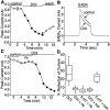
Application of serotonin causes a reduction of GABAA receptor currents in PFC pyramidal neurons.A, Time course of peak current evoked by GABA (100 μm) in the absence (control) or presence of serotonin (20 μm). The starting point for continuous application of serotonin is marked by the arrow. Application of serotonin caused an inhibition of GABAA receptor currents, whereas repeated application of GABA alone evoked a current that was stable during the whole-cell recording. B, Box plot summary of the percentage of reduction of peak GABAAcurrents produced by serotonin in a sample of 21 PFC pyramidal neurons.C, Plot of peak GABAA current as a function of time and ligand application. In the presence of the nonselective 5-HT receptor antagonist methysergide (meth; 10 μm), serotonin had little effect on GABAAcurrents; washing off the antagonist led to recovery of the serotonin inhibition (wash). The inset is a box plot summary showing the percentage of serotonin effect blocked by methysergide (n = 8). D, Representative current traces taken from the records used to constructC (at time points denoted by *).
Fig. 2.
Fig. 2.
Multiple 5-HT receptor mRNAs are coexpressed in single PFC pyramidal neurons. A, Photomicrograph of an ethidium bromide-stained gel showing the expression profile of 5-HT receptor mRNAs in PFC tissue by RT-PCR. B, Photomicrograph of an acutely isolated PFC pyramidal neuron. After recording, the cell was harvested by the patch electrode and subject to mRNA profiling. C, Expression profile of 5-HT receptor mRNAs in this PFC pyramidal neuron showing the coexpression of 5-HT1A, 5-HT2A, and 5-HT4 receptor mRNAs. C, Bar plot showing the coordinated expression of major 5-HT receptor mRNAs in a sample of 26 PFC pyramidal neurons. The extent of coexpression is indicated by the overlap of the bars.
Fig. 3.
Fig. 3.
Serotonergic modulation of GABAAcurrents is mediated by 5-HT2 receptors. A, Plot of peak GABAA current as a function of time and agonist application. The 5-HT2A/C agonist DOI (10 μm) caused an inhibition of GABAA currents.B, Representative current traces taken from the records used to construct A (at time points denoted by *).C, Plot of peak GABAA current as a function of time and ligand application. The 5-HT2 receptor antagonist risperidone (ris; 10 μm) blocked the effect of DOI on GABAA currents.D, Box plot summary of the percentage of reduction of peak GABAA currents produced by DOI or 5-HT in the absence and presence of different antagonists. The 5-HT2-class antagonist risperidone (ris; 10 μm) and ketanserin (ket; 20 μm) blocked the effect of DOI and 5-HT, whereas the 5-HT1-class antagonist cyanopindolol (cya; 20 μm) was ineffective.
Fig. 4.
Fig. 4.
5-HT2 modulation of GABAAcurrents is blocked by inhibition of PLCβ. A, Plot of peak GABAA current as a function of time and agonist application with U73122 (4 μm) or U74133 (4 μm) in the recording pipette. Dialysis with the PLCβ inhibitor U73122, but not its inactive analog U74133, blocked the DOI effect on GABAA currents. B, Box plot summary of the modulation of GABAA currents by DOI in the presence of U73122 (n = 5) or U74133(n = 5). C, D, Representative current traces taken from the records used to constructA (at time points denoted by *).
Fig. 5.
Fig. 5.
5-HT2 modulation of GABAAcurrents is dependent on activation of PKC. A, Time course of peak GABAA current in the presence of PMA (1 μm) or 4α-phorbol (1 μm). The starting point for continuous application of PMA or 4α-phorbol is marked by the arrow. The PKC activator PMA caused an irreversible reduction of GABAA receptor currents, whereas the inactive analog 4α-phorbol had little effect on GABAA currents.B, Box plot summary of the modulation of GABAA currents by PMA (n = 5) or 4α-phorbol (n = 5). C, Plot of peak GABAA current as a function of time and agonist application with or without heparin (10 U/ml) in the recording pipette. Blocking IP3-mediated Ca2+ release with heparin significantly attenuated the effect of DOI on GABAAcurrents. D, Box plot summary of the modulation of GABAA currents by DOI in the absence (control;n = 6) and presence of heparin (n = 5) or the high concentration (10 mm) of BAPTA (n = 5). E, Plot of peak GABAA current as a function of time and agonist application with or without PKC19–31 (20 μm) in the recording pipette. Dialysis with the PKC inhibitory peptide PKC19–31 blocked the effect of DOI on GABAA currents. F, Box plot summary of the modulation of GABAA currents by DOI in the absence (control; n = 5) and presence of PKC19–31 (n = 6).
Fig. 6.
Fig. 6.
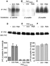
5-HT2 receptor activation increases PKC phosphorylation of GABAA receptor subunits.A, In vitro kinase activity of PKC immunoprecipitates toward a peptide derived from GABAAreceptor γ2 subunit. Brain slices containing PFC were incubated for 15 min in the absence (−) or presence of either DOI (20 μm) or PMA (1 μm). Lysates of these slices were used for immunoprecipitation with antibodies to PKCαβγ. PKC kinase activity of the immune complex was measured using a peptide derived from GABAA γ2 subunit as the substrate. Application of DOI or PMA enhanced PKC phosphorylation of the GABAA γ2 peptide. B, Including 1 μl of the PKC inhibitory peptide PKC19–31 (10 mg/ml) in thein vitro kinase reactions blocked PKC phosphorylation of the GABAA γ2 peptide induced by DOI or PMA.C, In vitro kinase activity of PKC immunoprecipitates toward a peptide derived from GABAAreceptor β2 subunit. Lysates of brain slices treated with or without DOI (20 μm) or PMA (1 μm) were immunoprecipitated with PKCαβγ antibodies, and PKC activity of the immune complex was measured using a GABAA β2 subunit peptide as the substrate. Application of DOI or PMA did not significantly alter PKC phosphorylation of the GABAA β2 peptide over the high basal level. D–F, Equal loading of PKC in the in vitro kinase assay. Half of the brain lysates used for in vitro kinase assay was immunoprecipitated with PKCαβγ antibodies and Western blotted with PKCαβγ antibodies. G, Histogram summary of the phosphorylation of GABAA receptor γ2 subunit-derived peptide and β2 subunit-derived peptide in PFC slices. Treatment with DOI or PMA significantly increased the phosphorylation of GABAA receptor γ2 subunit, which was blocked by the PKC inhibitory peptide PKC19–31 (left panel;n = 3; *p < 0.01), but did not significantly enhance the phosphorylation of GABAA receptor β2 subunit (right panel; n = 3).
Fig. 7.
Fig. 7.
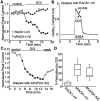
5-HT2 modulation of GABAAreceptor function requires anchoring of activated PKC to the channel by RACK1. A, Plot of peak GABAA current as a function of time and agonist application with RACK1-rVI peptide (40 μm) or the scrambled peptide sRACK1-rVI (40 μm) in the recording pipette. Disruption of the interaction between PKC and RACK1 with RACK1-rVI peptide, but not the scrambled peptide sRACK1-rVI, blocked the DOI effect on GABAA currents. B, Representative current traces taken from the records used to construct A (at time points denoted by *). C, Plot of peak GABAA current as a function of time and agonist application with AKAP[31–52] peptide (10 μm) in the recording pipette. D, Box plot summary of the modulation of GABAA currents by DOI in the presence of RACK1-rVI peptide (n = 11) or the scrambled peptide sRACK1-rVI (n = 5), or the AKAP[31–52] peptide (n = 5).

Similar articles

Cited by

References

    1. Abi-Dargham A, Laruelle M, Aghajanian GK, Charney D, Krystal J. The role of serotonin in the pathophysiology and treatment of schizophrenia. J Neuropsychiatry Clin Neurosci. 1997;9:1–17. - PubMed
    1. Andrade R. Regulation of membrane excitability in the central nervous system by serotonin receptor subtypes. Ann NY Acad Sci. 1998;861:190–203. - PubMed
    1. Araneda R, Andrade RA. 5-HT2 and 5-HT1A receptors mediate opposing responses on membrane excitability in rat association cortex. Neuroscience. 1991;40:399–412. - PubMed
    1. Battaini F, Pascale A, Paoletti R, Govoni S. The role of anchoring protein RACK1 in PKC activation in the aging rat brain. Trends Neurosci. 1997;20:410–415. - PubMed
    1. Baxter G, Kennett G, Blaney F, Blackburn T. 5-HT2 receptor subtypes: a family re-united? Trends Pharmacol Sci. 1995;16:105–110. - PubMed

Publication types

MeSH terms

LinkOut - more resources