. 2001 Sep 1;21(17):6553-60.
doi: 10.1523/JNEUROSCI.21-17-06553.2001.

Modulation of excitability by alpha-dendrotoxin-sensitive potassium channels in neocortical pyramidal neurons

Affiliations

Modulation of excitability by alpha-dendrotoxin-sensitive potassium channels in neocortical pyramidal neurons

J M Bekkers et al. J Neurosci. .

Abstract

Many neurons transduce synaptic inputs into action potentials (APs) according to rules that reflect their intrinsic membrane properties. Voltage-gated potassium channels, being numerous and diverse constituents of neuronal membrane, are important participants in neuronal excitability and thus in synaptic integration. Here we address the role of dendrotoxin-sensitive "D-type" potassium channels in the excitability of large pyramidal neurons in layer 5 of the rat neocortex. Low concentrations of 4-aminopyridine or alpha-dendrotoxin (alpha-DTX) dramatically increased excitability: the firing threshold for action potentials was hyperpolarized by 4-8 mV, and the firing frequency during a 1-sec-long 500 pA somatic current step was doubled. In nucleated outside-out patches pulled from the soma, alpha-DTX reversibly blocked a slowly inactivating potassium current that comprised approximately 6% of the total. This current first turned on at voltages just hyperpolarized to the threshold for spiking and activated steeply with depolarization. By assaying alpha-DTX-sensitive current in outside-out patches pulled from the axon and primary apical dendrite, it was found that this current was concentrated near the soma. We conclude that alpha-DTX-sensitive channels are present on large layer 5 pyramidal neurons at relatively low density, but their strategic location close to the site of action potential initiation in the axon may ensure that they have a disproportionate effect on neuronal excitability. Modulation of this class of channel would generate a powerful upregulation or downregulation of neuronal output after the integration of synaptic inputs.


Figures

Fig. 1.
Fig. 1.
Bath application of 100 μm 4-AP increases the excitability of large layer 5 cortical pyramidal neurons.A, Action potentials (APs) elicited by 1-sec-long 200 pA (left) or 500 pA (right) current steps in the same neuron before (top) or after (bottom) addition of 100 μm 4-AP to the external solution. For this neuron, 200 pA was just suprathreshold for the firing of an AP in control solution. Note that all membrane potentials have been corrected for the measured liquid junction potential (−7 mV; see Materials and Methods).B, Mean number of APs elicited by 1-sec-long current steps to the values given on the x-axis (●,Control; ○, 4-AP; n= 6 cells; ±SEM). 4-AP reduces the amount of current required to fire APs. C, Mean instantaneous AP frequency (i.e., reciprocal of interval between adjacent APs) versus the interval number, measured during a 1-sec-long 500 pA current step. Same data set and symbols as in B. 4-AP increases the firing rate of APs during a fixed current step.
Fig. 2.
Fig. 2.
Bath or puffer application of 1–2 μm α-dendrotoxin (α-DTX) increases excitability, like 100 μm 4-AP.A, Data recorded in the same neuron before (top) and after (bottom) puffing 2 μm α-DTX near the soma. B,C, Summary plots like those in Figure 1 (●,Control; ○, α-DTX;n = 4 cells; ±SEM). Like 4-AP, α-DTX reduces the current required to fire APs and increases AP firing rate.
Fig. 3.
Fig. 3.
4-AP (left panels) and α-DTX (right panels) both hyperpolarize the firing threshold for APs, but differ in their effects on AP half-width.A, APs near the start of a 1-sec-long 500 pA current step, recorded before (thin line) and after (thick line) application of 100 μm 4-AP (left) or 2 μm α-DTX (right). Each panel is from a different neuron.Horizontal dashed lines indicate the firing thresholds for later APs with and without drugs. B, Mean firing threshold for each AP in a train during a 1-sec-long 500 pA current step versus the AP number. ●, Control; ○, 100 μm 4-AP (left panel; n= 5 cells; ±SEM) or 2 μm α-DTX (right panel; n = 4 cells; ±SEM). Both 4-AP and α-DTX hyperpolarize the firing threshold throughout the train.C, Mean half-width for each AP in the train, for the same data set as in B. 4-AP significantly increases the half-width of all but the first AP (left panel), whereas α-DTX has no effect (right panel).Insets show the sixth AP in an illustrative train, recorded in control (thin line) or in drug solution (thick line).
Fig. 4.
Fig. 4.
Two micromolar α-DTX has a small inhibitory effect on K+ current recorded in voltage-clamped nucleated outside-out patches. A, Amplitudes of outward K+ current (top, ●) and inward Na+ current (bottom, ▪) measured simultaneously in a nucleated patch during voltage-clamp steps from −117 mV to +3 mV, plotted against time during the experiment. K+ current amplitude was averaged over a window 100–150 msec after the step onset, to avoid A-current; Na+ current was measured at the peak. Puffer application of 2 μm α-DTX (horizontal bar) caused a small inhibition of the K+current, whereas 0.5 μm tetrodotoxin (TTX) included in the puffer solution fully blocked the Na+ current, confirming that the toxins were bathing the patch. Insets show, on slow (top) and fast (bottom) time bases, the currents measured at the numbered time points. Unlabeled trace in the top inset is the α-DTX-sensitive current, obtained by subtraction. B, Mean normalized K+ current (●) and Na+ current (▪) measured in 11 experiments of this sort. The dashed line is drawn by eye to emphasize the small mean inhibition produced by 2 μm α-DTX: 6% on average.
Fig. 5.
Fig. 5.
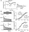
Bath application of TEA reduces the amount of the delayed rectifier current (IK) in nucleated patches, facilitating measurement of the activation properties of α-DTX-sensitive current. A, Averaged, normalized amplitudes of K+ current measured in nucleated patches bathed in external solution containing 30 mm TEA plus 0.4 μm TTX, plotted against time during the experiment. Currents were evoked by voltage-clamp steps from −117 to +43 mV, and their amplitudes were measured by averaging over a window 100–150 msec after the start of the step. Puffer application of TEA–TTX solution plus 2 μm α-DTX (horizontal bar) reversibly inhibited the K+ current by 28% on average. Insetshows typical currents recorded in one patch, averaged during the control and wash periods (trace a) and during the period of toxin application (trace b). Unlabeled trace is the subtraction of trace b froma, the α-DTX-sensitive current. B, Families of voltage-clamped K+ currents recorded in nucleated patches in TEA–TTX bath solution in the absence (top) and presence (bottom) of 2 μm α-DTX, applied by puffer. The pulse protocol is shown at the top. Square brackets over the current traces indicate the window over which the amplitude was measured for the activation plots (100–150 msec after the start of the test pulse). Note that the recordings were obtained from different patches. C, Averaged, normalized activation plots in the absence (●; n = 7 patches) and presence (○;n = 5 patches) of 2 μm α-DTX, all in TEA–TTX solution. Superimposed smooth curves are the results of fits of the Boltzmann function with the indicated fit parameters. D, Activation plot for the α-DTX-sensitive current, obtained by scaling down the + α-DTX data in C by 28%, subtracting this from the − α-DTX data, and renormalizing the result. Thesuperimposed continuous curve is a Boltzmann fit to the points, giving the indicated fit parameters. The dashed curve is the mean activation plot forIK measured in nucleated patches from the same neurons, reported in Bekkers (2000a).
Fig. 6.
Fig. 6.
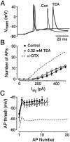
α-DTX-sensitive channels are functionally different from TEA-sensitive IK channels.A, APs near the start of a 500 pA current step, recorded in the same neuron before (Con, thin line) and after (TEA, thick line) perfusion of bath solution containing 0.32 mm TEA. This concentration of TEA blocks ∼6% ofIK, which is the same percentage of the total slow K+ current as is blocked by 2 μm α-DTX. The horizontal dashed lineindicates that the AP firing threshold is little affected by TEA (seeC); however, spike broadening is clearly apparent in TEA, probably because of its inhibition of the fast Ca2+-activated K+ current,IC. B, Mean number of APs elicited by 1-sec-long current steps to the indicated values (●,Control; ○, TEA; n= 7 cells). TEA (0.32 mm) has only a weak effect on the current required to fire APs. For comparison, the dashed line shows the effect of 2 μm α-DTX, from Figure 2B. C, Mean firing threshold for each AP in a train during a 1-sec-long 500 pA current step, plotted against the AP number. Same symbols and data set as inB. For comparison, the dashed line shows the effect of α-DTX, from Figure 3B. Unlike α-DTX, TEA has no significant effect on the firing threshold.
Fig. 7.
Fig. 7.
α-DTX-sensitive channels, assayed in conventional outside-out patches, are concentrated near the soma.A, Amplitude of K+ current measured during a voltage-clamp step from a prepulse (−117 mV for 500 msec) to a test pulse of +43 mV (repeated 5 times and averaged), plotted against time during the experiment. Amplitude was averaged over a window 100–150 msec after the step onset. α-DTX (2 μm) was applied by puffer (horizontal bar). Labeled traces in the inset are averages of the currents enclosed by square brackets in the time course plot; theunlabeled trace is the subtraction of trace b from a. This patch was pulled from the primary apical dendrite 105 μm from the soma. B, Amplitude of α-DTX-sensitive current (each point obtained from a separate outside-out patch using the averaged-subtraction method described in A) plotted against the distance from the soma that the patch was pulled. Patches were from either the axon (○) or the primary apical dendrite (●). The continuous straight line is given by I = 3.2 − 0.009d, where I is the mean amplitude of the α-DTX-sensitive current in picoamperes, and d is the distance from the soma in micrometers.

References

    1. Albert JL, Nerbonne JM. Calcium-independent depolarization-activated potassium currents in superior colliculus-projecting rat visual cortical neurons. J Neurophysiol. 1995;73:2163–2178. - PubMed
    1. Bekkers JM. Properties of voltage-gated potassium currents in nucleated patches from large Layer 5 cortical pyramidal neurons of the rat. J Physiol (Lond) 2000a;525:593–609. - PMC - PubMed
    1. Bekkers JM. Distribution and activation of voltage-gated potassium channels in cell-attached and outside-out patches from large Layer 5 cortical pyramidal neurons of the rat. J Physiol (Lond) 2000b;525:611–620. - PMC - PubMed
    1. Bekkers JM. Distribution of slow AHP channels on hippocampal CA1 pyramidal neurons. J Neurophysiol. 2000c;83:1756–1759. - PubMed
    1. Berger T, Larkum ME, Lüscher H-R. High Ih channel density in the distal apical dendrite of Layer V pyramidal cells increases bidirectional attenuation of EPSPs. J Neurophysiol. 2001;85:855–868. - PubMed

Publication types

MeSH terms

LinkOut - more resources