. 2001 Sep 1;21(17):6569-76.
doi: 10.1523/JNEUROSCI.21-17-06569.2001.

Recruitment of the mitochondrial-dependent apoptotic pathway in amyotrophic lateral sclerosis

Affiliations

Recruitment of the mitochondrial-dependent apoptotic pathway in amyotrophic lateral sclerosis

C Guégan et al. J Neurosci. .

Abstract

Molecular mechanisms of apoptosis may participate in motor neuron degeneration produced by mutant superoxide dismutase-1 (mSOD1), the only proven cause of amyotrophic lateral sclerosis (ALS). Consistent with this, here we show that the proapoptotic protein Bax translocates from the cytosol to the mitochondria, whereas cytochrome c translocates from the mitochondria to the cytosol in spinal cords of transgenic mSOD1 mice during the progression of the disease. Concomitantly, caspase-9 is activated in the spinal cord of transgenic mSOD1 mice. Only in end-stage transgenic mSOD1 mice is the downstream caspase-7 activated and the inhibitor of apoptosis, XIAP, cleaved. These results indicate a sequential recruitment of molecular elements of the mitochondrial-dependent apoptotic pathway in transgenic mSOD1 mice. We also provide immunohistochemical evidence that cytochrome c translocation occurs in the spinal cord of sporadic ALS patients. Collectively, these data suggest that the mitochondrial-dependent apoptotic pathway may contribute to the demise of motor neurons in ALS and that targeting key molecules of this cascade may prove to be neuroprotective.


Figures
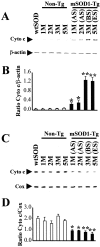
Fig. 1.
Fig. 1.
Western blot analysis of cytochrome c. Protein extracts from cytosol (A, B) and mitochondria (C, D) were obtained from spinal cords of wtSOD1 mice (3 months old), of transgenic mSOD1 mice at asymptomatic stage (AS; 1 and 2 months old), at the beginning of symptoms (BS; 3 months old), at end stage (ES; 5 months old), and of their age-matched nontransgenic mice littermates (Non-Tg). β-Actin and COX were used as internal controls for the cytosolic and mitochondrial fractions, respectively. The results of densitometric analysis are shown in B and D. The mean values (n = 4–6 per group; mean ± SEM) obtained for transgenic mSOD1 mice (black columns) were compared with the values of their age-matched littermates (white columns). *p < 0.05; **p < 0.01; Student's ttest.
Fig. 2.
Fig. 2.
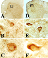
Cytochrome c immunostaining in spinal cord from age-matched littermate nontransgenic mice (A–C) and mSOD1 transgenic mice at end stage (D–F). In nontransgenic controls, the staining was faint and punctate (A–C), whereas in end-stage transgenic mSOD1 mice, it was robust and diffuse (D–F).
Fig. 3.
Fig. 3.
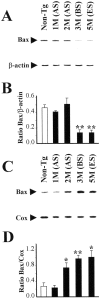
Subcellular localization of Bax. Bax expression was analyzed by Western blot in cytosolic (A,B) and mitochondrial (C,D) fractions of spinal cords from transgenic mSOD1 mice (black columns) at the asymptomatic stage (AS), at the beginning of symptoms (BS), and at the end stage (ES) and was compared with their age-matched nontransgenic littermates (Non-Tg; white columns). Represented values (n = 4–6 per group) correspond to mean ± SEM. *p < 0.05; **p < 0.01; Student's t test.
Fig. 4.
Fig. 4.
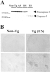
Activation of caspase-9 in spinal cords of transgenic mSOD1 mice. A, Using an antibody recognizing both forms of caspase-9, the expression of pro-caspase-9 (52 kDa) and its active fragment (35 kDa) was analyzed by Western blot in cytosolic fractions of spinal cords from transgenic mSOD1 mice at the asymptomatic stage (AS), at the beginning of symptoms (BS), and at the end stage (ES) and was compared with their age-matched nontransgenic littermates (Non-Tg). Each lane corresponds to a different animal. B, The immunolocalization of active caspase-9 was performed on spinal cord sections from transgenic (Tg) mSOD1 mice at end stage (ES) and their age-matched nontransgenic (Non-Tg) littermates.
Fig. 5.
Fig. 5.
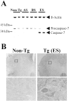
Activation of caspase-7 in spinal cords of transgenic mSOD1 mice. A, Using an antibody recognizing both forms of caspase-7, the expression of pro-caspase-7 (37 kDa) and its cleaved fragment (28 kDa) was analyzed by Western blot in cytosolic fractions of spinal cords from transgenic mSOD1 mice at the asymptomatic stage (AS), at the beginning of symptoms (BS), and at the end stage (ES) and was compared with their age-matched nontransgenic littermates (Non-Tg). The top panel corresponds to β-actin. Each lane corresponds to a different animal and is representative of four to six animals per group.B, The immunolocalization of active caspase-7 was performed on spinal cord sections from transgenic (Tg) mSOD1 mice at end stage (ES) and their age-matched nontransgenic (Non-Tg) littermates.
Fig. 6.
Fig. 6.
Cleavage of XIAP in spinal cords of transgenic mSOD1 mice. XIAP expression was analyzed by Western blot in cytosolic fractions of spinal cords from transgenic mSOD1 mice at the asymptomatic stage (AS), at the beginning of symptoms (BS), and at the end stage (ES) and was compared with their age-matched nontransgenic littermates (Non-Tg). The top panel corresponds to β-actin. Each lane corresponds to a different animal and is representative of four to six animals per group.
Fig. 7.
Fig. 7.
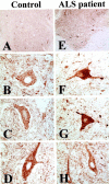
Immunostaining for cytochrome c of spinal cord from ALS patients and controls. The immunostaining for cytochrome c was processed on spinal cord sections obtained from control (A–D; n = 7) and from ALS (E–H; n = 6) patients. In controls, cytochrome c-positive neurons were faintly stained and punctuate (A–D). In ALS cases, some neurons exhibited a control-like appearance (H), whereas most others were much more intensely and evenly immunostained (F, G).

Similar articles

Cited by

References

    1. Adams JH, Corsellis JAN, Duchen LW. Greenfield's neuropathology. Butler and Tanner; London: 1984.
    1. Almer G, Vukosavic S, Romero N, Przedborski S. Inducible nitric oxide synthase upregulation in a transgenic mouse model of familial amyotrophic lateral sclerosis. J Neurochem. 1999;72:2415–2425. - PubMed
    1. Brown RH., Jr Superoxide dismutase in familial amyotrophic lateral sclerosis: models for gain of function. Curr Opin Neurobiol. 1995;5:841–846. - PubMed
    1. Bruijn LI, Cleveland DW. Mechanisms of selective motor neuron death in ALS: insights from transgenic mouse models of motor neuron disease. Neuropathol Appl Neurobiol. 1996;22:373–387. - PubMed
    1. Dal Canto MC, Gurney ME. Neuropathological changes in two lines of mice carrying a transgene for mutant human Cu,Zn SOD, and in mice overexpressing wild type human SOD: a model of familial amyotrophic lateral sclerosis (FALS). Brain Res. 1995;676:25–40. - PubMed

Publication types

MeSH terms