. 2001 Sep 1;21(17):6588-96.
doi: 10.1523/JNEUROSCI.21-17-06588.2001.

Chronic blockade of glutamate receptors enhances presynaptic release and downregulates the interaction between synaptophysin-synaptobrevin-vesicle-associated membrane protein 2

Affiliations

Chronic blockade of glutamate receptors enhances presynaptic release and downregulates the interaction between synaptophysin-synaptobrevin-vesicle-associated membrane protein 2

A Bacci et al. J Neurosci. .

Abstract

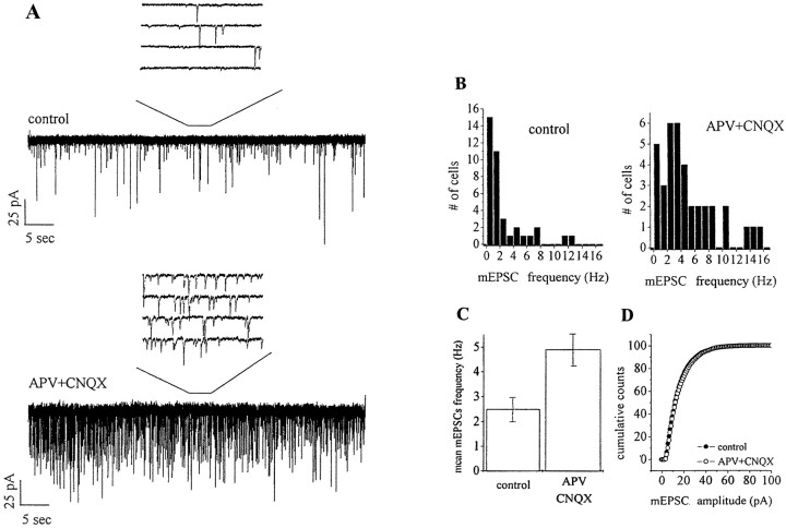
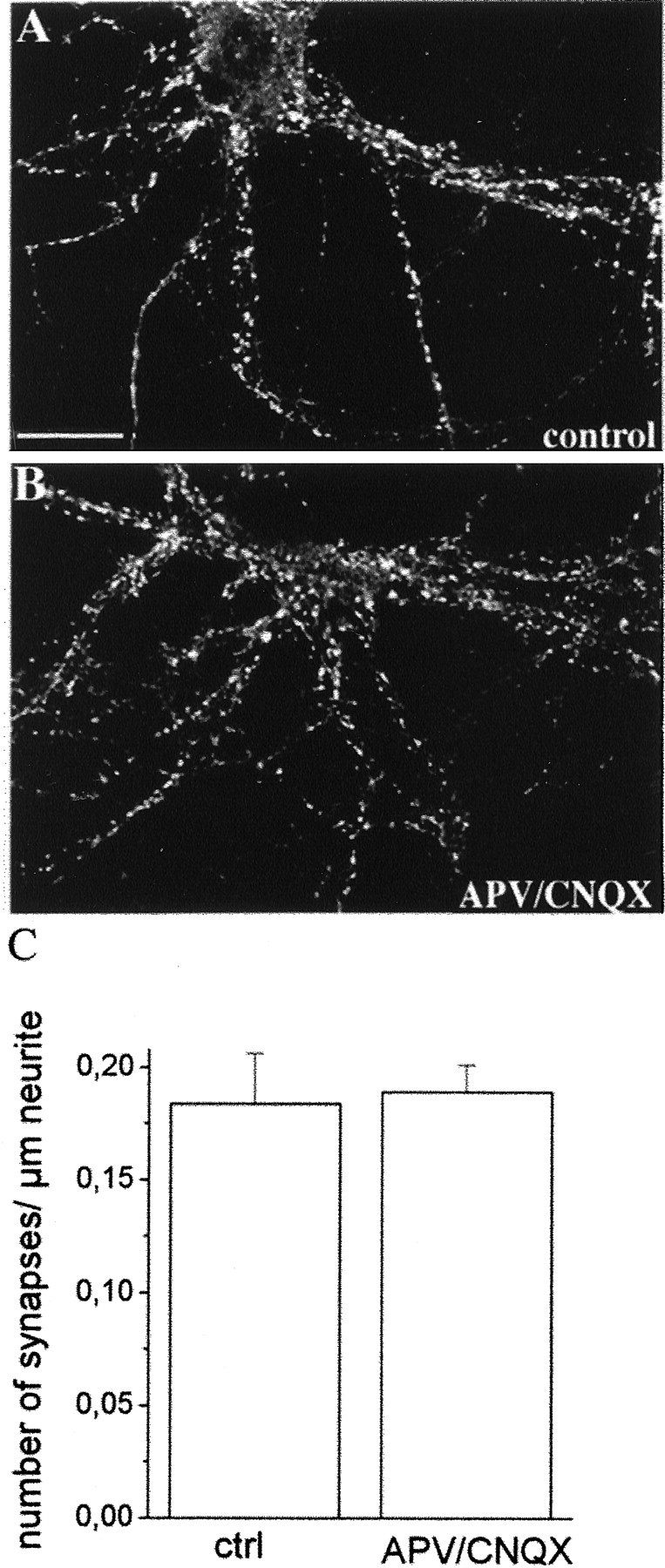
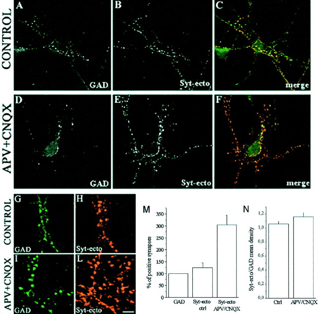
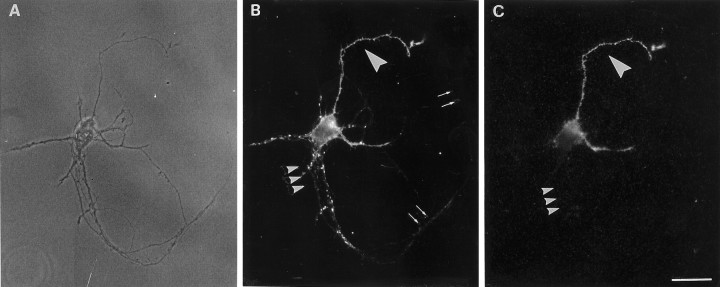
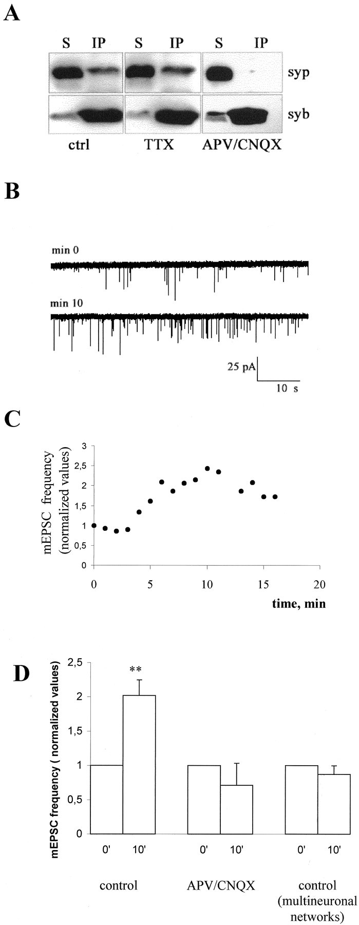
During development of neuronal circuits, presynaptic and postsynaptic functions are adjusted in concert, to optimize interneuronal signaling. We have investigated whether activation of glutamate receptors affects presynaptic function during synapse formation, when constitutive synaptic vesicle recycling is downregulated. Using primary cultures of hippocampal neurons as a model system, we have found that chronic exposure to both NMDA and non-NMDA glutamate receptor blockers during synaptogenesis produces an increase in miniature EPSC (mEPSC) frequency, with no significant changes in mEPSC amplitude or in the number of synapses. Enhanced synaptic vesicle recycling, selectively in glutamatergic nerve terminals, was confirmed by the increased uptake of antibodies directed against the lumenal domain of synaptotagmin. No increased uptake was detected in neuronal cultures grown in the chronic presence of TTX, speaking against an indirect effect caused by decreased electrical activity. Enhanced mEPSC frequency correlated with a reduction of synaptophysin-synaptobrevin-vesicle-associated membrane protein 2 (VAMP2) complexes detectable by immunoprecipitation. Intracellular perfusion with a peptide that inhibits the binding of synaptophysin to synaptobrevin-VAMP2 induced a remarkable increase of mEPSC frequency in control but not in glutamate receptor blocker-treated neurons. These findings suggest that activation of glutamate receptors plays a role in the downregulation of the basal rate of synaptic vesicle recycling that accompanies synapse formation. They also suggest that one of the mechanisms through which this downregulation is achieved is an increased interaction of synaptophysin with synaptobrevin-VAMP2.


Figures

Fig. 1.
Fig. 1.
Differential effect of glutamate receptor chronic blockade on mEPSC frequency and amplitude. A,Representative recordings from 15-d-old hippocampal neurons, maintained in control medium or in the presence of 100 μm APV and 20 μm CNQX. B, Distribution of mEPSC frequencies in control and APV–CNQX-treated cells. C, D, Analysis of the average mEPSC frequency (C) and cumulative amplitude distribution (D) in control and treated cells reveals the specific effect of glutamatergic chronic blockade on the frequency, but not the amplitude, of mEPSCs.
Fig. 2.
Fig. 2.
Glutamatergic block does not affect synapse number. Immunofluorescence stainings of control (A) and APV–CNQX-treated (B) cultures with antibodies directed against the synaptic vesicle protein SV2. Immunofluorescent puncta represent sites of synaptic contacts. Scale bar, 20.8 μm. C, Histogram showing the quantitative analysis of the number of synapses present for micrometer of neurite length. No significant difference is detectable between control and APV–CNQX-treated neurons.
Fig. 3.
Fig. 3.
Chronic treatment with glutamatergic blockers upregulates synaptic vesicle recycling. We incubated 15-d-old neurons from control (A–C,G–L) or APV–CNQX-treated (D–F, M–P) cultures for 5 min (A–H, M, and N, histogram in Q) or 25 min (histogram inQ) in the presence of Syt-ecto Abs in KRH containing glutamate receptor blockers. In a set of experiments, incubation was performed for 3 min in the presence of 55 mm KCl (I,L, O,P, histogram in Q). After this incubation, neurons were washed, fixed, detergent-permeabilized, reacted with fluorescein-conjugated goat anti-rabbit IgGs (B, E, G,I, M,O), and counterstained with antibodies against SV2 followed by rhodamine-conjugated goat anti-mouse IgGs (A,D, H, L, N,P). Puncta of immunoreactivity represent presynaptic nerve terminals that outline perikarya and dendrites. Syt-ecto Abs are internalized only at few synaptic contacts in control cultures (B, G) and in most synaptic contacts in APV–CNQX-treated neurons (E, M).C,F, Fluorescein- and rhodamine-merged images. Incubation in the presence of KCl results in Syt-ecto Ab internalization in virtually all synaptic contacts, both in control (I) and treated (O) cultures. Scale bars: A–C, 20.8 μm;D–F, 25 μm; G–P, 3.5 μm.Q, Histogram showing the quantitative evaluation of Syt-ecto-positive synapses in control and in APV–CNQX-treated neurons.
Fig. 4.
Fig. 4.
Blockade of glutamate receptors affect glutamatergic, but not GABAergic, presynaptic nerve terminals.A–F, Immunofluorescence images of control (A–C) and APV–CNQX-treated (D–F) 15-d-old hippocampal neurons incubated for 5 min in the presence of Syt-ecto Abs (B,E) and counterstained with antibodies against the synthetic enzyme GAD (A, D). In control neurons, the few synapses labeled by Syt-ecto Abs are generally GAD positive (A, B, see also merged image inC). In APV–CNQX-treated neurons, the synapses positive for Syt-ecto Abs (E) largely exceed GABAergic terminals (D) (see merged image inF). G–L, Details of control (G, H) and APV–CNQX-treated (I, L) neurons stained for Syt-ecto Abs (H, L) and for GAD (G,I). Scale bar: A–F, 30.7 μm;G–L, 10.2 μm. M, Histogram showing the quantitative analysis of the percentages of Syt-ecto Ab-positive synapses in control and APV–CNQX-treated neurons, normalized to the number of GAD-positive synapses. N, Histogram showing that the amount of internalized Syt-ecto Abs is not significantly different in GABAergic terminals of control and treated neurons. Syt-ecto Ab intensity values are normalized to GAD immunoreactivity.
Fig. 5.
Fig. 5.
Basal rate of exo-endocytosis is downregulated after contact with a postsynaptic cell. A–C, Synaptic vesicle exo-endocytosis occurs at different basal rates, in distinct compartments of a same 14-d-old neuron grown in a microisland, depending on the contact with the postsynaptic target. An efficient internalization of Syt-ecto Abs takes place in basal conditions in the isolated axon (C, large arrowhead) but not at autaptic contacts (C, small arrowheads) of the same neuron, shown as a bright field in A. B, Double immunolabeling of the same neuron for the synaptic vesicle protein SV2. Scale bar, 18.75 μm.
Fig. 6.
Fig. 6.
Impairment in the formation of the synaptophysin-synaptobrevin–VAMP2 complex is detectable in APV–CNQX-treated neurons and is responsible for the increase in mEPSC frequency. A, Triton X-100 extracts of control and APV–CNQX-treated 12-d-old neurons were immunoprecipitated using the monoclonal antibody against synaptobrevin–VAMP2 (syb). Immunoprecipitates (IP) and their corresponding supernatants (S) were analyzed using polyclonal antibodies against synaptophysin (syp). Note that synaptophysin is efficiently immunoprecipitated from control cultures and is almost completely detectable in the supernatant of APV–CNQX- treated cultures. B, Representative recordings from a single 12-d-old hippocampal neuron forming autaptic contacts, intracellularly perfused via the patch pipette with a peptide corresponding to the 32-residue-long N-terminal segment of synaptobrevin–VAMP2, which inhibits complex formation. C, Time course of the increase in the frequency of mEPSCs recorded from the same neuron as inB. D, Histogram showing the increase in mEPSC frequency occurring in control neurons 10–11 min after the beginning of recordings (p < 0.002). No effect is detectable in either single neurons maintained in APV–CNQX (p > 0.1) or in neurons grown in polyneural networks (p > 0.1). Values are normalized to the first or second minute of recording.

References

    1. Ahmari SE, Buchanan J, Smith SJ. Assembly of presynaptic active zones from cytoplasmic transport packets. Nat Neurosci. 2000;3:445–451. - PubMed
    1. Bartlett WP, Banker GA. An electron microscopic study of the development of axons and dendrites by hippocampal neurons in culture. I. Cells which develop without intercellular contacts. J Neurosci. 1984;4:1944–1953. - PMC - PubMed
    1. Becher A, Drenckhahn A, Pahner I, Margittai M, Jahn R, Ahnert-Hilger G. The synaptophysin-synaptobrevin complex: a hallmark of synaptic vesicle maturation. J Neurosci. 1999a;19:1922–1931. - PMC - PubMed
    1. Becher A, Drenckhahn A, Pahner I, Ahnert-Hilger G. The synaptophysin-synaptobrevin complex is developmentally upregulated in cultivated neurons but is absent in neuroendocrine cells. Eur J Cell Biol. 1999b;78:650–656. - PubMed
    1. Benson DL, Watkins FH, Stewaed O, Banker GA. Characterization of GABAergic neurons in hippocampal cell cultures. J Neurocytol. 1994;23:279–295. - PubMed

Publication types

MeSH terms

LinkOut - more resources