. 2001 Aug 28;98(18):10386-91.
doi: 10.1073/pnas.151257998. Epub 2001 Aug 21.

The nuclear factor kappa B (NF-kappa B): a potential therapeutic target for estrogen receptor negative breast cancers

Affiliations

The nuclear factor kappa B (NF-kappa B): a potential therapeutic target for estrogen receptor negative breast cancers

D K Biswas et al. Proc Natl Acad Sci U S A. .

Abstract

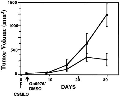
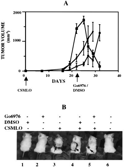
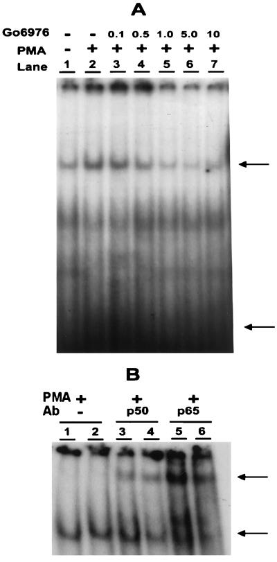
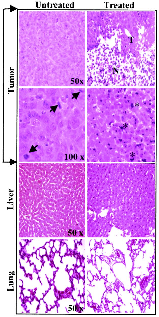
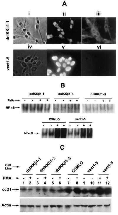
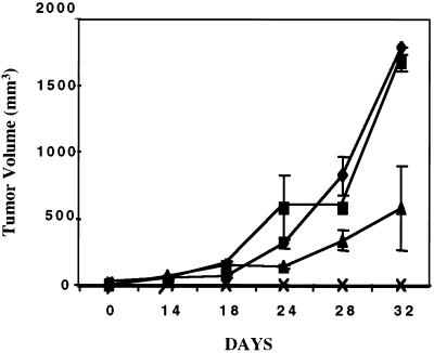
The effect of a kinase inhibitor Go6796 on growth of epidermal growth factor (EGF)-stimulated estrogen receptor negative (ER-) breast cancer cells in vivo and role of nuclear factor kappa B (NF-kappaB) on tumorogenesis have been investigated. This was studied in an animal model by implanting ER- mouse mammary epithelial tumor cells (CSMLO) in syngeneic A-J mice. (i) Local administration of Go6976 an inhibitor of protein kinases C alpha and beta inhibited growth of tumors and caused extensive necrotic degeneration and regression of the tumors without causing any microscopically detectable damage to the vital organs liver and lung. (ii) Stable expression of dominant-negative mutants of the beta subunit (dnIkkbeta) of the inhibitory kappa B (IkappaB) kinase (dnIkk) that selectively blocked activation of NF-kappaB caused loss of tumorigenic potential of CSMLO cells. Stable expression of dnIkkbeta also blocked phorbol 12-myristate 13-acetate (PMA)-induced activation of NF-kappaB and overexpression of cyclin D1, concomitantly with the loss or reduced tumorigenic potential of these cells. Thus, results from in vivo and in vitro experiments strongly suggest the involvement of NF-kappaB in ER- mammary epithelial cell-mediated tumorigenesis. We propose that blocking NF-kappaB activation not only inhibits cell proliferation, but also antagonizes the antiapoptotic role of this transcription factor in ER- breast cancer cells. Thus, NF-kappaB is a potential target for therapy of EGFR family receptor-overexpressing ER- breast cancers.


Figures

Figure 1
Figure 1
Inhibition of growth of tumors by Go6976. Tumors were generated in female A-J mice by implantation of CSMLO cells (106) on day 0 (indicated by the arrow). Go6976 (0.5 ml of 0.05 mM solution in 0.1% DMSO) was administered 48 h later to a group of five animals, twice a week locally at the site of implantation of cells (♦). Control group (five animals) received 0.5 ml of 0.1% DMSO similarly (□). Growth of tumors was monitored by measurements of tumor volume at regular intervals (32).
Figure 2
Figure 2
Regression of tumors by Go6976. (A) CSMLO cells were implanted on day 0 (indicated by the arrow) as for Fig. 1. Go6976 was administered on day 21 (indicated by the arrow), locally under the tumors of the five animals with comparatively larger tumors (■). Another five animals received the same volume of 0.1% DMSO (♦) on the same day, and the control group of five animals received nothing (□). (B) One representative of five animals from each group. Animals 1 and 2 are DMSO- and Go6976-treated non-tumor-bearing animals, respectively. Animal 3 is tumor-bearing and without treatment. Animals 4 and 5 represent tumor-bearing groups treated with DMSO and Go6976, respectively. Animal 6 is an A-J mouse that received nothing.
Figure 3
Figure 3
Active NF-κB complex in CSMLO cells: Stimulation by PMA and inhibition by Go6976. (A) Nuclear extracts (5 μg protein) from control and PMA-treated (20 ng/ml for 18 h) CSMLO cells were incubated in a standard EMSA reaction mixture containing [γ-32P]-labeled double-stranded oligonucleotide plus Go6976 at the indicated μM concentrations for 48 h and subjected to nondenaturing PAGE (7, 31). The autoradiographic signals of the retarded NF-κB–[32P]DNA complex is indicated by the upper arrow and the free [γ-32P]-labeled probe by the lower arrow. (B) The NF-κB–[32P]DNA complex was characterized by supershift assay with anti-p50 (lanes 3 and 4) or p65 (B, lanes 5 and 6) antibodies. Nuclear extracts were incubated with specific antibodies for 15 min at room temperature, followed by incubation for an additional 30 min in the presence of [γ-32P] double-stranded NF-κB oligonucleotide, and subjected to EMSA as described (7, 31). The supershifted complexes are indicated by the upper arrow.
Figure 4
Figure 4
Histology of tissues from untreated and Go6976-treated tumor-bearing animals. Tumor growth and treatment conditions are the same as described for Fig. 1. Tumor, liver, and lung tissues from untreated and treated tumor bearing animals were dissected 11 days after the initiation of treatment and 32 days after implantation of the cells. Tissues were processed for hematoxylin/eosin (H&E) staining, examined under a light microscope, and photographed at the indicated magnifications. Arrows show mitotic cells in untreated tumor tissue. Residual tumor (T) and necrotic cells (N) of the treated tumor are shown. Stars in the treated block indicate pycnotic cells with apparent fragmentation and clumping of nuclear DNA. Treated and untreated liver and lung tissues did not show significant microscopically detectable damages and were not different from liver and lung tissues of normal mice without tumors (not shown).
Figure 5
Figure 5
Inhibition of NF-κB activation and down stream events by stable expression of dominant-negative IκB-kinase β (dnIkkβ). (A Upper) dnIkkβ-Expressing transfectant cells by light microscopy (i), 4′,6-diamidino-2-phenylindole (DAPI)-stained nuclei (ii), and immunofluorescence with anti-FLAG antibody in the presence of the secondary antibody (iii). The positive signals show dnIkkβ-conjugated FLAG protein in the cytoplasm. (Lower) Processed vector-control plasmid-transfected CSMLO cells in which no FLAG protein could be detected (vi). (B Upper) Active NF-κB was determined by its [γ-32P]DNA binding activity by EMSA, in three dnIkkβ-expressing stable transfectants (dnIkkβ1-1, dnIkkβ1-3, and dnIkk-β1-5) and (Lower) in parent CSMLO cells and vector control plasmid expressing transfectant (vect1-5). (C Upper) The level of ccD1 in the same three-dnIkkβ-expressing CSMLO and vector control transfected cells, as measured by Western blot analysis. (Lower) Actin analyzed similarly by immunodetection with anti-β-actin antibody that serves as a loading control.
Figure 6
Figure 6
Loss of tumorigenic potential of CSMLO cells by the stable expression of pdnIkkβ in A-J mice. Tumors were generated in female A-J mice by implanting either CSMLO cells (five animals), or two vector control plasmid transfected clones (vect1-3 and vect1-5, three animals per clone) or two pdnIkkβ-expressing clones (pdnIkkβ1-1 and pdnIkkβ-1-3, three animals per clone). Average with standard deviations of CSMLO-administered animals (♦), vector-control-administered animals (■, six animals), and one of the two pdnIkkβ-expressing-clones (pdnIkkβ1-3)-administered animals (▵, three animals) are plotted. The other pdnIkkβ1-1 clone did not form any tumor even 32 days after implantation of the cells in any one of the three animals (ρ).

References

    1. Jordan V C. Breast Cancer Res Treat. 1995;36:267–285. - PubMed
    1. Hedden A, Muller V, Jensen E V. Ann NY Acad Sci. 1995;761:109–120. - PubMed
    1. Nakshatri H, Bhat-Nakshatri P, Martin D A, Goulet R J, Jr, Sledge G W. Mol Cell Biol. 1997;17:3629–3639. - PMC - PubMed
    1. Bhat-Nakshatri P, Newton T R, Goulet R, Jr, Nakshatri H. Proc Natl Acad Sci USA. 1998;95:6971–6976. - PMC - PubMed
    1. Sovak M A, Bellas R E, Kim D W, Zaneiski G J, Rogers A E, Traish A M, Sonenshein G E. J Clin Invest. 1997;100:2952–2960. - PMC - PubMed

Publication types

MeSH terms