. 2001 Aug 28;98(18):10085-9.
doi: 10.1073/pnas.181347998. Epub 2001 Aug 21.

Human DNA replication initiation factors, ORC and MCM, associate with oriP of Epstein-Barr virus

Affiliations

Human DNA replication initiation factors, ORC and MCM, associate with oriP of Epstein-Barr virus

B Chaudhuri et al. Proc Natl Acad Sci U S A. .

Abstract

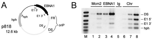
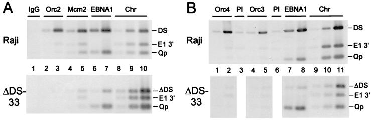
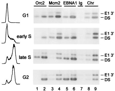
The 165-kb chromosome of Epstein-Barr virus (EBV) is replicated by cellular enzymes only once per cell cycle in human cells that are latently infected. Here, we report that the human origin recognition complex, ORC, can be detected in association with an EBV replication origin, oriP, in cells by using antibodies against three different subunits of human ORC to precipitate crosslinked chromatin. Mcm2, a subunit of the MCM replication licensing complex, was found to associate with oriP during G(1) and to dissociate from it during S phase. The detection of ORC and Mcm2 at oriP was shown to require the presence of the 120-bp replicator of oriP. Licensing and initiation of replication at oriP of EBV thus seem to be mediated by ORC. This is an example of a virus apparently using ORC and associated factors for the propagation of its genome.


Figures

Figure 1
Figure 1
Detection of Mcm2 at DS of oriP by ChIP. (A) Diagram of p818, a 12.6-kb plasmid carrying oriP and flanking sequences (light gray), the EBNA1 gene and flanking sequences (black), and hygromycin B-resistance cassette (hph, dark gray). The regions that were amplified by PCR for ChIP are indicated inside the circle. The distal end of the control region, E1 3′, is 560 bp from the edge of FR and 2.1 kb from DS, whereas E1 5′ is 2.1 kb farther away from oriP. hph is about 6 kb from oriP. (B) ChIP of chromatin from 293 cells carrying p818. Chromatin (30 μg) was precipitated by using antibodies to Orc2, Mcm2, or EBNA1 or by using nonimmune rabbit IgG, as indicated. Either 1/10th (lanes 2, 4, and 6) or 1/50th (lanes 1, 3, and 5) of the recovered DNA was amplified by PCR to detect the four indicated regions of the plasmid. DNA from the chromatin preparation was tested in lanes 6–8, by using 6.4, 32, and 160 ng, respectively. PCR products were separated by agarose-gel electrophoresis and detected by staining with SYBR Green. A reverse image is shown. M, marker. The specific PCR products ranged from 184 bp (hph) to 435 bp (DS).
Figure 2
Figure 2
The association of ORC and MCM with oriP requires the replicator, DS. ChIP analysis of 30 μg of chromatin from Raji cells or BL30 cells carrying the EBV mutant P3-ΔDS-33. Preimmune sera (PI) corresponding to the antisera against Orc4 and Orc3 were used for B, lanes 3 and 6, respectively. The amounts of ChIP DNA used for PCR were 1/250th (lanes 1, 3, 5, and 7) and 1/1,250th (lanes 2, 4, and 6) (A) and 1/250th (lanes 2, 3, 5, 6, and 8) and 1/1,250th (lanes 1, 4, and 7) (B). The amounts of DNA tested from input chromatin were for Raji, 0.06, 0.3, and 1.5 ng (lanes 8–10 in A, lanes 9–11 in B) and for P3-ΔDS-33, 0.1, 0.5, and 2.4 ng (A) and 0.05, 0.25, and 1.25 ng (B).
Figure 3
Figure 3
Cell cycle dependence of the association of Mcm2 with DS of oriP. DG75 cells carrying p818 were separated by centrifugal elutriation into four populations enriched for cells in G1, early S, late S, and G2; 30 μg of chromatin from each was analyzed by ChIP with antibodies against Orc2, Mcm2, or EBNA1, or with nonimmune rabbit IgG, as indicated. Flow cytometric analysis of the cells for DNA content is shown (Left) in the conventional manner. For this experiment, a 254-bp region bordering DS was amplified to detect the presence of DS, rather than the 435 bp spanning DS as done for Figs. 1 and 2. 1/50th (lanes 2, 4, and 6) or 1/250th (lanes 1, 3, and 5) of the recovered DNA were tested. For lanes 8 and 9, 2.4 ng and 12 ng of DNA from input chromatin were tested.

References

    1. Rickinson A B, Kieff E. In: Fields Virology. Fields B N, Knipe D M, Howley P M, editors. Philadelphia: Lippincott; 1996. pp. 2397–2446.
    1. Young L S. Semin Cancer Biol. 1996;7:163–164. - PubMed
    1. Adams A. J Virol. 1987;61:1743–1746. - PMC - PubMed
    1. Yates J, Warren N, Reisman D, Sugden B. Proc Natl Acad Sci USA. 1984;81:3806–3810. - PMC - PubMed
    1. Yates J L, Warren N, Sugden B. Nature (London) 1985;313:812–815. - PubMed

Publication types

Substances

LinkOut - more resources