. 2001 Sep 11;98(19):10886-91.
doi: 10.1073/pnas.191260798. Epub 2001 Aug 21.

An acquired and a native penicillin-binding protein cooperate in building the cell wall of drug-resistant staphylococci

Affiliations

An acquired and a native penicillin-binding protein cooperate in building the cell wall of drug-resistant staphylococci

M G Pinho et al. Proc Natl Acad Sci U S A. .

Abstract

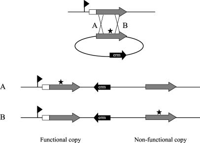
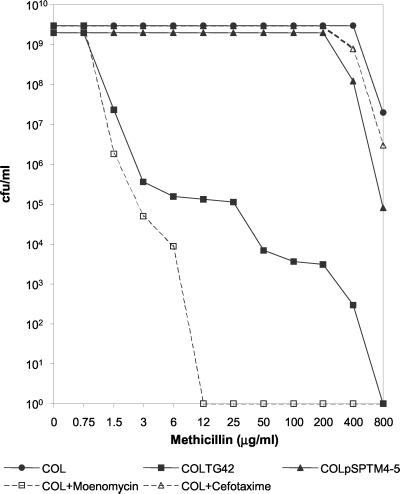
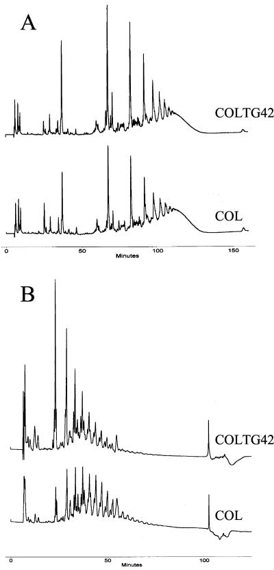
The blanket resistance of methicillin-resistant Staphylococcus aureus to all beta-lactam antibiotics--which had such a devastating impact on chemotherapy of staphylococcal infections--is related to the properties of the key component of this resistance mechanism: the "acquired" penicillin-binding protein (PBP)-2A, which has unusual low affinity for all beta-lactam antibiotics. Until now, the accepted model of resistance implied that in the presence of beta-lactam antibiotics in the surrounding medium, PBP2A must take over the biosynthesis of staphylococcal cell wall from the four native staphylococcal PBPs because the latter become rapidly acylated and inactivated at even low concentrations of the antibiotic. However, recent observations indicate that this model requires revision. Inactivation of the transglycosylase domain, but not the transpeptidase domain, of PBP2 of S. aureus prevents expression of beta-lactam resistance, despite the presence of the low-affinity PBP2A. The observations suggest that cell-wall synthesis in the presence of beta-lactam antibiotics requires the cooperative functioning of the transglycosylase domain of the native staphylococcal PBP2 and the transpeptidase domain of the PBP2A, a protein imported by S. aureus from an extra species source.


Figures

Figure 1
Figure 1
Strategy for mutagenesis of PBP2 domains. A fragment of pbp2 that does not include its promoter (indicated by flag) or its membrane anchor (□) was cloned into the integrational plasmid pSP64E, generating plasmid pPBP2M1, which was mutagenized by site-directed mutagenesis (★). The mutated plasmid was then introduced into the methicillin-susceptible strain RN4220 by electroporation. After insertion in the chromosome by a Campbell-type mechanism, the plasmid should generate a strain that has a functional copy of pbp2 followed by the integrational vector and by a nonfunctional copy of pbp2 (without its promoter and membrane anchor). If the single crossover recombination that occurs during transformation takes place before the mutated codon (A), the end result will be a strain with the mutation in the functional copy of pbp2, whereas if the recombination point is located after the mutated codon (B) the end result will be a strain with a native functional copy and a mutated nonfunctional copy, which should have a phenotype similar to the wild type.
Figure 2
Figure 2
Methicillin PAPs. The TPase mutation (COLpSPTM4–5) did not affect methicillin resistance, whereas the TGase mutation (COLTG42) caused more than 100-fold decrease in MIC to methicillin. The control strains COLpSPTM4–10 and COLTG2-1 had the same resistance profiles as strain COL. For the synergy assay, PAPs were done in the presence of 0.5 μg/ml moenomycin (an inhibitor of transglycosylation) or 200 μg/ml cefotaxime (a β-lactam with selective affinity for PBP2).
Figure 3
Figure 3
HPLC cell-wall analysis of the TGase mutant COLTG42 and in the parental strain COL. Muropeptide composition (A) was not significantly altered, whereas the glycan profile (B) was found to shift in the direction of peaks with shorter retention times, corresponding to shorter glycan strands with fewer disaccharide units. Control strain COLTG2-1 had both the muropeptide and the glycan profile identical to parental strain COL.
Figure 4
Figure 4
Model for the cooperative functioning of the TGase domain of PBP2 and the TPase activity of PBP2A in methicillin-resistant S. aureus. (Upper) In the absence of antibiotic it is assumed that both the TPase and TGase domains of PBP2 participate in the biosynthesis of staphylococcal peptidoglycan. Whether or not the TPase activity of PBP2A (present in methicillin-resistant strains) also functions in the crosslinking of the peptidoglycan in the absence of antibiotic in the medium is not clear at the present time. (Lower) When antibiotic (▾) is added to the medium, the TPase domain of PBP2 is acylated and is no longer capable of performing its peptide crosslinking activity. However, the observations described in this paper demonstrate that the penicillin-insensitive TGase domain of PBP2 remains functional and cooperates with the TPase activity of the acquired PBP2A and is actually essential for cell-wall synthesis and bacterial growth in the presence of β-lactam antibiotics in the surrounding medium.

References

    1. Beck W D, Berger-Bachi B, Kayser F H. J Bacteriol. 1986;165:373–378. - PMC - PubMed
    1. Kuhl S A, Pattee P A, Baldwin J N. J Bacteriol. 1978;135:460–465. - PMC - PubMed
    1. Song M D, Wachi M, Doi M, Ishino F, Matsuhashi M. FEBS Lett. 1987;221:167–171. - PubMed
    1. Gaisford W C, Reynolds P E. Eur J Biochem. 1989;185:211–218. - PubMed
    1. Goffin C, Ghuysen J M. Microbiol Mol Biol Rev. 1998;62:1079–1093. - PMC - PubMed

Publication types

MeSH terms