. 2001 Sep 11;98(19):10863-8.
doi: 10.1073/pnas.191284198. Epub 2001 Aug 21.

Loss of presenilin 1 is associated with enhanced beta-catenin signaling and skin tumorigenesis

Affiliations

Loss of presenilin 1 is associated with enhanced beta-catenin signaling and skin tumorigenesis

X Xia et al. Proc Natl Acad Sci U S A. .

Abstract

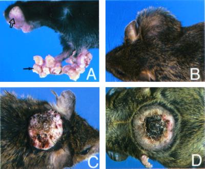
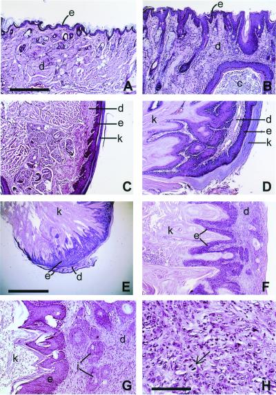
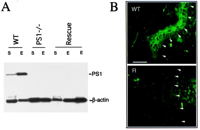
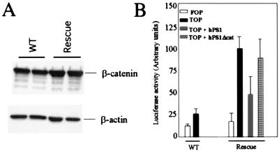
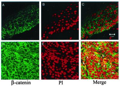
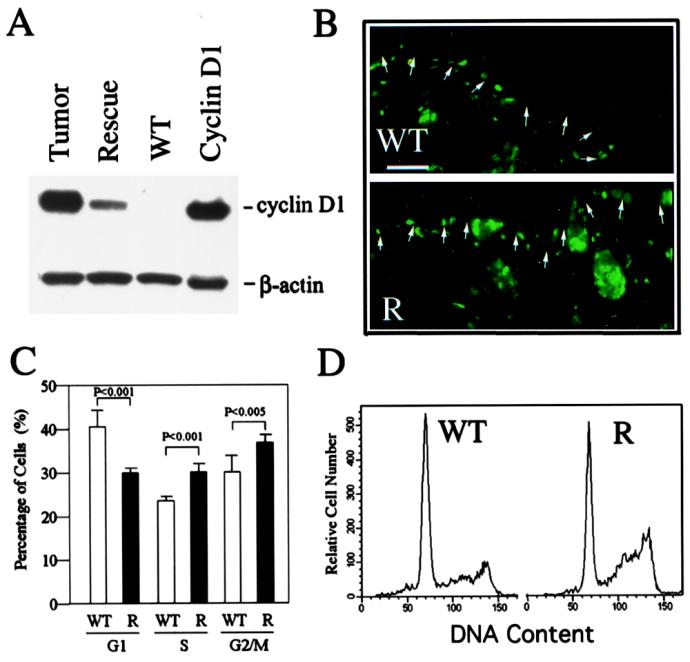
Presenilin 1 (PS1) is required for the proteolytic processing of Notch and the beta-amyloid precursor protein (APP), molecules that play pivotal roles in cell-fate determination during development and Alzheimer's disease pathogenesis, respectively. In addition, PS1 interacts with beta-catenin and promotes its turnover through independent mechanisms. Consistent with this activity, we report here that PS1 is important in controlling epidermal cell proliferation in vivo. PS1 knockout mice that are rescued through neuronal expression of human PS1 transgene develop spontaneous skin cancers. PS1-null keratinocytes exhibit higher cytosolic beta-catenin and beta-catenin/lymphoid enhancer factor-1/T cell factor (beta-catenin/LEF)-mediated signaling. This effect can be reversed by reintroducing wild-type PS1, but not a PS1 mutant active in Notch processing but defective in beta-catenin binding. Nuclear beta-catenin protein can be detected in tumors. Elevated beta-catenin/LEF signaling is correlated with activation of its downstream target cyclin D1 and accelerated entry from G(1) into S phase of the cell cycle. This report demonstrates a function of PS1 in adult tissues, and our analysis suggests that deregulation of beta-catenin pathway contributes to the skin tumor phenotype.


Figures

Figure 1
Figure 1
Representative skin lesions in hPS1 rescue mice. (A) Formation of cystic dermal nodules in perioral skin and front paws (open and solid arrows). (B–D) Non-ulcerated (B) and ulcerated (C and D) cutaneous masses present in hPS1 rescue mice.
Figure 2
Figure 2
Histopathology of hPS1 rescue mice. (A and C) Sections of perioral skin and paws of WT mice, respectively. (B and D) The same areas of hPS1 rescue mice, showing epidermal hyperplasia and hyperkeratosis. The epidermal layers (e, darker stain) were thickened and expanded with numerous projections of stratified squamous epithelium that were covered by massive amounts of dense, layered keratin (k in D). A large keratin-filled epidermal cyst (c) is embedded in dermal layer in B. D also shows that the two layers of hyperplastic epithelium were folded on one another, sharing a common dermal layer and resulting in a keratoacanthoma-like appearance. The same pathology is also detected in a cutaneous tumor (E and F, lower and higher power view, respectively). (G and H) The expanded dermis with infiltrating clusters of neoplastic squamous epithelial cells (i in G), which are characteristic of locally invasive squamous cell carcinoma. The arrow in H indicates a dividing neoplastic cell in a disorganized cluster of invading epithelial cells. e, epidermis; d, dermis; c, epidermal cyst; k, keratin; i, islands of infiltrating epidermal cells. A–D, F, and G are of the same magnification; scale bar in A = 275 μm. Scale bars in E and H represent 1.5 mm and 70 μm, respectively.
Figure 3
Figure 3
PS1 deficiency in newborn hPS1 rescue mouse skin. (A) Western blot analysis of PS1 expression. PS1 protein can be detected from both whole skin (S) and epidermis (E) of WT mouse but was found below the level of detection in either PS1 null (PS1−/−) or mPS−/−, hPS1 rescue (Rescue) newborns. PS1NT antibody was used, which detected a 28-kDa PS1-NTF fragment (top bands). β-Actin staining serves as loading control. (B) Immunofluorescence staining of newborn mouse skin using PS1NT antibody. In WT skin (Upper), PS1 expression is enriched in the epidermis (epidermis and dermis separated by arrows). PS1 protein can be detected in both the cytoplasm and plasma membrane in the basal cell layer of the epidermis (highlighted by arrows) but is mostly concentrated at the intercellular contacts toward more differentiated suprabasal layers (layers distal to arrows). No specific PS1 staining can be detected in hPS1 rescue skin (R, Lower). (Scale bar = 100 μm.)
Figure 4
Figure 4
Accumulation of soluble β-catenin and elevation of β-catenin/LEF signaling in the absence of PS1. (A) Higher levels of soluble β-catenin in hPS1 rescue primary keratinocytes (lanes labeled as Rescue) as compared with controls (WT). β-Actin staining was used as loading control. Shown are representative results from six independent experiments, and the average increase is ≈2-fold. (B) Effects of PS1 on β-catenin/LEF signaling. Primary keratinocytes isolated from WT or hPS1 rescue neonates were transfected with either the FOP-flash control construct containing the mutated LEF binding sites (FOP) or the TOP-flash reporter construct containing the LEF-responsive elements (TOP). In addition, the rescue culture was cotransfected with the TOP-flash vector plus wild-type hPS1 (TOP + hPS1) or the TOP-flash vector plus β-catenin binding-defective hPS1 (TOP + hPS1Δcat). The cells were assayed for luciferase activity 24 h posttransfection.
Figure 5
Figure 5
Nuclear localization of β-catenin in tumors. Immunostaining and confocal image analysis shows that in WT epidermis (Upper), β-catenin is predominately localized on the plasma membranes (A), and there is no overlapping staining between β-catenin (green) and nucleus (visualized by staining with propidium iodode, red) (C). In a representative tumor sample (Lower), there is a dramatic increase of β-catenin protein in the cytoplasm (D). In addition, nuclear β-catenin can be readily detected as shown by overlapping staining (highlighted by arrows) between β-catenin (green) and nucleus (red) (F). (Scale bar = 20 μM.)
Figure 6
Figure 6
Activation of cyclin D1 and altered cell-cycle control in PS1-deficient epidermis. (A) Overexpression of cyclin D1 in the skin of hPS1 rescue neonates and tumor samples. WT and Rescue, newborn epidermis from WT and rescue pups, respectively; Cyclin D1, 293 cells transfected with cyclin D1; Tumor, tumor sample taken from a representative adult hPS1 rescue mouse. β-Actin staining serves as loading control. (B). BrdUrd incorporation in WT (Upper) and hPS1 rescue (R, Lower) newborn skin. BrdUrd-positive cells are mostly localized to the hair follicles and basal cell layer (highlighted by arrows) of the epidermis, with rescue epidermis showing a higher labeling index. Scale bar = 100 μm. Cumulative (C) and representative (D) FACS analysis shows that there is accelerated G1 to S and G2/M phase transition in the absence of PS1. Keratinocytes isolated from WT (open bars) and hPS1 rescue (R, filled bars) were subjected to FACS analysis, and the percentages of cells in G1, S, and G2/M phases (mean ± SD of eight independent measurements) were plotted.

Comment in

References

    1. Sherrington R, Rogaev E I, Liang Y, Rogaeva E A, Levesque G, Ikeda M, Chi H, Lin C, Li G, Holman K, et al. Nature (London) 1995;375:754–760. - PubMed
    1. Selkoe D J. Trends Cell Biol. 1998;8:447–453. - PubMed
    1. De Strooper B, Beullens M, Contreras B, Levesque L, Craessaerts K, Cordell B, Moechars D, Bollen M, Fraser P, George-Hyslop P S, Van Leuven F. J Biol Chem. 1997;272:3590–3598. - PubMed
    1. Annaert W G, Levesque L, Craessaerts K, Dierinck I, Snellings G, Westaway D, George-Hyslop P S, Cordell B, Fraser P, De Strooper B. J Cell Biol. 1999;147:277–294. - PMC - PubMed
    1. Georgakopoulos A, Marambaud P, Efthimiopoulos S, Shioi J, Cui W, Li H C, Schutte M, Gordon R, Holstein G R, Martinelli G, et al. Mol Cell. 1999;4:893–902. - PubMed

Publication types

MeSH terms