. 2001:2:16.
doi: 10.1186/1471-2121-2-16. Epub 2001 Aug 2.

Non-enzymatic glycosylation of a type I collagen matrix: effects on osteoblastic development and oxidative stress

Affiliations

Non-enzymatic glycosylation of a type I collagen matrix: effects on osteoblastic development and oxidative stress

A D McCarthy et al. BMC Cell Biol. 2001.

Abstract

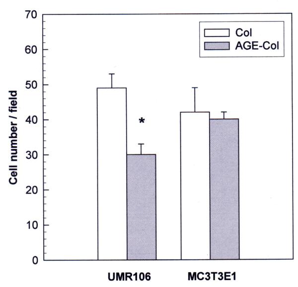
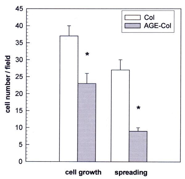
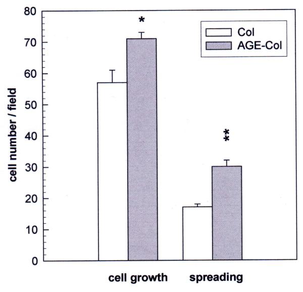
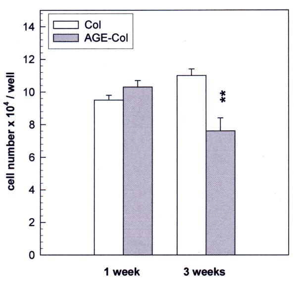
Background: The tissue accumulation of protein-bound advanced glycation endproducts (AGE) may be involved in the etiology of diabetic chronic complications, including osteopenia. The aim of this study was to investigate the effect of an AGE-modified type I collagen substratum on the adhesion, spreading, proliferation and differentiation of rat osteosarcoma UMR106 and mouse non-transformed MC3T3E1 osteoblastic cells. We also studied the role of reactive oxygen species (ROS) and nitric oxide synthase (NOS) expression on these AGE-collagen mediated effects.

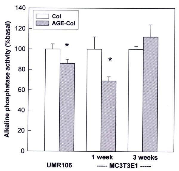
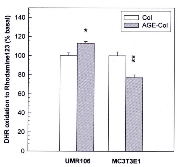
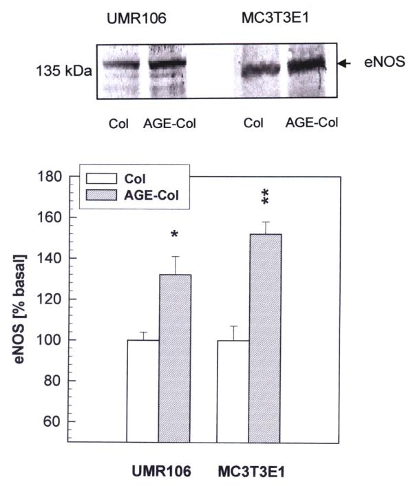
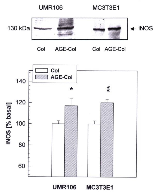
Results: AGE-collagen decreased the adhesion of UMR106 cells, but had no effect on the attachment of MC3T3E1 cells. In the UMR106 cell line, AGE-collagen also inhibited cellular proliferation, spreading and alkaline phosphatase (ALP) activity. In preosteoblastic MC3T3E1 cells (24-hour culture), proliferation and spreading were significantly increased by AGE-collagen. After one week of culture (differentiated MC3T3E1 osteoblasts) AGE-collagen inhibited ALP activity, but had no effect on cell number. In mineralizing MC3T3E1 cells (3-week culture) AGE-collagen induced a decrease in the number of surviving cells and of extracellular nodules of mineralization, without modifying their ALP activity. Intracellular ROS production, measured after a 48-hour culture, was decreased by AGE-collagen in MC3T3E1 cells, but was increased by AGE-collagen in UMR106 cells. After a 24-hour culture, AGE-collagen increased the expression of endothelial and inducible NOS, in both osteoblastic cell lines.

Conclusions: These results suggest that the accumulation of AGE on bone extracellular matrix could regulate the proliferation and differentiation of osteoblastic cells. These effects appear to depend on the stage of osteoblastic development, and possibly involve the modulation of NOS expression and intracellular ROS pathways.


Figures

Figure 1
Figure 1
Effect of an AGE-modified type I collagen matrix on cell adhesion. UMR106 and MC3T3E1 osteoblast-like cells were incubated on an unmodified collagen matrix or an AGE-collagen matrix for 1 hour at 37°C. Cells adhering to the matrices were fixed, stained with Giemsa and counted microscopically at a magnification of 400X. Results are given as the number of cells counted per field, and are expressed as the mean ± SEM. Differences between collagen vs. AGE-collagen are as follows: * p < 0.001
Figure 2
Figure 2
Effect of an AGE-modified type I collagen matrix on UMR106 cell proliferation and spreading. UMR106 osteosarcoma cells were cultured on an unmodified collagen matrix or an AGE-collagen matrix for 24 hours at 37°C. Cell proliferation was evaluated by microscopically counting cells stained with Giemsa, at a magnification of 400X. Cellular spreading was also estimated microscopically, as the number of cells possessing at least two comers. Results are given as the number of cells counted per field, and are expressed as the mean ± SEM. Differences between collagen vs. AGE-collagen are as follows: * p < 0.001
Figure 3
Figure 3
Effect of an AGE-modified type I collagen matrix on MC3T3E1 cell proliferation and spreading. MC3T3E1 osteoblastic cells were cultured on an unmodified collagen matrix or an AGE-collagen matrix for 24 hours at 37°C. Cell proliferation was evaluated by microscopically counting cells stained with Giemsa, at a magnification of 400X. Cellular spreading was also estimated microscopically, as the number of cells possessing at least two corners. Results are given as the number of cells counted per field, and are expressed as the mean ± SEM. Differences between collagen vs. AGE-collagen are as follows: * p<0.01; ** p<0.001.
Figure 4
Figure 4
Effect of an AGE-modified type I collagen matrix on MC3T3E1 cell growth. MC3T3E1 calvaria-derived osteoblastic cells were cultured on control collagen or AGE-modified collagen at 37°C for one week (differentiated osteoblasts) or three weeks (mineralizing cultures). The quantity of cells growing on the corresponding collagenous matrices at each timepoint, was evaluated by trypsinization and cell counting with a haemocytometer. Results are given as the number of cells counted in each case, and are expressed as the mean ± SEM. Differences between collagen vs. AGE-collagen are as follows: ** p<0.001.
Figure 5
Figure 5
Effect of an AGE-modified type I collagen matrix on the ALP activity of UMR106 and MC3T3E1 cells. Osteoblastic cells were cultured on control or AGE-modified type I collagen at 37°C either for 24 hours (UMR106 cells), or for one and three weeks (MC3T3E1 cells). At the end of all incubations, the cell monolayer was lysated in 0.1 % Triton X-100. Alkaline phosphatase activity was determined in the lysates using p-nitro-phenyl-phosphate as a substrate, and was normalized for cellular protein content. Results are given as a percentage of the corresponding basal condition (unmodified collagen), and are expressed as the mean ± SEM. Differences between collagen vs. AGE-collagen are as follows: * p<0.01. Basal ALP activity was 168 ± 8 nmol pNP / min x mg protein for UMR106 cells; 2.77 ± 0.33 nmol pNP / min x mg protein for a one-week culture of MC3T3E1 cells; and 3.05 ± 0.23 nmol pNP / min x mg protein for a three-week culture of MC3T3E1 cells.
Figure 6
Figure 6
Effect of an AGE-modified type I collagen substratum on the generation of intracellular reactive oxygen species by MC3T3E1 and UMR106 cells. Osteoblasts were cultured at 37°C either on control collagen or on AGE-modified collagen. After 48 hours, media were replaced by phenol red-free DMEM with 10 μM dihydro-rhodamine, and cells were incubated for an additional 4 hours. At the end of this incubation the cell monolayers were lysated with 0.1 % Triton X-100, and the levels of the oxidation product rhodamine were measured in the lysates by the determination of fluorescence intensity (excitation wavelength 495 nm, emission wavelength 532 nm). Results are given as a percentage of the corresponding basal condition (unmodified collagen), and are expressed as the mean ± SEM. Differences between collagen vs. AGE-collagen are as follows: * p<0.01; ** p<0.001
Figure 7
Figure 7
Effect of AGE-modified type I collagen on the expression of eNOS in osteoblast-like cells. UMR106 and MC3T3E1 cells were cultured on control or AGE-modified type I collagen matrices at 37ºC for 24 hours. After this incubation, cell monolayers were lysated in Laemmli's buffer and electrophoresed under reducing conditions on an 8 % SDS-PAGE. Western blotting was performed, and specific bands were detected using a rabbit polyclonal antibody against eNOS. A representative blot from three independent experiments is presented (top figure). Images were scanned and analyzed by densitometry. The relative intensity of each band is shown (bottom figure) as a percentage of the corresponding basal condition (unmodified collagen), and represents the mean ± SEM. Differences between collagen vs. AGE-collagen are as follows: * p<0.02; ** p<0.002
Figure 8
Figure 8
Effect of AGE-modified type I collagen on the expression of iNOS in osteoblast-like cells. UMR106 and MC3T3E1 cells were cultured on control or AGE-modified type I collagen matrices at 37°C for 24 hours. After this incubation, cell monolayers were lysated in Laemmli's buffer and electrophoresed under reducing conditions on an 8 % SDS-PAGE. Western blotting was performed, and specific bands were detected using a rabbit polyclonal antibody against iNOS. A representative blot from three independent experiments is presented (top figure). Images were scanned and analyzed by densitometry. The relative intensity of each band is shown (bottom figure) as a percentage of the corresponding basal condition (unmodified collagen), and represents the mean ± SEM. Differences between collagen vs. AGE-collagen are as follows: * p<0.05; ** p<0.002

References

    1. Vlassara H, Bucala R, Striker L. Pathogenic effects of advanced glycosylation: biochemical, biologic and clinical implications for diabetes and ageing. Lab Invest. 1994;70:138–151. - PubMed
    1. Baynes JW, Thorpe SR. Role of oxidative stress in diabetic complications: a new perspective on an old paradigm. Diabetes. 1999;48:1–9. - PubMed
    1. Thornalley PJ. Cell activation by glycated proteins. Age receptors, receptor recognition factors and functional classification of AGEs. Cell Mol Biol (Noisy-le-Grand) 1998;44:1013–1033. - PubMed
    1. Chakravarthy U, Hayes RG, Stitt AW, McAuley E, Archer DB. Constitutive nitric oxide synthase expression in retinal vascular endothelial cells is supported by high glucose and advanced glycation end products. Diabetes. 1998;47:945–952. - PubMed
    1. Rojas A, Caveda L, Romay C, López E, Valdés S, Padrón J, Glarái L, Martínez 0, Delgado R. Effect of advanced glycation end products on the induction of nitric oxide synthase in murine macrophages. Biochem Biophys Res Comm. 1996;225:358–362. doi: 10.1006/bbrc.1996.1180. - DOI - PubMed

Publication types

LinkOut - more resources