. 2001 Aug;108(4):527-36.
doi: 10.1172/JCI12153.

A role for mitogen-activated protein kinase activation by integrins in the pathogenesis of psoriasis

Affiliations

A role for mitogen-activated protein kinase activation by integrins in the pathogenesis of psoriasis

I Haase et al. J Clin Invest. 2001 Aug.

Abstract

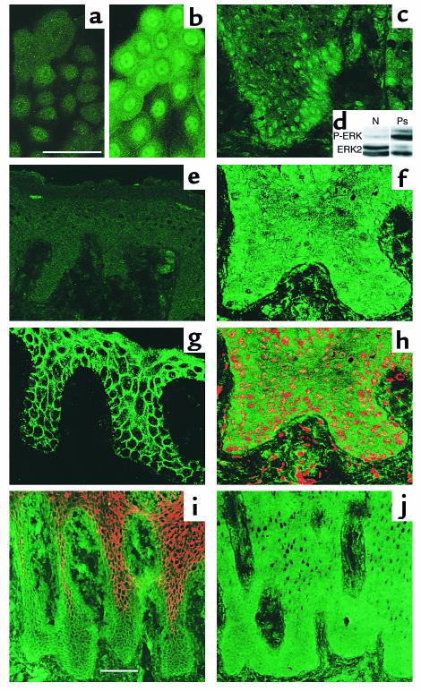
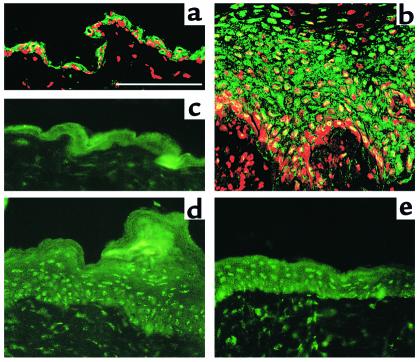
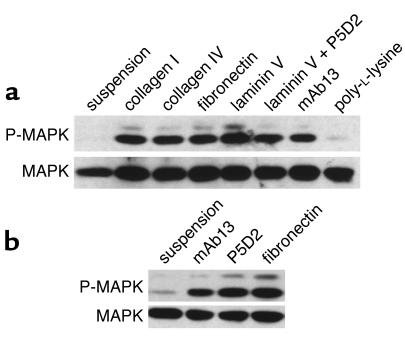
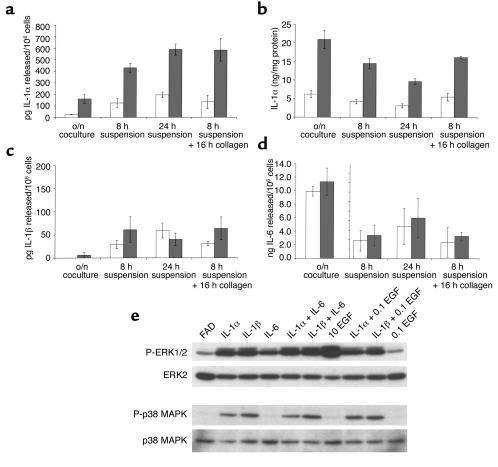
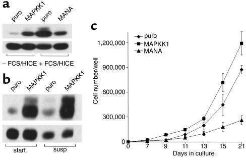
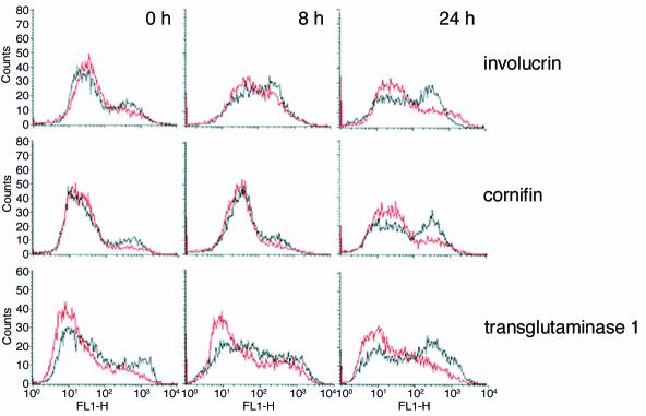
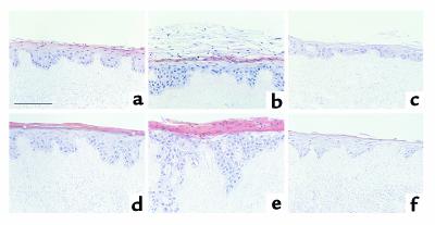
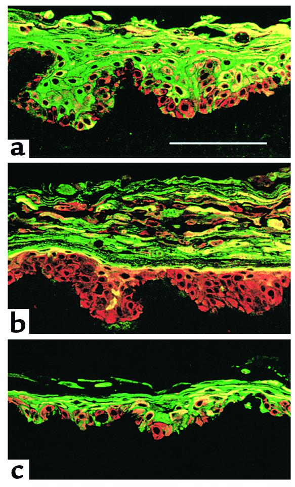
In normal epidermis, beta1 integrin expression is confined to the basal layer, whereas in hyperproliferative epidermis, integrins are also expressed in the suprabasal layers. Transgenic mice in which integrins are expressed suprabasally via the involucrin promoter have a sporadic psoriatic phenotype; however, the mechanism by which integrins contribute to the pathogenesis of psoriasis is unknown. We observed activation of mitogen-activated protein kinase (MAPK) in basal and suprabasal keratinocytes of human and transgenic mouse psoriatic lesions and healing mouse skin wounds, correlating in each case with suprabasal integrin expression. Phenotypically normal human and transgenic mouse epidermis did not contain activated MAPK. Transgene-positive keratinocytes produced more IL-1alpha than controls did, and keratinocyte MAPK could be activated by ligation of suprabasal integrins or treatment with IL-1alpha. Constitutive activation of MAPK increased the growth rate of human keratinocytes and delayed the onset of terminal differentiation, recreating many of the histological features of psoriatic epidermis. We propose that activation of MAPK by integrins, either directly or through increased IL-1alpha production, is responsible for epidermal hyperproliferation in psoriasis and wound healing, and that the sporadic phenotype of the transgenic mice may reflect the complex mechanisms by which IL-1 release and responsiveness are controlled in skin.


Figures

Figure 1
Figure 1
Distribution and activation of p42mapk in cultured keratinocytes and in normal and hyperproliferative human epidermis. Confocal immunofluorescence microscopy of growth factor–starved cultured keratinocytes (a) or keratinocytes treated with FCS + HICE for 15 minutes (b); psoriatic (c, f, and hj) or normal human epidermis (e and g). Green staining in ac, eh, and j is for MAPK with antibody specific for activated (ac and e) or total MAPK (fh and j); red staining in h is nuclear dye, Topro III. The field shown in h is part of the field shown in f. (i) Staining for β1 integrins (green) and involucrin (red). Scale bars: 50 μm (a, b, and fh), 100 μm (c, e, i, and j). (d) Western blot of shaved biopsies of normal human skin (N) or psoriatic lesion (Ps) probed with phosphorylation-specific antibody to ERK1/2 (P-ERK) or total ERK2.
Figure 2
Figure 2
Distribution and activation of p42mapk in phenotypically normal and hyperproliferative transgenic mouse epidermis. (a and b) Antibody to total MAPK. (ce) Antibody to activated MAPK. Hyperproliferative β1 transgenic mouse epidermis 7 days after wounding (b and e) or unwounded transgenic mouse skin (a). Phenotypically normal epidermis (c) and psoriatic lesion (d) of α6β1 transgenic mouse. Red staining in a and b is nuclear dye, Topro III. Scale bar: 50 μm.
Figure 3
Figure 3
Integrin-mediated MAPK phosphorylation. Western blot analysis of MAPK phosphorylation: upper panels show threonine/tyrosine phosphorylation of ERK1/2 visualized with a phosphorylation-specific antibody; lower panels show the same blots incubated with an antibody against total ERK2 as a loading control. (a) Human keratinocytes in FAD medium containing 10 ng/ml EGF were held in suspension or plated for 30 minutes onto dishes coated with poly-L-lysine or the ECM proteins and antibodies shown. (b) Invβ1 mouse keratinocytes were cultured in suspension for 12 hours to induce terminal differentiation and transgene expression and then held in suspension or plated for 30 minutes in the presence of 10 ng/ml EGF onto fibronectin or the human β1–specific antibodies mAb13 and P5D2.
Figure 4
Figure 4
Proinflammatory cytokine production and cytokine-induced MAPK phosphorylation. Secretion of IL-1α (a), IL-1β (c), and IL-6 (d) by nontransgenic (open bars) and α2β1 transgene-expressing (filled bars) mouse keratinocytes. Intracellular IL-1α levels are shown in b. In the case of overnight keratinocyte/feeder cocultures (o/n coculture), cytokine secreted represents the total produced by both cell types, whereas intracellular levels were measured in keratinocytes alone. (e) Western blot analysis of ERK1/2 and p38 MAPK phosphorylation (P-ERK1/2 and P-p38 MAPK, respectively) and total ERK2 and p38 MAPK. Preconfluent human keratinocytes had feeders removed and were starved overnight before a 15-minute stimulation with medium alone (FAD) or medium supplemented with IL-1α/β, IL-6, or EGF alone or in combinations. IL-1 α/β and IL-6 were added to a final concentration of 10 ng/ml while EGF was added to 10 ng/ml when the only cytokine present (10 EGF) or 0.1 ng/ml when used in combination with other cytokines (0.1 EGF).
Figure 5
Figure 5
Effect of MAPK activity on keratinocyte proliferation. (a and b) Western blot detection of MAPK phosphorylation (upper panels) and total MAPK (lower panels) as described in the legend to Figure 3. (a) Adherent keratinocytes infected with empty vector puro, MAPKK1, or MANA were starved overnight and lysed 10 minutes after addition of fresh medium with or without FCS/HICE. (b) Keratinocytes infected with puro or MAPKK1 were trypsinized and held in suspension for 15 minutes (start) or 24 hours (susp). (c) Growth curves: 500 (puro, MAPKK1) or 1,000 (MANA) keratinocytes were plated per 35-mm dish (to achieve equal numbers of adherent cells; ref. 4) and cultured for 21 days. Triplicate dishes were harvested on the days shown. Error bars = SD.
Figure 6
Figure 6
Effect of constitutive MAPK activation on suspension-induced terminal differentiation. Flow cytometry profiles of keratinocytes fixed immediately after trypsinization (0 hours) or after 8 or 24 hours in suspension. Cells were labeled with antibodies to the differentiation markers indicated. Black lines: Cells expressing empty vector puro; red lines: cells expressing MAPKK1.
Figure 7
Figure 7
Effects of constitutive activation or inhibition of MAPK on reconstituted epidermis. Human keratinocytes infected with puro (a and d), MAPKK1 (b and e), or MANA (c and f) were cultured for 11 (ac) or 14 (df) days on dead, de-epidermized dermis. Asterisks in d indicate cells in granular layer. Scale bar: 100 μm.
Figure 8
Figure 8
Effects of constitutive activation or inhibition of MAPK on involucrin expression in reconstituted epidermis. Keratinocytes infected with empty vector puro (a), MAPKK1 (b), or MANA (c) were cultured on dead, de-epidermized dermis for 11 days and stained with antibodies against keratin 14 (expressed by all cells; red) and involucrin (green). Scale bar: 100 μm.

References

    1. Watt FM. Epidermal stem cells: markers, patterning and the control of stem cell fate. Philos Trans R Soc Lond B Biol Sci. 1998;353:1–7. - PMC - PubMed
    1. Fuchs E. Epidermal differentiation. Curr Opin Cell Biol. 1990;2:1028–1035. - PubMed
    1. Jones PH, Watt FM. Separation of human epidermal stem cells from transit amplifying cells on the basis of differences in integrin function and expression. Cell. 1993;73:713–724. - PubMed
    1. Zhu AJ, Haase I, Watt FM. Signaling via β1 integrins and mitogen-activated protein kinase determines human epidermal stem cell fate in vitro. Proc Natl Acad Sci USA. 1999;96:6728–6733. - PMC - PubMed
    1. Levy L, Broad S, Diekmann D, Evans RD, Watt FM. β1 integrins regulate keratinocyte adhesion and differentiation by distinct mechanisms. Mol Biol Cell. 2000;11:453–466. - PMC - PubMed

Publication types

MeSH terms