

doi: 10.1093/embo-reports/kve177.
Epub 2001 Aug 23.
Extracellular signal regulated kinase 5 (ERK5) is required for the differentiation of muscle cells
Affiliations
- PMID: 11520859
- PMCID: PMC1084032
- DOI: 10.1093/embo-reports/kve177
Item in Clipboard
Extracellular signal regulated kinase 5 (ERK5) is required for the differentiation of muscle cells
EMBO Rep.
2001 Sep.
Abstract
Extracellular signal regulated kinase 5 (ERK5) is a novel member of the mitogen-activated protein kinase (MAPK) family with a poorly defined physiological function. Since ERK5 and its upstream activator MEK5 are abundant in skeletal muscle we examined a function of the cascade during muscle differentiation. We show that ERK5 is activated upon induction of differentiation in mouse myoblasts and that selective activation of the pathway results in promoter activation of differentiation-specific genes. Moreover, myogenic differentiation is completely blocked when ERK5 expression is inhibited by antisense RNA. Thus, we conclude that the MEK5/ERK5 MAP kinase cascade is critical for early steps of muscle cell differentiation.
Figures
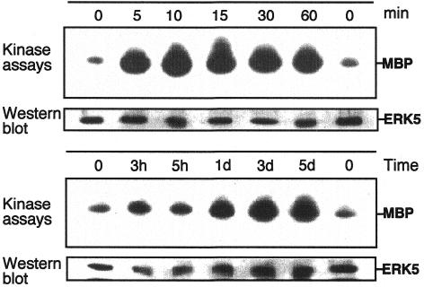
Fig. 1. ERK5 is activated upon differentiation in C2C12 cells. C2C12 myoblasts were plated at a density of 2.5 × 10⁵ on 10 cm dishes in DMEM/10%FBS. Twenty-four hours later cells were shifted to DMEM/2%HS to induce differentiation. Cell lysates were prepared at different time points as indicated. ERK5 was immunoprecipitated from the lysates using a polyclonal goat anti-ERK5 antiserum. ERK5 activity was measured by an immune complex kinase assay using MBP as a substrate. Kinase assays were performed essentially as described (Ludwig et al., 1996). Equal loading of kinases was assessed by immunoblotting with an ERK5-specific antiserum.
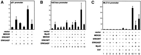
Fig. 2. MEK5 and ERK5 induce the expression of promoters upregulated upon muscle differentiation. A p21 promoter-luciferase construct (A), a 4× E-box-containing promoter construct (B) or a full-length MLC1A promoter luciferase plasmid (C) were cotransfected with expression vectors encoding constitutively active MEK5 (MEK5D), wild-type ERK5 (ERK5WT), a dominant-negative version of ERK5 (ERK5AEF), MyoD or E47, as indicated. Twenty hours after transfection cells were shifted to DMEM/2%HS for another 24 h and then harvested to measure luciferase activity as described. Fold induction is the ratio of luciferase activity in kinase-transfected cells versus cells transfected with the empty vectors.
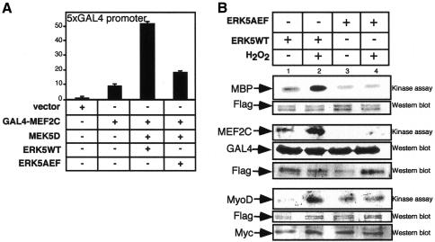
Fig. 3. MEF2C is activated by ERK5 in C2C12 cells under differentiation conditions. (A) C2C12 cells were transfected with a Gal4–MEF2C fusion construct, and transactivation of MEF2C under differentiation conditions induced by coexpressed MEK5D, ERK5WT and ERK5AEF was monitored by luciferase expression from a 5× Gal4 luciferase construct. (B) Flag-ERK5WT and flag-ERK5AEF were immunoprecipitated with an anti-flag antiserum from transfected 293 cells, which were either left untreated or treated with H2O2 (200 µM, 60 min) for activation of the kinase. Immunocomplexes were incubated in a kinase reaction with either MBP (upper panel), Gal4MEF2C (middle panel) or Myc-MyoD (lower panel), which were immunopurified from transfected 293 lysates with anti-Gal4 and anti-Myc antibodies.
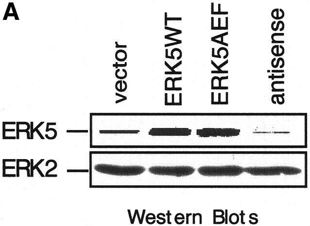
Fig. 4. Block of ERK5 expression results in the inhibition of myogenic differentiation. (A) ERK5 expression in stably transduced C2C12 cell lines harbouring empty vector, ERK5WT, ERK5AEF or antisense-ERK5. (B) Cells were seeded at a density of 2 × 10⁵ cells per 10 cm in DMEM/10%FBS and shifted 24 h later to DMEM/2%HS to induce differentiation. Pictures at indicated time points correspond to distinct stages of differentiation. Expression of GFP (last column) is indicative of successfully transduced cells. Bar, 65 µm. (C) Cells were lysed in TLB after 1 and 3 days post-differentiation induction and protein lysates were separated on SDS–PAGE gels. After subsequent immunoblotting, blots were analysed for p21, MyoD and myogenin with the respective antisera described in Methods. Equal protein load of the different samples was controlled with an anti-ERK2 antiserum.
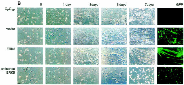
Fig. 4. Block of ERK5 expression results in the inhibition of myogenic differentiation. (A) ERK5 expression in stably transduced C2C12 cell lines harbouring empty vector, ERK5WT, ERK5AEF or antisense-ERK5. (B) Cells were seeded at a density of 2 × 10⁵ cells per 10 cm in DMEM/10%FBS and shifted 24 h later to DMEM/2%HS to induce differentiation. Pictures at indicated time points correspond to distinct stages of differentiation. Expression of GFP (last column) is indicative of successfully transduced cells. Bar, 65 µm. (C) Cells were lysed in TLB after 1 and 3 days post-differentiation induction and protein lysates were separated on SDS–PAGE gels. After subsequent immunoblotting, blots were analysed for p21, MyoD and myogenin with the respective antisera described in Methods. Equal protein load of the different samples was controlled with an anti-ERK2 antiserum.
Fig. 4. Block of ERK5 expression results in the inhibition of myogenic differentiation. (A) ERK5 expression in stably transduced C2C12 cell lines harbouring empty vector, ERK5WT, ERK5AEF or antisense-ERK5. (B) Cells were seeded at a density of 2 × 10⁵ cells per 10 cm in DMEM/10%FBS and shifted 24 h later to DMEM/2%HS to induce differentiation. Pictures at indicated time points correspond to distinct stages of differentiation. Expression of GFP (last column) is indicative of successfully transduced cells. Bar, 65 µm. (C) Cells were lysed in TLB after 1 and 3 days post-differentiation induction and protein lysates were separated on SDS–PAGE gels. After subsequent immunoblotting, blots were analysed for p21, MyoD and myogenin with the respective antisera described in Methods. Equal protein load of the different samples was controlled with an anti-ERK2 antiserum.
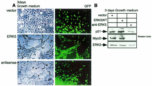
Fig. 5. ERK5WT overexpression is sufficient for the commitment of muscle cells to differentiate. (A) The C2C12 cell lines indicated were grown for 3 days in DMEM/10% FBS and were then examined morphologically. (B) Cells shown in (A) were lysed and cell lysates were subjected to SDS–PAGE. After blotting, proteins were detected with antibodies against p21, MyoD, myogenin (not shown) and ERK2 as a loading control.
References
-
- Abe J., Kusuhara, M., Ulevitch, R.J., Berk, B.C. and Lee, J.D. (1996) Big mitogen-activated protein kinase 1 (BMK1) is a redox-sensitive kinase. J. Biol. Chem., 271, 16586–16590. - PubMed
-
- Choi J., Costa, M.L., Mermelstein, C.S., Chagas, C., Holtzer, S. and Holtzer, H. (1990) MyoD converts primary dermal fibroblasts, chondroblasts, smooth muscle, and retinal pigmented epithelial cells into striated mononucleated myoblasts and multinucleated myotubes. Proc. Natl Acad. Sci. USA, 87, 7988–7992. - PMC - PubMed
-
- Cox R.D. and Buckingham, M.E. (1992) Actin and myosin genes are transcriptionally regulated during mouse skeletal muscle development. Dev. Biol., 149, 228–234. - PubMed
-
- Cox R.D., Garner, I. and Buckingham, M.E. (1990) Transcriptional regulation of actin and myosin genes during differentiation of a mouse muscle cell line. Differentiation, 43, 183–191. - PubMed
Publication types
MeSH terms
Substances
LinkOut - more resources
Full Text Sources
Other Literature Sources
Molecular Biology Databases
Miscellaneous
