. 2001 Sep 1;29(17):E89-9.
doi: 10.1093/nar/29.17.e89.

Linear 2' O-Methyl RNA probes for the visualization of RNA in living cells

Affiliations

Linear 2' O-Methyl RNA probes for the visualization of RNA in living cells

C Molenaar et al. Nucleic Acids Res. .

Abstract

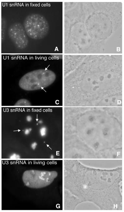
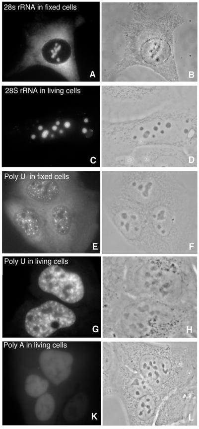
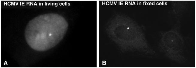
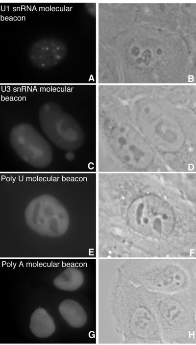
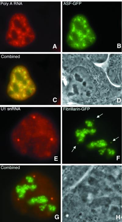
U1snRNA, U3snRNA, 28 S ribosomal RNA, poly(A) RNA and a specific messenger RNA were visualized in living cells with microinjected fluorochrome-labeled 2' O-Methyl oligoribonucleotides (2' OMe RNA). Antisense 2' OMe RNA probes showed fast hybridization kinetics, whereas conventional oligodeoxyribonucleotide (DNA) probes did not. The nuclear distributions of the signals in living cells were similar to those found in fixed cells, indicating specific hybridization. Cytoplasmic ribosomal RNA, poly(A) RNA and mRNA could hardly be visualized, mainly due to a rapid entrapment of the injected probes in the nucleus. The performance of linear probes was compared with that of molecular beacons, which due to their structure should theoretically fluoresce only upon hybridization. No improvements were achieved however with the molecular beacons used in this study, suggesting opening of the beacons by mechanisms other than hybridization. The results show that linear 2' OMe RNA probes are well suited for RNA detection in living cells, and that these probes can be applied for dynamic studies of highly abundant nuclear RNA. Furthermore, it proved feasible to combine RNA detection with that of green fluorescent protein-labeled proteins in living cells. This was applied to show co-localization of RNA with proteins and should enable RNA-protein interaction studies.


Figures

Figure 1
Figure 1
Similar localization patterns of 2′ OMe RNA oligonucleotides hybridized to U1 snRNA and U3 snRNA in fixed and living U2OS cells (A, C, E and G). The distinct localization patterns observed for U1 snRNA (C) and U3 snRNA (G) detected in living cells after microinjection of the antisense oligonucleotides are similar to those observed in fixed cells (A and E), indicating a specific hybridization to RNA in living cells. Corresponding phase contrast images (B, D, F and H). The intense spots in (C) and (E), indicated by arrows, are Cajal bodies, the faint structures in the nucleoplasm in (C) are speckles.
Figure 2
Figure 2
Hybridization of linear 2′ OMe RNA probes to abundant RNA targets in living cells. Probes for 28S rRNA (A and C) and poly(A) RNA (E and G) hybridized to fixed and living cells show a similar nuclear hybridization pattern but a different cytoplasmic localization pattern. Probes to 28S rRNA and poly(A) RNA accumulate in the nucleus within minutes of injection and the level of hybridization to the RNA present in the cytoplasm is negligible. The (A)18 probe shows no specific hybridization pattern (K). Corresponding phase contrast images (B, D, F, H and L).
Figure 3
Figure 3
Detection of the transcription site of CMV RNA, transcribed from the integrated HCMV IE 1 gene in R9G cells. (A) Transcription sites and cytoplasmic mRNA are visible. Detection of transcription sites in living cells. The small bright spot shows transcription from the integration sites. A high background was observed in the nucleus and nucleoli. The signal in the cytoplasm was very weak. (B) Representative example of an in situ hybridization with three linear 2′ OMe RNA oligonucleotides on fixed R9G cells.
Figure 4
Figure 4
Hybridization of molecular beacons in living cells. U1 snRNA (A) and U3 snRNA (C) molecular beacons show similar hybridization patterns as the linear probes (Fig. 1), but have a lower signal-to-noise ratio. A molecular beacon targeted to poly(A) RNA consisting of a 22 base loop and a 7 bp stem showed only a very vague specific staining (E), compared to the linear probe (Fig. 2G). The overall intensity was lower for molecular beacons. For none of the RNAs that were targeted, a molecular beacon showed hybridization with an improved contrast. The control poly(A) molecular beacon probe does not show a specific hybridization pattern (G). Corresponding phase contrast images (B, D, F and H).
Figure 5
Figure 5
No specific hybridization visible after microinjection of DNA oligonucleotides. Two examples of microinjected cells where RNA was targeted with DNA oligonucleotides. Thirty-five and 40 base antisense DNA probes for 28S ribosomal RNA (A) and the poly(A) tail of mRNA (C) were microinjected into living U2OS cells. The probes accumulated in the nucleus with time as was expected but did not show any further specific localization as was observed with the 2′ OMe RNA probes (as in Figs 1 and 2). (B and D) Corresponding phase contrast images.
Figure 6
Figure 6
Combined detection of RNA and expression of GFP in living cells. (AD) Co-localization of poly(A) RNA and ASF–GFP in living U2OS cells in speckles and nucleoplasm. (EH) Co-localization of U1 snRNA and fibrillarin–GFP in coiled bodies. Arrows in (F) indicate Cajal Bodies containing fibrillarin–GFP. Cells expressing ASF–GFP (B) were microinjected with a probe for poly(A) RNA (A) and cells expressing fibrillarin–GFP (F) with a probe for U1 snRNA (E).

References

    1. Lamond A.I. and Earnshaw,W.C. (1998) Structure and function in the nucleus. Science, 280, 547–553. - PubMed
    1. Verschure P.J., van Der Kraan,I., Manders,E.M. and van Driel,R. (1999) Spatial relationship between transcription sites and chromosome territories. J. Cell Biol., 147, 13–24. - PMC - PubMed
    1. Leonhardt H., Rahn,H.P., Weinzierl,P., Sporbert,A., Cremer,T., Zink,D. and Cardoso,M.C. (2000) Dynamics of DNA replication factories in living cells. J. Cell Biol., 149, 271–280. - PMC - PubMed
    1. Dirks R.W., Hattinger,C.M., Molenaar,C. and Snaar,S.P. (1999) Synthesis, processing, and transport of RNA within the three-dimensional context of the cell nucleus. Crit. Rev. Eukaryot. Gene Expr., 9, 191–201. - PubMed
    1. Misteli T. (2000) Cell biology of transcription and pre-mRNA splicing: nuclear architecture meets nuclear function. J. Cell Sci., 113, 1841–1849. - PubMed

Publication types

MeSH terms