. 2001 Oct;69(4):673-84.
doi: 10.1086/323610. Epub 2001 Aug 27.

Mutations in a novel gene with transmembrane domains underlie Usher syndrome type 3

Affiliations

Mutations in a novel gene with transmembrane domains underlie Usher syndrome type 3

T Joensuu et al. Am J Hum Genet. 2001 Oct.

Erratum in

  • Am J Hum Genet 2001 Nov;69(5):1160

Abstract

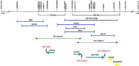
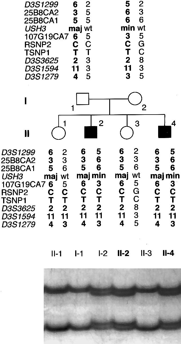
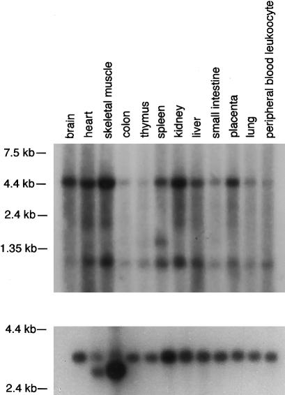
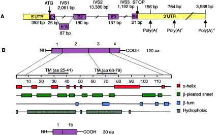
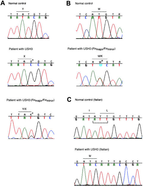
Usher syndrome type 3 (USH3) is an autosomal recessive disorder characterized by progressive hearing loss, severe retinal degeneration, and variably present vestibular dysfunction, assigned to 3q21-q25. Here, we report on the positional cloning of the USH3 gene. By haplotype and linkage-disequilibrium analyses in Finnish carriers of a putative founder mutation, the critical region was narrowed to 250 kb, of which we sequenced, assembled, and annotated 207 kb. Two novel genes-NOPAR and UCRP-and one previously identified gene-H963-were excluded as USH3, on the basis of mutational analysis. USH3, the candidate gene that we identified, encodes a 120-amino-acid protein. Fifty-two Finnish patients were homozygous for a termination mutation, Y100X; patients in two Finnish families were compound heterozygous for Y100X and for a missense mutation, M44K, whereas patients in an Italian family were homozygous for a 3-bp deletion leading to an amino acid deletion and substitution. USH3 has two predicted transmembrane domains, and it shows no homology to known genes. As revealed by northern blotting and reverse-transcriptase PCR, it is expressed in many tissues, including the retina.


Figures

Figure  1
Figure 1
Schematic representation of physical and transcript maps covering the USH3 region, depicting the initial 250-kb USH3 region and the extended 133-kb USH3 region, with partly overlapping BAC clones. Polymorphic markers and sequence-tagged sites are indicated by vertical lines. Previously mapped BAC clones are shown in dark blue, and the 207-kb sequence contig assembled is indicated by a purple line. Database-derived and partly assembled BAC sequences from clones RP11-385G14 and RP11-251C9 are indicated by green dashed arrows. The centromere is on the left. A pair of previously known genes—KIAA0001 and H963—and a group of three novel genes—NOPAR, UCRP, and USH3—are indicated by yellow and light-blue arrows, respectively, showing the orientation of transcription. ESTs representing parts of the 3′ UTRs of the transcripts are indicated by vertical arrows. H963, NOPAR, UCRP, and USH3 were subjected to mutation analyses in this study.
Figure  2
Figure 2
Pedigree of a Finnish family with USH3, segregating a paternal Finmajor mutation (maj)—c.300T→G—and a maternal Finminor mutation (min)—c.131T→A. Alleles at seven microsatellite markers, two SNPs, and USH3 are shown; wt = wild type. The chromosomes assumed to carry the disease allele are in boldface type. The father carries a conserved ancestral haplotype, whereas the mother’s disease-associated haplotype shares alleles at the RSNP2–D3S1594 segment only. A recombination in the maternal chromosome of individual II-1 excludes the segment RSNP2–D3S1279 as the site of the USH3 mutation. At the bottom is the result of an SSCP analysis used in the detection of the Finminor mutation. A mobility shift caused by the mutation was detected in individuals I-2, II-2, and II-4.
Figure  3
Figure 3
USH3 expression. Northern analysis of an adult-human multiple-tissue blot was performed by use of a probe comprising the coding region of USH3. Two signals, of ∼4.5 kb and ∼1.0 kb, are detected in all tissues. In addition, a weak signal, of ∼1.5 kb, is detected in spleen mRNA. The blot was hybridized with a β-actin probe as a control (bottom).
Figure  4
Figure 4
Predicted schematic structure and regions of secondary structure in the human USH3 gene and in USH3 protein. A, The five exons of USH3, depicted by numbered boxes (E1–E4). The lengths of the exons are shown below the boxes, and those of the three separating introns are shown above the lines. The translation-initiation site and the first stop codon are indicated. Three polyadenylation signals (Poly(A)) and their predicted locations downstream of the termination codon are indicated by arrows. B, One-hundred-twenty-amino-acid protein, encoded by USH3, with two predicted transmembrane domains, at residues 25–41 and 63–79. The combination of putative secondary structures, such as α-helices, β-pleated sheets, and β-turns, features possible functional protein domains. An alternatively spliced transcript (bottom) of USH3 predicts a 30-amino-acid protein.
Figure  5
Figure 5
Detection of three different mutations segregating in families with USH3. A, Sequence analysis of a genomic PCR fragment comprising exon 3 in a normal control, in a patient with USH3 who is homozygous for the ancestral Finmajor mutation—c.300T→G (resulting in Y100X)—in a heterozygous carrier, and in a compound heterozygous patient (Finmajor/Finminor). Positions of mutated nucleotides are indicated by arrows and asterisks. B, Finnish mutation Finminor—c.131T→A (resulting in M44K) in exon 2: sequence chromatograms of a control and of a compound-heterozygous patient with USH3 (Finmajor/Finminor). C, Sequence chromatograms of a control, of a homozygous patient, and of a heterozygous carrier, representing the Italian mutation—a 3-bp deletion, c.231–233delATT—in exon 3. The deletion results in the substitution of one methionine for isoleucine and leucine. The deleted nucleotides are indicated below the normal control sequence.

References

Electronic-Database Information

    1. Baylor College of Medicine Search Launcher, http://searchlauncher.bcm.tmc.edu/ (for MatInspector/TRANSFAC and Neural Network Promoter Input programs)
    1. BLAST, http://www.ncbi.nlm.nih.gov/BLAST/
    1. Celera Publication Site, http://publication.celera.com/ (for Assembled and Annotated Mouse Genome)
    1. Expressed Sequence Tags Database, http://www.ncbi.nlm.nih.gov/dbEST/
    1. GenBank, http://www.ncbi.nlm.nih.gov/Genbank/ (for 207-kb contig sequence [accession number AF388363], BAC clones RP11-385G14 [accession number AC011103] and RP11-251C9 [accession number AC020636], human NOPAR coding sequence [accession number AF388364], human NOPAR2 coding sequence [accession number AF388365], human UCRP putative pseudogene sequence [accession number AF388367], human HOPA coding sequence [accession number AF132033], human H963 coding sequence [accession number AF002986], human TRAP230 mRNA [accession number NM_005120], human KIAA0001 coding sequence [accession number NM_014879], EST 35f2 sequence [accession number W27577], and cDNA of USH3 [accession number AF388366] and of a splice variant, USH3 isoform b [accession number AF388368], of USH3)

References

    1. Adato A, Kalinski H, Weil D, Chaib H, Korostishevsky M, Bonne-Tamir B (1999) Possible interaction between USH1B and USH3 gene products as implied by apparent digenic deafness inheritance. Am J Hum Genet 65:261–265 - PMC - PubMed
    1. Ahmed ZM, Riazuddin S, Bernstein SL, Ahmed Z, Khan S, Griffith AJ, Morell RJ, Friedman TB, Riazuddin S, Wilcox ER (2001) Mutations of the protocadherin gene PCDH15 cause Usher syndrome type 1F. Am J Hum Genet 69:25–34 - PMC - PubMed
    1. Alagramam KN, Murcia CL, Kwon HY, Pawlowski KS, Wright CG, Woychik RP (2001) The mouse Ames Waltzer hearing-loss mutant is caused by mutation of Pcdh15, a novel protocadherin gene. Nat Genet 27:99–102 - PubMed
    1. Altschul SF, Madden TL, Schäffer AA, Zhang J, Zhang Z, Miller W, Lipman DJ (1997) Gapped BLAST and PSI-BLAST: a new generation of protein database search programs. Nucleic Acids Res 25:3389–3402 - PMC - PubMed
    1. Bassam BJ, Caetano-Anolles G, Gresshoff PM (1991) Fast and sensitive silver staining of DNA in polyacrylamide gels. Anal Biochem 196:80–83 - PubMed

Publication types

MeSH terms