

Salmonella enterica serovar Typhimurium DT104 displays a rugose phenotype
- PMID: 11526004
- PMCID: PMC93128
- DOI: 10.1128/AEM.67.9.4048-4056.2001
Salmonella enterica serovar Typhimurium DT104 displays a rugose phenotype
Abstract
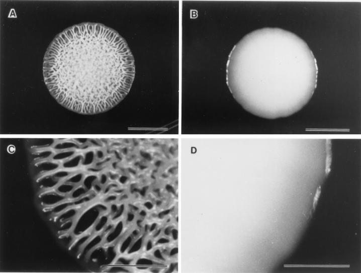
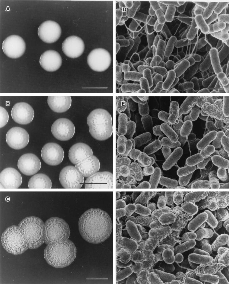
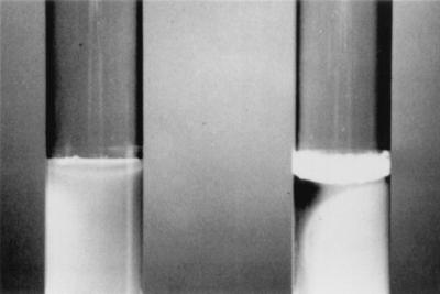
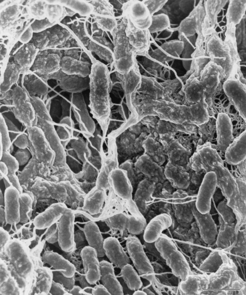
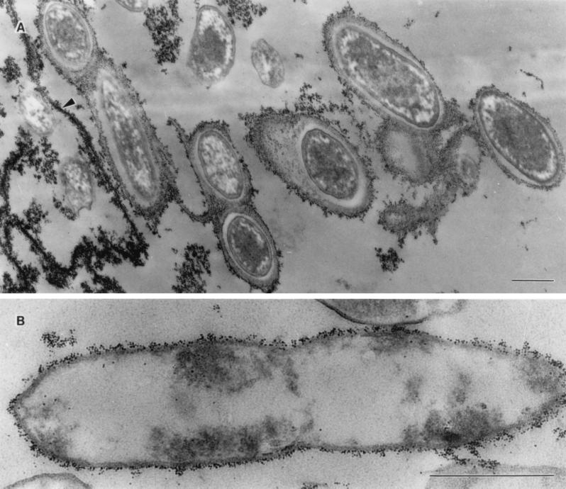
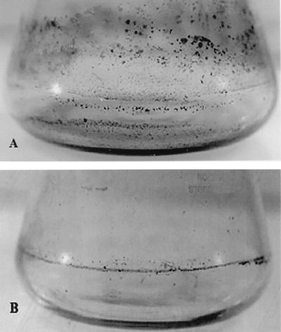
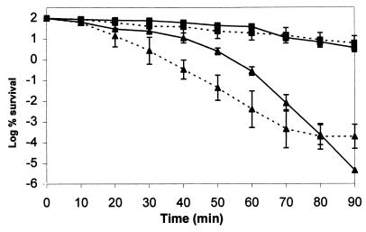
Rugose phenotypes, such as those observed in Vibrio cholerae, have increased resistance to chlorine, oxidative stress, and complement-mediated killing. In this study we identified and defined a rugose phenotype in Salmonella enterica serovar Typhimurium DT104 and showed induction only on certain media at 25 degrees C after 3 days of incubation. Incubation at 37 degrees C resulted in the appearance of the smooth phenotype. Observation of the ultrastructure of the rugose form and a stable smooth variant (Stv), which was isolated following a series of passages of the rugose cells, revealed extracellular substances only in cells from the rugose colony. Observation of the extracellular substance by scanning electron microscopy (SEM) was correlated with the appearance of corrugation during development of rugose colony morphology over a 4-day incubation period at 25 degrees C. In addition, the cells also formed a pellicle in liquid broth, which was associated with the appearance of interlacing slime and fibrillar structures, as observed by SEM. The pellicle-forming cells were completely surrounded by capsular material, which bound cationic ferritin, thus indicating the presence of an extracellular anionic component. The rugose cells, in contrast to Stv, showed resistance to low pH and hydrogen peroxide and an ability to form biofilms. Based on these results and analogy to the rugose phenotype in V. cholerae, we propose a possible role for the rugose phenotype in the survival of S. enterica serovar Typhimurium DT104.
Figures
References
-
- Besser T E, Gay C C, Gay J M, Hancock D D, Rice D, Pritchett L C, Erickson E D. Salmonellosis associated with S. typhimurium DT104 in the USA. Vet Rec. 1997;140:75. - PubMed
-
- Boyd A, Chakrabarty A M. Pseudomonas aeruginosa biofilms: role of the alginate exopolysaccharide. J Ind Microbiol. 1995;15:162–1688. - PubMed
-
- Cole G T. Preparation of microfungi for scanning electron microscopy. In: Aldrich H C, Todd W J, editors. Ultrastructure techniques for microorganisms. New York, N.Y: Plenum Press; 1986. pp. 1–38.
Publication types
MeSH terms
Substances
LinkOut - more resources
Full Text Sources
