. 2001 Sep 11;98(19):10910-7.
doi: 10.1073/pnas.181354698. Epub 2001 Aug 28.

Adult-generated hippocampal and neocortical neurons in macaques have a transient existence

Affiliations

Adult-generated hippocampal and neocortical neurons in macaques have a transient existence

E Gould et al. Proc Natl Acad Sci U S A. .

Abstract

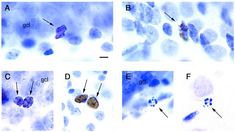
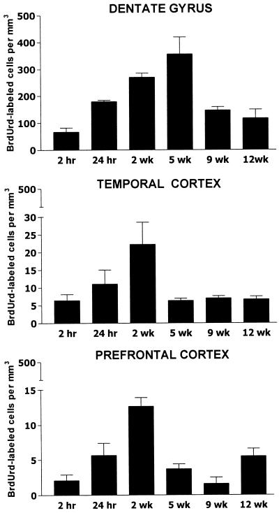
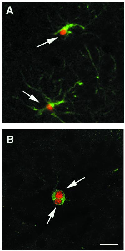
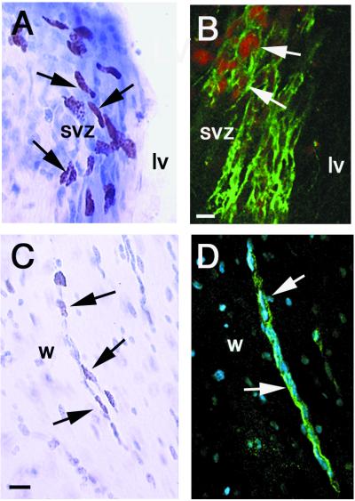
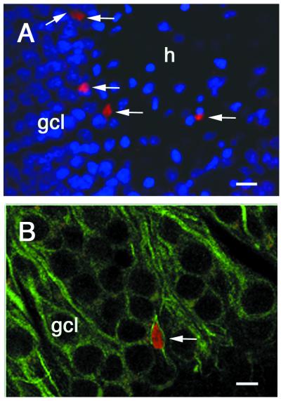
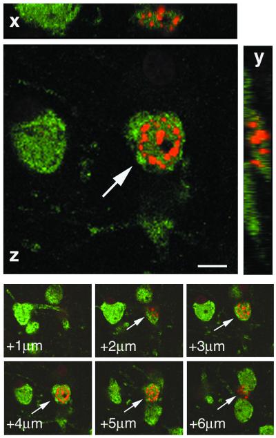
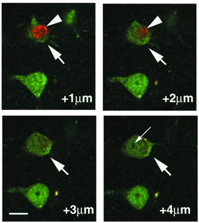
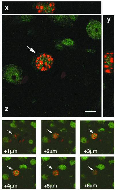
Previously we reported that new neurons are added to the hippocampus and neocortex of adult macaque monkeys. Here we compare the production and survival of adult-generated neurons and glia in the dentate gyrus, prefrontal cortex, and inferior temporal cortex. Twelve adult macaques were injected with the thymidine analogue BrdUrd, and the phenotypes of labeled cells were examined after 2 h, 24 h, 2 wk, 5 wk, 9 wk, and 12 wk by using the following immunocytochemical markers: for immature and mature neurons, class III beta-tubulin (TuJ1); for mature neurons, neuronal nuclei; for astrocytes, glial fibrillary acidic protein; and for oligodendrocytes, 2',3'-cyclic nucleotide 3' phosphodiesterase. We found that the dentate gyrus had many more BrdUrd-labeled cells than either neocortical area. Furthermore, a greater percentage of BrdUrd-labeled cells expressed a neuronal marker in the dentate gyrus than in either neocortical area. The number of new cells in all three areas declined by 9 wk after BrdUrd labeling, suggesting that some of the new cells have a transient existence. BrdUrd-labeled cells also were found in the subventricular zone and in the white matter between the lateral ventricle and neocortex; some of the latter cells were double-labeled for BrdUrd and TuJ1. Adult neocortical neurogenesis is not restricted to primates. Five adult rats were injected with BrdUrd, and after a 3-wk survival time, there were cells double-labeled for BrdUrd and either TuJ1 or neuronal nuclei in the anterior neocortex as well as the dentate gyrus.


Figures

Figure 1
Figure 1
Evidence for new cells and dying cells in adult macaque brains. Light photomicrographs of cresyl violet-stained tissue. (A and B) BrdUrd-labeled mitotic figures (arrows) in the (A) dentate gyrus and (B) svz 24 h after BrdUrd injection. Cells appear to be in anaphase. (C and D) Closely adjacent BrdUrd-labeled cells with the nuclear morphology of neurons (arrows) in (C) dentate gyrus and (D) prefrontal cortex 2 wk after BrdUrd injection. (E and F) Pyknotic cells (arrows) in (E) dentate gyrus and (F) prefrontal cortex. gcl, granule cell layer. Note that the new cells in the dentate gyrus and prefrontal cortex are similar to each other as are the dying cells in the two areas. (Scale bar = 10 μm.)
Figure 2
Figure 2
Density of BrdUrd-labeled cells (number of BrdUrd-labeled cells per mm3) in the dentate gyrus (Top), inferior temporal cortex (Middle), and prefrontal cortex (Bottom) at different survival times after a single injection of BrdUrd. There were two adult monkeys at each time point.
Figure 3
Figure 3
Confocal laser scanning microscope images of glia in prefrontal cortex of a macaque 2 wk after BrdUrd injection. (A) Two astrocytes (arrows) double-labeled with BrdUrd (red nuclear stain) and GFAP (green cytoplasmic stain). (B) Two adjacent oligodendrocytes double-labeled with BrdUrd (red) and CNP (green cytoplasmic stain). (Scale bar = 5 μm.)
Figure 4
Figure 4
Immature cells that appear to be migrating in the svz and white matter between the lateral ventricle and prefrontal cortex of macaques. (A) Light photomicrograph of BrdUrd-labeled cells (arrows) in the svz 2 h post-BrdUrd injection. lv, lateral ventricle. (B) Confocal image of cells (arrows) double-labeled with BrdUrd (red) and TuJ1 (green cytoplasmic stain) in the svz 2 h post-BrdUrd injection. (C) BrdUrd-labeled cells (arrow) that appear to be migrating in the white matter (w) 2 wk after BrdUrd injection. (D) Confocal image of a stream of TuJ1 (green)-positive cells (arrows) that appear to be migrating in the white matter of a prefrontal section. The blue nuclear stain is the DNA dye Hoechst 44323. (Scale bars = 5 μm.)
Figure 5
Figure 5
Confocal images of new neurons in dentate gyrus of adult macaques 5 wk after BrdUrd injection. (A) Granule cells (arrows) labeled with BrdUrd (red). Other granule cells are labeled with the DNA dye Hoechst 44323 (blue). (B) Granule cell double-labeled with BrdUrd (red) and TuJ1 (green). [Scale bars = 20 μm (A) and 5 μm (B).]
Figure 6
Figure 6
Confocal image of a cell in prefrontal cortex of a macaque double-labeled with NeuN (green nuclear and cytoplasmic stain) and BrdUrd (red) rotated in orthogonal planes (x,y,z) and z-sectioned to verify double-labeling throughout its extent. Note the double-labeled cell begins at +2 μm. The monkey was perfused 2 wk after the BrdUrd injection. (Scale bar = 5 μm.)
Figure 7
Figure 7
Confocal z-sections through two closely adjacent cells in prefrontal cortex of a macaque 2 wk after BrdUrd injection. The neuron (arrow) is labeled with NeuN (green), and the satellite cell (arrowhead) is labeled with BrdUrd (red). If only the +1-μm and +2-μm sections were examined they might have been incorrectly interpreted as containing a cell double-labeled with NeuN and BrdUrd. The nucleolus (thin arrow) in a section that does not have BrdUrd labeling indicates that these are two separate cells. (Scale bar = 10 μm.)
Figure 8
Figure 8
Confocal image of a cell in anterior cortex of an adult rat double-labeled with NeuN (green) and BrdUrd (red) rotated in orthogonal planes and z-sectioned to verify double-labeling throughout its extent. The rat was perfused 3 wk after the BrdUrd injection. (Scale bar = 5 μm.)

References

    1. Hastings N B, Tanapat P, Gould E. Neuroscientist. 2000;6:315–325.
    1. Goldman S A. J Neurobiol. 1998;36:267–286. - PubMed
    1. Privat A, Leblond C P. J Comp Neurol. 1972;146:277–302. - PubMed
    1. Lopez-Garcia C, Molowny A, Garcia-Verdugo J M, Ferrer I. Brain Res. 1988;471:167–174. - PubMed
    1. Altman J. Anat Rec. 1963;145:573–591. - PubMed

Publication types

MeSH terms

LinkOut - more resources