. 2001 Sep 11;98(19):10608-13.
doi: 10.1073/pnas.191321998. Epub 2001 Aug 28.

Human mitochondrial topoisomerase I

Affiliations

Human mitochondrial topoisomerase I

H Zhang et al. Proc Natl Acad Sci U S A. .

Abstract

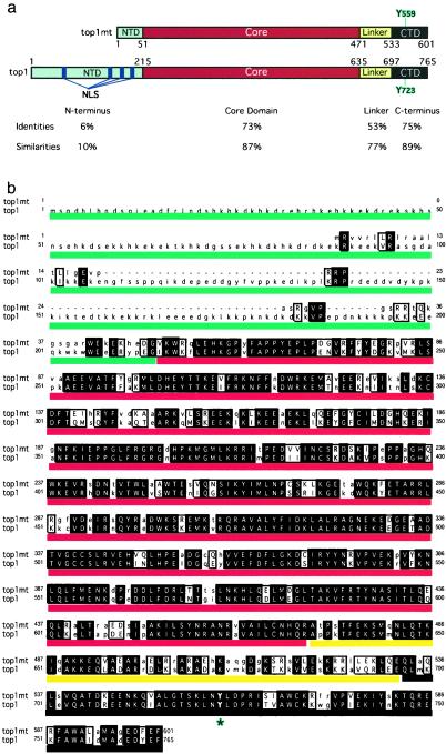
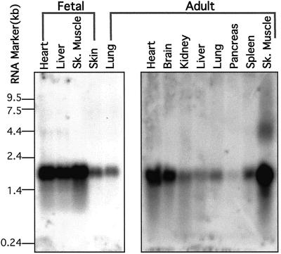
Tension generated in the circular mitochondrial genome during replication and transcription points to the need for mtDNA topoisomerase activity. Here we report a 601-aa polypeptide highly homologous to nuclear topoisomerase I. The N-terminal domain of this novel topoisomerase contains a mitochondrial localization sequence and lacks a nuclear localization signal. Therefore, we refer to this polypeptide as top1mt. The pattern of top1mt expression matches the requirement for high mitochondrial activity in specific tissues. top1mt is a type IB topoisomerase that requires divalent metal (Ca(2+) or Mg(2+)) and alkaline pH for optimum activity. The TOP1mt gene is highly homologous to the nuclear TOP1 gene and consists of 14 exons. It is localized on human chromosome 8q24.3.


Figures

Figure 1
Figure 1
Comparison between the novel human top1 (top1mt) and the previously known top1 (top1) polypeptides. (a) Domains are defined according to the nuclear top1 (33, 47). The positions of catalytic tyrosines are outlined in blue. The NTD, core domain, linker, and C-terminal domain are colored in blue, red, yellow, and black, respectively. Amino acid identities and homologies are indicated for each domain. There is no homology between the two NTDs, whereas the other domains are highly conserved. (b) Polypeptide sequence alignment for top1mt (upper lines) and the previously known human top1 (top1, lower lines). Identical matches are in uppercase, bold, and shaded; similar amino acids are in uppercase but not shaded. The unmatched amino acids are in lowercase. The catalytic tyrosines (Y723 for top1 and Y559 for top1mt) are indicated by an asterisk. Colored lines correspond to a: blue for the NTD, red for the core, yellow for the linker, and black for the C-terminal domain.
Figure 2
Figure 2
Mitochondrial localization of top1mt. a shows the constructs used: top1mt-GFP, NTD-GFP, and ΔNTD-top1mt-GFP. b and c show fluorescence microscopy images 24 h after transient transfection of human glioblastoma M059J cells with the constructs indicated above each. Mitochondria were localized by using MitoTracker red CM-H2Xros.
Figure 3
Figure 3
Biochemical characterization of top1mt. (a) Recombinant top1mt was expressed as a His-tagged protein, purified through nickel column and Q-Sepharose FPLC chromatography (Amersham Pharmacia Biotech). Coomassie blue staining shows that recombinant top1mt was homogeneous (≈72 kDa). (b) top1mt is a type I topoisomerase whose relaxation activity is inhibited by CPT, and stimulated by Ca2+ or Mg2+ in combination with a higher pH optimum than that of top1. Parallel experiments were performed with top1mt (Upper) and top1 (Lower). Experimental conditions are indicated above lanes. (c) Schematic representation of the covalent intermediates formed by types IA and IB topoisomerases. Type IA enzymes link to the 5′ terminus of the broken DNA, whereas type IB enzymes link to the 3′ terminus (3). (d) SDS/PAGE gel: both top1mt (72 kDa, His-tagged) and top1(68 kDa, His-tagged, N-terminal truncated) formed a DNA–protein complex (DPC) with 5′ end-labeled DNA (Left). No DPC was detected with 3′end-labeled DNA (Right). (e) Sequencing gel: both top1mt and top1 release a 3′ end-labeled oligonucleotide (Right); cleavage is not detectable when the DNA is 5′ end-labeled (Left).
Figure 4
Figure 4
Northern blot for TOP1mt expression. Blots were probed with our full-length TOP1mt cDNA. Two membranes are shown (Invitrogen). The four lanes on the Left are from human fetal tissues. The nine lanes on the Right are from human adult tissues. The size markers (in kb) are indicated Left.
Figure 5
Figure 5
FISH localization of TOP1mt. Metaphase chromosome spreads were derived from methotrexate-synchronized normal human peripheral leukocytes after hybridization with a genomic DNA probe and 4′,6-diamidino-2-phenylindole (DAPI) counterstaining. Both chromosomes with symmetrical FITC signals on sister chromatids (arrows) were identified as chromosome 8 on DAPI-inverted G-banded spreads, and fluorescent signals were localized at band 8q24.3.

References

    1. Wang J C. J Mol Biol. 1971;55:523–533. - PubMed
    1. Wang J C. J Biol Chem. 1991;266:6659–6662. - PubMed
    1. Wang J C. Annu Rev Biochem. 1996;65:635–692. - PubMed
    1. Gellert M. Annu Rev Biochem. 1981;50:879–910. - PubMed
    1. Clayton D A. Int Rev Cytol. 1992;141:217–232. - PubMed

MeSH terms