. 2001 Aug 28;98(18):10398-403.
doi: 10.1073/pnas.171321298.

AML1-ETO expression is directly involved in the development of acute myeloid leukemia in the presence of additional mutations

Affiliations

AML1-ETO expression is directly involved in the development of acute myeloid leukemia in the presence of additional mutations

Y Yuan et al. Proc Natl Acad Sci U S A. .

Abstract

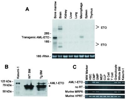
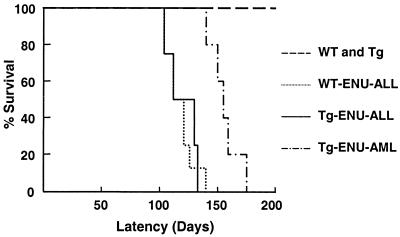
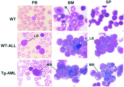
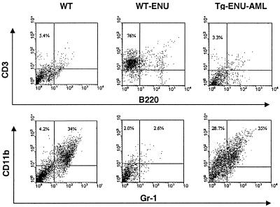
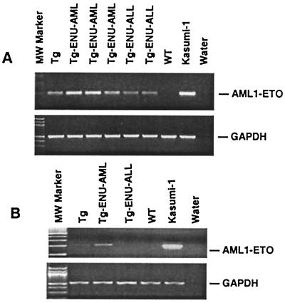
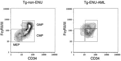
The t(8;21) is one of the most frequent chromosomal abnormalities associated with acute myeloid leukemia (AML). The translocation, which involves the AML1 gene on chromosome 21 and the ETO gene on chromosome 8, generates an AML1-ETO fusion transcription factor. To examine the effect of the AML1-ETO fusion protein on leukemogenesis, we made transgenic mice in which expression of AML1-ETO is under the control of the human MRP8 promoter (hMRP8-AML1-ETO). AML1-ETO is specifically expressed in myeloid cells, including common myeloid progenitors of hMRP8-AML1-ETO transgenic mice. The transgenic mice were healthy during their life spans, suggesting that AML1-ETO alone is not sufficient for leukemogenesis. However, after treatment of newborn hMRP8-AML1-ETO transgenic mice and their wild-type littermates with a strong DNA-alkylating mutagen, N-ethyl-N-nitrosourea, 55% of transgenic mice developed AML and the other 45% of transgenic mice and all of the wild-type littermates developed acute T lymphoblastic leukemia. Our results provide direct evidence that AML1-ETO is critical for causing myeloid leukemia, but one or more additional mutations are required for leukemogenesis. The hMRP8-AML1-ETO-transgenic mice provide an excellent model that can be used to isolate additional genetic events and to further understand the molecular pathogenesis of AML1-ETO-related leukemia.


Figures

Figure 1
Figure 1
Specific expression of AML1-ETO in hMRP8-AML1-ETO transgenic mice. (A) AML1-ETO mRNA is specifically expressed in bone marrow cells and macrophages in line no. 28. Approximately 10 μg of total RNA from different tissues was separated electrophoretically in a 1% agarose gel, transferred to nylon membrane, and hybridized with an ETO cDNA probe. The ethidium bromide staining of the 18S ribosomal RNA is presented to show the loading of the RNA samples. The positions of transcripts of endogenous ETO in the brain and transgenic AML1-ETO are marked. (B) AML1-ETO fusion protein is expressed in the bone marrow cells in hMRP8-AML1-ETO transgenic mice. Cell lysates from 4 × 10⁶ bone marrow (BM) cells from wild-type mice (WT) or transgenic mice (Tg) were separated by electrophoresis on an SDS/8% polyacrylamide gel, transferred to a nitrocellulose membrane, and probed with a 1:500 dilution of an anti-ETO polyclonal antibody. Lysate from Kasumi-1 cells was used as a positive control. The position of AML1-ETO is marked. The asterisk indicates nonspecific signals. (C) AML1-ETO mRNA is specifically expressed in the myeloid cells of hMRP8-AML1-ETO transgenic mice. Bone marrow cells from hMRP8-AML1-ETO transgenic mice were double sorted into different populations according to their surface marker expression. One thousand hematopoietic stem cells (HSC), common myeloid progenitors (CMP), granulocyte/monocyte progenitors (GMP), megakaryocyte/erythroid progenitors (MEP), common lymphoid progenitors (CLP), B cells, T cells, granulocytes, and whole bone marrow (BM) cells were subjected to RT-PCR analysis. RNA from Kasumi-1 cells was used as a positive control. PCRs without reverse transcription were used as negative controls.
Figure 2
Figure 2
Survival curves of transgenic mice (Tg) and wild- type mice (WT) after ENU treatment. Wild-type mice developed acute lymphoblastic leukemia (WT-ENU-ALL, n = 8). Transgenic mice developed acute lymphoblastic leukemia (Tg-ENU-ALL, n = 4) and AML (Tg-ENU-AML, n = 5). Untreated transgenic mice and wild-type mice (WT and Tg, n = 5, respectively) are shown as controls.
Figure 3
Figure 3
Development of leukemia in transgenic mice and wild-type littermates after ENU treatment. Wright–Giemsa staining of peripheral blood smears (PB), bone marrow cytospins (BM), and spleen cytospins (SP) from representative leukemic and wild-type mice. N, neutrophil; LB, lymphoblast; MB, myeloblast. (Original magnification, ×1,000.)
Figure 4
Figure 4
Flow cytometry analysis of bone marrow cells from leukemic transgenic mice. Healthy wild-type mice (WT), wild type with signs of disease (WT-ENU), and AML1-ETO-transgenic mice with signs of disease (Tg-ENU) were killed and single-cell suspensions were made from the bone marrow. Cells were double stained with anti-CD3 [phycoerythrin (PE)-labeled] and anti-B220 (FITC-labeled) or anti-CD11b-1 (PE) and anti-Gr-1 (FITC) antibodies. The percentages of CD3+/B220 cells, CD11b+/Gr-1, and CD11b+/Gr-1+ double-positive cells are indicated. Consistent results were obtained from the analysis of four WT-ENU mice and six Tg-ENU-AML mice.
Figure 5
Figure 5
Detection of AML1-ETO expression in the spleen and bone marrow cells from transgenic mice without ENU treatment (Tg), leukemic transgenic mice (Tg-ENU-AML or Tg-ENU-ALL), and wild-type (WT) mice. Representative results are shown. Kasumi-1 cell line RNA was used as a positive control. (A) RT-PCR analysis of bone marrow cells from three AML and two ALL transgenic mice. (B) RT-PCR analysis of spleen cells from one AML and one ALL transgenic mice. PCR fragments were not detectable in the absence of an RT reaction (data not shown). GAPDH, glyceraldehyde-3-phosphate dehydrogenase; MW, molecular weight.
Figure 6
Figure 6
Identification of myeloid progenitor populations in transgenic mice. FACS analysis of bone marrow cells from a non-ENU-treated healthy transgenic mouse (Left) and an ENU-treated AML transgenic mouse (Right). The distribution of Lin/Sca-1/c-Kit+ bone marrow myeloid progenitors is presented based on the expression of CD34 and Fcγ receptors II/III (FcγRII/III). CMP, common myeloid progenitors; GMP, granulocyte/monocyte progenitors; MEP, megakaryocyte/erythroid progenitors.

References

    1. Downing J R, Higuchi M, Lenny N, Yeoh A E. Semin Cell Dev Biol. 2000;11:347–360. - PubMed
    1. Friedman A D. Leukemia. 1999;13:1932–1942. - PubMed
    1. Lutterbach B, Hiebert S W. Gene. 2000;245:223–235. - PubMed
    1. Miyoshi H, Shimizu K, Kozu T, Maseki N, Kaneko Y, Ohki M. Proc Natl Acad Sci USA. 1991;88:10431–10434. - PMC - PubMed
    1. Meyers S, Lenny N, Hiebert S W. Mol Cell Biol. 1995;15:1974–1982. - PMC - PubMed

Publication types

MeSH terms

LinkOut - more resources