Review
. 2001 Sep;65(3):463-79, table of contents.
doi: 10.1128/MMBR.65.3.463-479.2001.

Autophagy in yeast: mechanistic insights and physiological function

Affiliations
Review

Autophagy in yeast: mechanistic insights and physiological function

H Abeliovich et al. Microbiol Mol Biol Rev. 2001 Sep.

Abstract

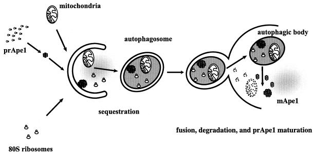
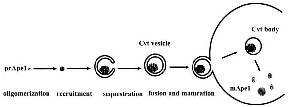
Unicellular eukaryotic organisms must be capable of rapid adaptation to changing environments. While such changes do not normally occur in the tissues of multicellular organisms, developmental and pathological changes in the environment of cells often require adaptation mechanisms not dissimilar from those found in simpler cells. Autophagy is a catabolic membrane-trafficking phenomenon that occurs in response to dramatic changes in the nutrients available to yeast cells, for example during starvation or after challenge with rapamycin, a macrolide antibiotic whose effects mimic starvation. Autophagy also occurs in animal cells that are serum starved or challenged with specific hormonal stimuli. In macroautophagy, the form of autophagy commonly observed, cytoplasmic material is sequestered in double-membrane vesicles called autophagosomes and is then delivered to a lytic compartment such as the yeast vacuole or mammalian lysosome. In this fashion, autophagy allows the degradation and recycling of a wide spectrum of biological macromolecules. While autophagy is induced only under specific conditions, salient mechanistic aspects of autophagy are functional in a constitutive fashion. In Saccharomyces cerevisiae, induction of autophagy subverts a constitutive membrane-trafficking mechanism called the cytoplasm-to-vacuole targeting pathway from a specific mode, in which it carries the resident vacuolar hydrolase, aminopeptidase I, to a nonspecific bulk mode in which significant amounts of cytoplasmic material are also sequestered and recycled in the vacuole. The general aim of this review is to focus on insights gained into the mechanism of autophagy in yeast and also to review our understanding of the physiological significance of autophagy in both yeast and higher organisms.


Figures

FIG. 1
FIG. 1
Overview of membrane trafficking in yeast macroautophagy. During autophagy, various components of the cytoplasm, such as mitochondria, ribosomes, specific cargo such as prApe1 (see Fig. 2), and soluble cytoplasmic material (depicted as diffuse gray), are sequestered into autophagosomes, which are large (300- to 900-nm) double-bilayer vesicles. Once formed, autophagosomes fuse with the vacuole, releasing a single-bilayer-bound autophagic body into the lumen of this organelle. Vacuolar hydrolases act on the limiting membrane of the autophagic body, releasing its contents, which are degraded into biosynthetic building blocks.
FIG. 2
FIG. 2
Discrete steps in cytoplasm-to-vacuole targeting of aminopeptidase I. Aminopeptidase I (Ape1), a resident vacuolar hydrolase, is synthesized as a soluble zymogen on cytoplasmic ribosomes. It then undergoes homo-oligomerization to form dodecamers, which further assemble into large, membrane-associated complexes. The membrane-associated form is sequestered into an autophagosome-like structure called the Cvt vesicle (140 to 160 nm diameter). Cvt vesicles fuse with the vacuole, giving rise to an intravacuolar Cvt body that is degraded, releasing prApe1 (61 kDa) into the lumen, where it is processed to the mature form (mApe1; 50 kDa).
FIG. 3
FIG. 3
Autophagosomal sequestration is analogous to a reverse fenestration. It is thought that during fenestration, as occurs in Golgi lamellae in higher eukaryotes (bottom panel), the lumenal faces of the limiting lamellar membrane fuse, creating a pore that then expands to form a fenestration. In the formation of the autophagosome, a similar process is occurring but does so in the opposite direction (top panel). Here, a circular pore is being closed to form a closed lamellar sheet, with the added complication that this sheet is spherical.
FIG. 4
FIG. 4
Apg12 undergoes a ubiquitin-like conjugation to Apg5. Apg12 is activated by the E1-like protein, Apg7, by formation of a labile thioester bond between the carboxyl group of the Apg12 C-terminal glycine residue and a thiol group from an internal cysteine (Cys 507) of Apg7. The activated Apg12 is transferred to a cysteine group on Apg10 in a fashion reminiscent of ubiquitin transfer from E1 proteins to E2. Apg10 then transfers Apg12 to lysine 149 of Apg5 by the formation of a more stable amide bond. The Apg12-Apg5 conjugate recruits Apg16 dimers. The divalent nature of Apg16 results in the formation of large multimeric complexes, and these are thought to play a role in the nucleation of both autophagosomes and Cvt vesicles. In addition, it has been shown that the Apg12-Apg5 complex is required for the recruitment of Aut7 to membranes (see Fig. 5).
FIG. 5
FIG. 5
Aut7 undergoes ubiquitin-like conjugation to phosphatidylethanolamine. Aut7 is synthesized with a C-terminal arginine residue encoded in the reading frame of the gene (i). The cysteine protease Aut2/Apg4 hydrolyzes the peptide bond to the C-terminal arginine to create form (ii), which has a C-terminal glycine residue. Form (ii) is activated by forming a thioester link with cysteine 507 of Apg7 (form iii) and is then transferred to a cysteine residue on an E2 analog, Aut1 (form iv). Aut1 then transfers Aut7 to phosphatidylethanolamine (PE), via the formation of an amide bond, to create the tightly membrane bound form (v). Aut7 is released from the membrane by hydrolysis of the amide bond, a reaction again catalyzed by Aut2, to regenerate form (ii). In the absence of Aut2, ectopic expression of form (ii) does not completely rescue the Cvt and autophagy defects of the aut2Δ mutant, indicating that a complete cycle is required for Aut7 function.
FIG. 6
FIG. 6
Apg1 activity is modulated by different regulatory proteins under different physiological conditions. In logarithmically growing cells, Apg13 is highly phosphorylated and can be weakly coprecipitated with Apg1. Under these conditions, Vac8 can be coprecipitated with Apg13. In addition, two-hybrid data show an interaction of Apg1 with Apg17 and Cvt9. On induction of autophagy, Apg13 undergoes partial dephosphorylation, concomitant with an increase in its coprecipitation efficiency with Apg1 and an increase in in vitro Apg1 protein kinase activity. However, Vac8 and Cvt9 are dispensible for autophagy, suggesting that they may be lost from the complex upon induction of autophagy. Likewise, Apg17 is not required for Cvt trafficking and thus may require activation of autophagy to bind tightly to the complex. Thus, in the complex on the left, Apg17 is designated by a dashed circle, as are Vac8 and Cvt9 in the complex on the right. Proteins are indicated by number as follows: Apg1, 1; Apg13, 13; Apg17, 17; Cvt9, 9; Vac8, 8. Additional proteins may also be present in this complex. Phosphorylation is indicated by “p”. The phosphorylation state of Apg17 is not known. SD-N, minimal medium lacking nitrogen. Modified from reference with permission of the publisher.
FIG. 7
FIG. 7
Induction of autophagy can be divided into nucleation and expansion steps. Tor activity prevents dephosphorylation of Apg13, by an unknown mechanism. Inhibition of Tor by the rapamycin-RBP complex then results in rapid dephosphorylation of Apg13. Dephosphorylated Apg13 acts on Apg1 to mediate autophagosome nucleation. In the absence of new protein synthesis, this results in abnormally small autophagosomes. Under normal conditions, however, inhibition of Tor also up-regulates the transcription of starvation- and autophagy-specific genes, and the ensuing increase in levels of specific proteins, such as Aut7, allows the expansion of autophagosomes. Reprinted from reference with permission of the publisher.

References

    1. Abeliovich H, Darsow T, Emr S D. A t-SNARE/Sec1p complex composed of Tlg2p and Vps45p is required for cytoplasm to vacuole targeting of aminopeptidase I. EMBO J. 1999;18:6005–6016. - PMC - PubMed
    1. Abeliovich H, Dunn W A, Jr, Kim J, Klionsky D J. Dissection of autophagosome biogenesis into distinct nucleation and expansion steps. J Cell Biol. 2000;151:1025–1033. - PMC - PubMed
    1. Abeliovich H, Grote E, Novick P, Ferro-Novick S. Tlg2p, a yeast syntaxin homolog that resides on the Golgi and endocytic structures. J Biol Chem. 1998;273:11719–11727. - PubMed
    1. Anglade P, Vyas S, Javoy-Agid F, Herrero M T, Michel P P, Marquez J, Mouatt-Progent A, Ruberg M, Hirsch E C, Agid Y. Apoptosis and autophagy in nigral neurons of patients with Parkinson's disease. Histol Histopathol. 1997;12:25–31. - PubMed
    1. Ashford T P, Porter K R. Cytoplasmic components in hepatic cell lysosomes. J Cell Biol. 1962;12:198–202. - PMC - PubMed

Publication types

LinkOut - more resources