

Review
doi: 10.1093/emboj/20.17.4629.
Lysosomal cysteine proteases: facts and opportunities
Affiliations
- PMID: 11532926
- PMCID: PMC125585
- DOI: 10.1093/emboj/20.17.4629
Item in Clipboard
Review
Lysosomal cysteine proteases: facts and opportunities
EMBO J.
.
Abstract
From their discovery in the first half of the 20th century, lysosomal cysteine proteases have come a long way: from being the enzymes non-selectively degrading proteins in lysosomes to being those responsible for a number of important cellular processes. Some of the features and roles of their structures, specificity, regulation and physiology are discussed.
Figures
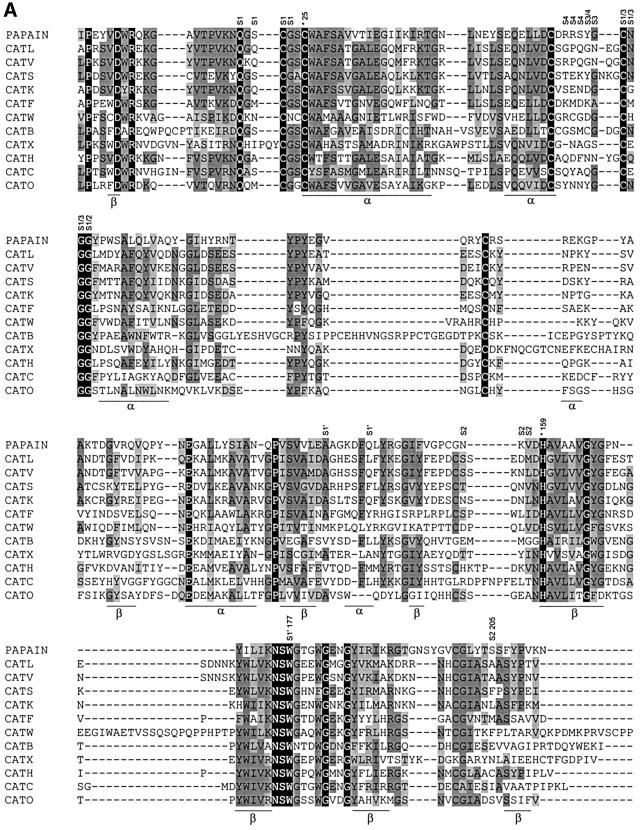
Fig. 1. (A) Structure-based amino acid sequence alignment of mature parts of papain and related human lysosomal cysteine proteases was performed by the CLUSTAL_W program as described previously (Turk et al., 2000). The sequences were taken from the SWISS-PROT or GenBank databases. The active site residues Cys25 and His159 are marked with asterisks and numbered. (B) Fold of cathepsin L, a typical human lysosomal cysteine protease (Gunčar et al., 1999).
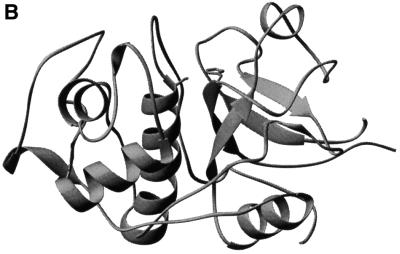
Fig. 1. (A) Structure-based amino acid sequence alignment of mature parts of papain and related human lysosomal cysteine proteases was performed by the CLUSTAL_W program as described previously (Turk et al., 2000). The sequences were taken from the SWISS-PROT or GenBank databases. The active site residues Cys25 and His159 are marked with asterisks and numbered. (B) Fold of cathepsin L, a typical human lysosomal cysteine protease (Gunčar et al., 1999).
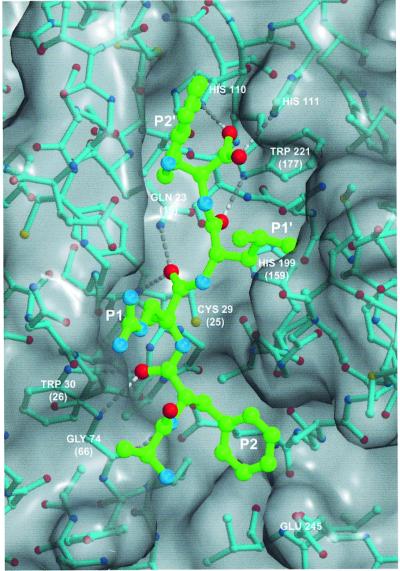
Fig. 2. A substrate model APRLW bound along the active site of cathepsin B (Turk et al., 1995; 1CSB). Bonds of substrate and cathepsin B structure are shown in cyan and green, respectively. Non-hydrogen atoms are shown as small colored spheres: oxygens are red, sulfurs yellow and nitrogens blue, whereas carbons are shown in cyan and green, corresponding to the coloring code of covalent bonds of cathepsin B and substrate models. The GRASP generated surface (Nicholls et al., 1991) of cathepsin B is gray. Crucial residues involved in the binding of substrate main chain atoms are labeled with their residue name and sequence ID. Papain numbering is shown in parentheses for the contacts between essential enzyme residues and substrate. Hydrogen bonds along the substrate main chain are shown as white broken lines. The figure was prepared with MAIN (Turk, 1992) and rendered with RENDER (Merritt and Bacon, 1997).
References
-
- Barrett A.J., Rawlings,N.D. and Woessner,J.F.,Jr (eds) (1998) Handbook of Proteolytic Enzymes. Academic Press, London, UK.
-
- Brocklehurst K. (1994) A sound basis for pH-dependent kinetic studies on enzymes. Protein Eng., 7, 291–299. - PubMed
-
- Brömme D., Li,Z., Barnes,M. and Mehler,E. (1999) Human cathepsin V functional expression, tissue distribution, electrostatic surface potential, enzymatic characterization and chromosomal localization. Biochemistry, 38, 2377–2385. - PubMed
-
- Chapman H.A., Riese,R.J. and Shi,G.-P. (1997) Emerging roles for cysteine proteases in human biology. Annu. Rev. Physiol., 59, 63–88. - PubMed
Publication types
MeSH terms
Substances
LinkOut - more resources
Full Text Sources
Other Literature Sources
