. 2001 Sep 3;20(17):4639-47.
doi: 10.1093/emboj/20.17.4639.

Activation of integrins in endothelial cells by fluid shear stress mediates Rho-dependent cytoskeletal alignment

Affiliations

Activation of integrins in endothelial cells by fluid shear stress mediates Rho-dependent cytoskeletal alignment

E Tzima et al. EMBO J. .

Abstract

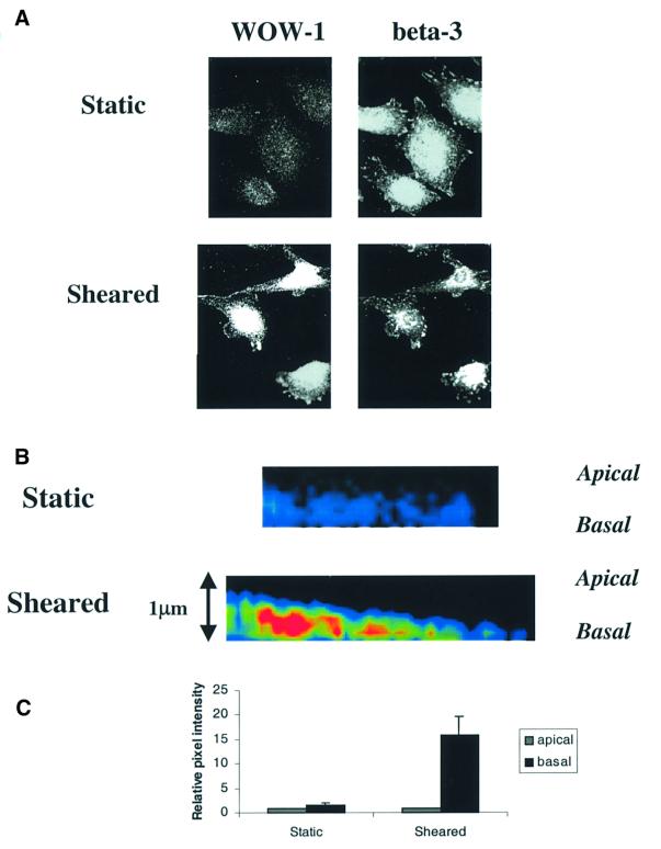
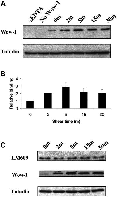
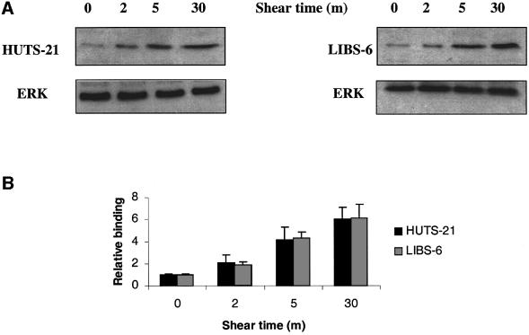
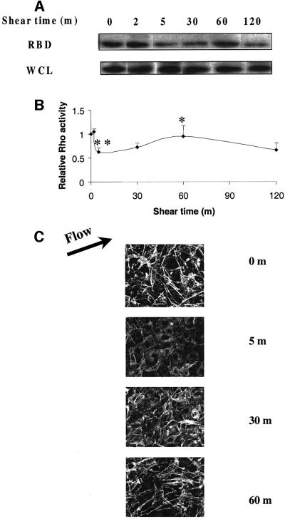
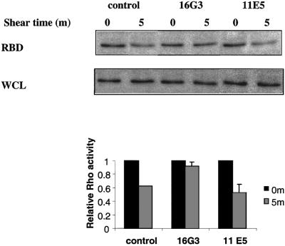
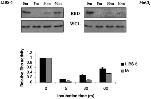
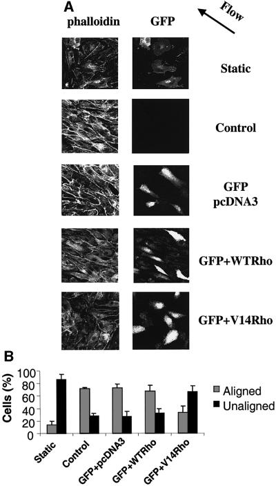
Fluid shear stress is a critical determinant of vascular remodeling and atherogenesis. Both integrins and the small GTPase Rho are implicated in endothelial cell responses to shear but the mechanisms are poorly understood. We now show that shear stress rapidly stimulates conformational activation of integrin alpha(v)beta3 in bovine aortic endothelial cells, followed by an increase in its binding to extracellular cell matrix (ECM) proteins. The shear-induced new integrin binding to ECM induces a transient inactivation of Rho similar to that seen when suspended cells are plated on ECM proteins. This transient inhibition is necessary for cytoskeletal alignment in the direction of flow. The results therefore define the role of integrins and Rho in a pathway leading to endothelial cell adaptation to flow.


Figures

None
Fig. 1. Integrin αvβ3 activation by shear stress. (A) BAECs under static conditions or after shear stress at 12 dynes/cm2 for 5 min were fixed and stained with WOW-1 to label high-affinity unoccupied αvβ3 and double labeled with a polyclonal antibody that recognizes all αvβ3 integrins. (B) Color intensity profiles of representative XZ reconstructions of static and sheared cells that have been stained with WOW-1. Images show portions of single cells. The apical and basal surfaces are labeled. Red represents high and blue is low WOW-1 staining. (C) Histogram showing relative intensity of WOW-1 staining of basal compared with apical Z-sections of static and sheared cells. Cell edges were defined by TRITC– wheatgerm agglutinin (WGA) staining and were used as reference for quantitation of pixel intensity of WOW-1 staining; 45 cells were scored per condition.
None
Fig. 2. Quantitation of high affinity αvβ3 integrin. (A) BAECs were subjected to shear for the indicated times and incubated with WOW-1 to label activated αvβ3. Bound antibody was quantitated by western blotting. As a control, EDTA was added to block specific binding. Blots were stripped and reprobed for tubulin to assess protein loading. (B) Histogram showing WOW-1 binding normalized to tubulin. Values are means ± SEM (n = 4 independent experiments) relative to cells at time 0, values are statistically significant as defined by a student’s t-test (P <0.01). (C) Coverslips were incubated with WOW-1 or LM609 and bound antibody detected by western blotting. Results are representative of three independent experiments. Blots were stripped and reprobed for tubulin to assess protein loading.
None
Fig. 3. Quantitation of integrin occupancy. (A) BAECs plated on VN or FN were sheared for the indicated times and incubated with 5 µg/ml LIBS-6 or HUTS-21, respectively. Bound antibody was assessed by western blotting. Blots were stripped and reprobed for extracellular signal regulator kinase (ERK) to assess protein loading. (B) Results from three independent experiments were quantitated. Values are means ± SEM.
None
Fig. 4. Regulation of Rho activity by shear stress. (A) BAECs were subjected to shear stress for the indicated times. Rho activity is indicated by the amount of RBD-bound Rho, normalized to total Rho in whole cell lysates (WCL). (B) Quantitation of Rho activity relative to cells at time 0. Values are means ± SEM from three independent experiments, each of which was performed in duplicate. For the statistical analysis 5 min was compared with 0 min (**P <0.01) and 60 min was compared with 5 min (*P <0.05). (C) Time course of F-actin organization. Cells were subjected to shear stress, fixed and stained with rhodamine–phalloidin to detect actin stress fibers.
None
Fig. 5. Blocking free FN sites. BAECs were plated on FN-coated slides for 2 h in the absence of serum and then incubated with anti-FN Fab fragments for 15 min. Cells were either sheared (5 min, 12 dynes/cm2) or kept as static control and Rho assayed. Values are means ± SEM (n = 3, each in duplicate) relative to static samples.
None
Fig. 6. Integrin activation mimics the shear-induced Rho activation. BAECs were incubated with 0.5 mM MnCl2 or 15 µg/ml LIBS-6 antibody for the indicated times and Rho activity was determined. Values are means ± SEM (n = 3, each in duplicate) and represent Rho activity relative to cells at time 0.
None
Fig. 7. Constitutively activated Rho blocks cell alignment. (A) Cells transiently transfected with empty vector, WT Rho or V14Rho plus GFP were subjected to shear stress or kept under static conditions for 16 h. The cells were fixed, stained with rhodamine–phalloidin and observed by confocal microscopy. The direction of flow is indicated by an arrow. (B) Quantification of cell alignment with the direction of flow. Values are means ± SEM, n = 3; >100 cells were scored per condition.

References

    1. Allen W.E., Zicha,D., Ridley,A.J. and Jones,G.E. (1998) A role for Cdc42 in macrophage chemotaxis. J. Cell Biol., 141, 1147–1157. - PMC - PubMed
    1. Arthur W.T., Petch,L.A. and Burridge,K. (2000) Integrin engagement suppresses RhoA activity via a c-Src-dependent mechanism. Curr. Biol., 10, 719–722. - PubMed
    1. Avnur Z. and Geiger,B. (1981) The removal of extracellular fibronectin from areas of cell–substrate contact. Cell, 25, 121–132. - PubMed
    1. Barbee K.A., Davies,P.F. and Lal,R. (1994) Shear stress-induced reorganization of the surface topography of living endothelial cells imaged by atomic force microscopy. Circ. Res., 74, 163–171. - PubMed
    1. Bazzoni G. and Hemler,M.E. (1998) Are changes in integrin affinity and conformation overemphasized? Trends Biochem. Sci., 23, 30–34. - PubMed

Publication types

MeSH terms