. 2001 Sep 3;20(17):4704-16.
doi: 10.1093/emboj/20.17.4704.

Mutation in Brca2 stimulates error-prone homology-directed repair of DNA double-strand breaks occurring between repeated sequences

Affiliations

Mutation in Brca2 stimulates error-prone homology-directed repair of DNA double-strand breaks occurring between repeated sequences

A Tutt et al. EMBO J. .

Abstract

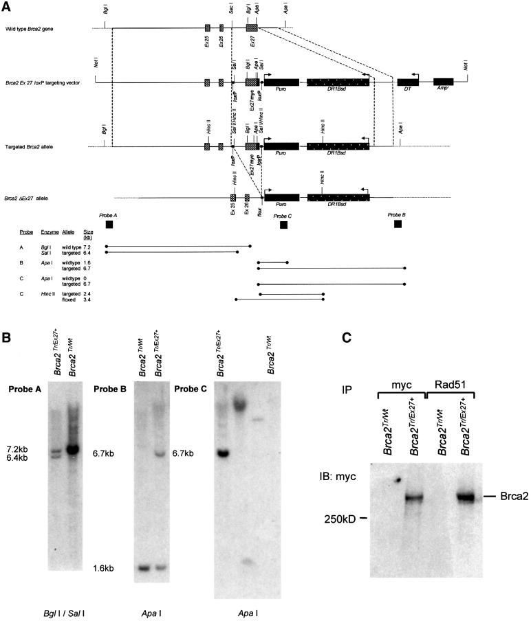
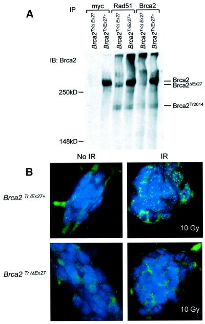
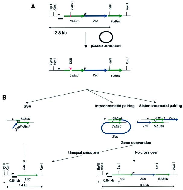
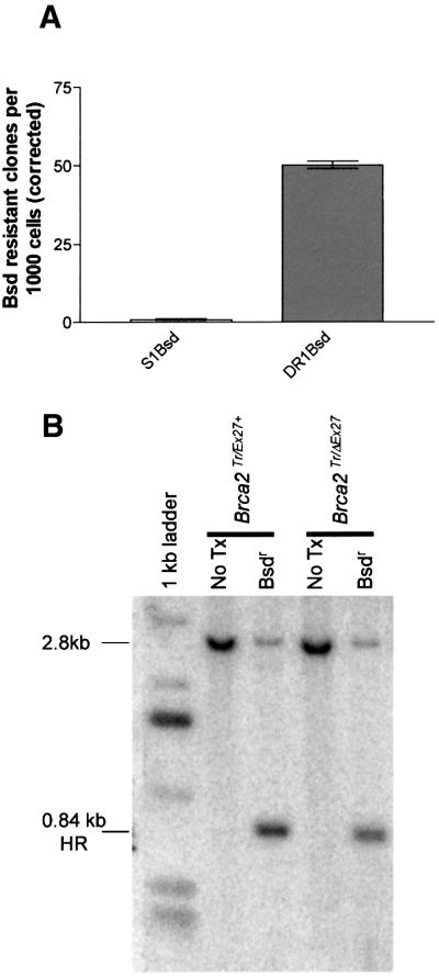
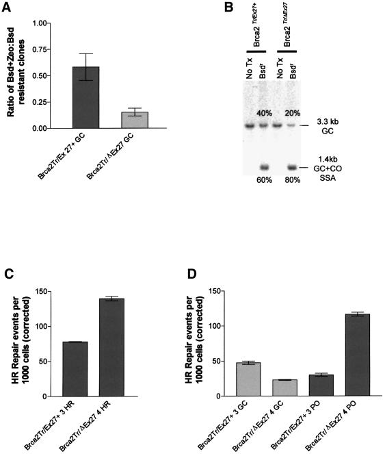
Mutation of BRCA2 causes familial early onset breast and ovarian cancer. BRCA2 has been suggested to be important for the maintenance of genome integrity and to have a role in DNA repair by homology- directed double-strand break (DSB) repair. By studying the repair of a specific induced chromosomal DSB we show that loss of Brca2 leads to a substantial increase in error-prone repair by homology-directed single-strand annealing and a reduction in DSB repair by conservative gene conversion. These data demonstrate that loss of Brca2 causes misrepair of chromosomal DSBs occurring between repeated sequences by stimulating use of an error-prone homologous recombination pathway. Furthermore, loss of Brca2 causes a large increase in genome-wide error-prone repair of both spontaneous DNA damage and mitomycin C-induced DNA cross-links at the expense of error-free repair by sister chromatid recombination. This provides insight into the mechanisms that induce genome instability in tumour cells lacking BRCA2.


Figures

None
Fig. 1. Brca2 exon 27 ‘knock in’ strategy and analysis of clones. (A) Structure of the 3′ end of the mouse Brca2 locus and targeting vector containing HR repair substrate. The upper line represents the wild-type allele. The second and third lines represent the linearized targeting vector and the targeted allele, respectively. Exons 25, 26 and 27 are shown as grey boxes. The positions of relevant restriction enzyme sites are marked. 5′ and 3′ regions of homology between the targeting vector and the wild-type allele are enclosed between dashed lines flanking the ‘knock in’ region. This includes a mutated exon 27 with an in-frame 9E10 myc epitope tag shown as a black box. loxP sites flanking exon 27 myc are shown as small black squares. The lower line demonstrates the effect of Cre-mediated recombination between the labelled loxP sites to produce a Flox site. This deletes the intervening sequence including exon 27myc. Large black boxes represent the puromycin (Puro) selectable marker gene, the diphtheria toxin (DT) negative selection marker and the ampicillin (Amp) resistance gene. The HR repair substrate DR1Bsd is represented by a black speckled box. Regions of hybridization to the three probes (A, B and C) used in Southern blot analyses are indicated by large dark squares. The restriction enzymes and the probes used for Southern analysis of the targeted allele, and the positions and sizes of the fragments detected are shown at the bottom of the figure. (B) Southern blot analysis. The targeted allele was termed Brca2Ex27+ and the targeted cell line termed Brca2Tr/Ex27+. Southern blots of genomic DNA from parental cells Brca2Tr/Wt and a Brca2Tr/Ex27+-targeted clone subjected to restriction digestion and hybridization with the marked probe. Probe A is a flanking probe 5′ to the 5′ homology. Probe B is 3′ to the 3′ homology. Probe C is a fragment of Puro. In the left panel, BglI–SalI digestion shows the 7.2 kb wild-type fragment in Brca2Tr/Wt ES cells, and both the 7.2 kb wild-type fragment and the predicted 6.4 kb targeted fragment in Brca2Tr/Ex27+ cells. In the middle panel, ApaI digestion shows the 1.6 kb wild-type fragment and predicted 6.7 kb targeted fragment. These confirm correct integration into the Brca2 locus. The right panel shows ApaI digests probed with Puro, confirming the absence of additional random integrants in Brca2Tr/Ex27+ cells. Brca2Tr/Wt is a negative control. The two middle lanes are clones containing non-targeted random integrants. (C) Correct integration of the targeting construct into the wild-type allele of Brca2Tr/Wt ES cells was confirmed by confirmation of production of full-length myc-tagged Brca2 protein. Immunoblot analysis of whole-cell lysates of Brca2Tr/Wt and Brca2Tr/Ex27+ ES cells immunoprecipated (IP) with anti-myc and anti-Rad51 antibodies and immunoblotted (IB) with an anti-myc antibody.
None
Fig. 2. Cre-mediated deletion of the C-terminus of Brca2 in ES cells. (A) Immunoblotting analysis of whole-cell extract of Brca2Tr/Ex27+ and Brca2Tr/ΔEx27 clonal cell lines immunoprecipated (IP) with anti-myc, anti-Rad51 and N-terminal anti-Brca2 antibodies, and immunoblotted (IB) with the anti-Brca2 antibody. The positions of full-length Brca2, the exon 27-deleted (Brca2ΔEx27) and the exon 11-truncated (Brca2Tr2014) proteins are indicated. (B) Failure of normal induction of Rad51 foci in Brca2Tr/ΔEx27 ES cells. Brca2Tr/Ex27+ and Brca2Tr/ΔEx27 ES cells mock irradiated (left upper and lower panels) or irradiated with 10 Gy (right upper and lower panels) were fixed and analysed by immunofluorescent microscopy. DNA is labelled with DAPI and appears blue. Rad51 was detected with anti-Rad51 antibody and a secondary FITC-conjugated antibody. The right upper panel shows induction of Rad51-containing nuclear foci in Brca2Tr/Ex27+ cells. The right lower panel shows failure of induction of Rad51 foci in Brca2Tr/ΔEx27 ES cells.
None
Fig. 3. HR repair substrate DR1Bsd. (A) The repair substrate is represented in a 5′ to 3′ orientation. Relevant restriction endonuclease recognition sites are marked. The 5′ mutated Bsd repeat is shown as a green line and is labelled S1Bsd. The upstream TK promoter sequence from pMCINeo is marked with an arrow. The site of mutation of the wild-type SalI site by insertion of the 18 bp recognition sequence of the I-SceI endonuclease is shown as a red bar. The central Zeo antibiotic selection marker is shown as a blue line with its upstream PGK promoter marked with an arrow. The downstream promoterless direct repeat 5′ΔBsd is marked as a green line. The position of the wild-type SalI site is marked. A black bar indicates the position of a TK promoter probe that can hybridize in all repair products (B, lower panels) equally. The effect of transient expression of I-SceI from the pCAGGS expression vector is illustrated. The upper line represents the undamaged repair substrate. The I-SceI expression vector is shown as a circle. The lower line demonstrates the site of induction of a DNA DSB at the I-SceI recognition sequence in S1Bsd. (B) Mechanisms by which wild-type Bsd may be created by HR repair of the I-SceI DSB in DR1Bsd. Repair by use of the SSA pathway is depicted in the left panels. This involves 5′–3′ resection of one strand on either side of the DSB, leaving a 3′ tail. When complementary Bsd sequences from S1Bsd and 5′ΔBsd on either side of the DSB are exposed, they can anneal. This is indicated by thin vertical lines. The single-stranded tails are resected by a nuclease, gaps are filled in and nicks ligated. This process deletes all sequence between S1Bsd and 5′ΔBsd, and thus results in the creation of wild-type Bsd and the deletion of Zeo. The repair product and the size of predicted restriction fragments are marked in the left lower panel. Repair of S1Bsd by use of the GC pathway (right panels) involves similar 5′–3′ resection to leave 3′ single-stranded tails. These invade and pair with homologous 5′ΔBsd sequence on either the same chromatid (central panel) or sister chromatid (right panel). The break may thus be repaired using wild-type sequence as the template. Regions of pairing are indicated with a cross and may be resolved either with or without a crossover (CO) event. If the substrate is repaired without CO, the repair product contains wild-type Bsd, Zeo and 5′ΔBsd. The repair product and the size of predicted restriction fragments are marked in the right lower panel. If an unequal CO event takes place, the central Zeo is removed. This product, referred to as the ‘Pop out’ repair product, is identical whether repair is by SSA or CO (left lower panel). Equal CO events recreate S1Bsd and are, therefore, not recovered.
None
Fig. 4. Repair of DR1Bsd leads to blasticidin resistance by homology-directed repair. (A) Bar graph showing the number of blasticidin-resistant colonies per thousand cells plated (corrected for transfection and cloning efficiencies). The left column represents Brca2Tr/Wt cells containing S1Bsd alone and the right column Brca2Tr/Ex27+ cells that contain both the S1Bsd and the homologous donor repeat 5′ΔBsd in DR1Bsd. There is very little induction of blasticidin resistance in S1Bsd; therefore, the frequency of repair of a DSB in S1Bsd relative to wild-type Bsd by NHEJ is extremely low. Error bars represent ± 1 SEM. (B) A representative Southern blot of BglII–SalI-digested genomic DNA from Brca2Tr/Ex27+ or Brca2Tr/ΔEx27 ES cells before (marked No Tx) and after I-SceI-induced DSB repair and subsequent blasticidin selection (marked Bsdr). A TK promoter fragment (indicated in Figure 3) was used as a probe. A dominant 2.8 kb restriction fragment is seen in both cell lines before transfection of pCAGGS 3 × nls I-SceI and is from the unbroken DR1Bsd substrate. After DSB induction, repair and selection of blasticidin-resistant colonies, the predicted 0.84 kb HR fragment is dominant in all cell lines. This arises due to HR with 5′ΔBsd and transfer of the wild-type SalI-containing sequence from 5′ΔBsd to S1Bsd, to create Bsd.
None
Fig. 5. Brca2 truncation affects choice of HR repair pathway. (A) The proportion of blasticidin-resistant clones that are also resistant to zeocin is plotted for Brca2Tr/Ex27+ and Brca2Tr/ΔEx27 ES cells. The ratio expresses the proportion of all HR events due to GC without CO. The experiment was performed three times on three independent sets of Brca2Tr/Ex27+ and Brca2Tr/ΔEx27 ES cell clones. Error bars indicate ± 1 SEM. (B) A representative Southern blot analysis of KpnI-digested genomic DNA from Brca2Tr/Ex27+ or Brca2Tr/ΔEx27 ES cells before (marked No Tx) and after I-SceI-induced DSB repair and subsequent blasticidin selection (marked Bsdr). A TK promoter fragment was used as a probe (Figure 3). The untransfected control DNA has a single 3.3 kb fragment. After DSB induction, repair and selection of blasticidin-resistant colonies, the predicted 3.3 kb GC conservative HR product and the 1.4 kb SSA/CO ‘Pop out’ deletion product are both seen. The relative proportions of these products within each cell line were analysed by phosphoimager and are annotated adjacent to each fragment. DNA from three independent experiments was analysed. Brca2Tr/ΔEx27 ES cells show a reduced proportion of HR repair due to GC. (C) Absolute frequencies of overall HR repair events (GC, CO and SSA) are compared in Brca2Tr/ΔEx27 ES cells and compared with Brca2Tr/Ex27+ control. A successful HR repair event will produce a blasticidin-resistant daughter clone. The frequency of these events is expressed per 1000 cells, corrected for both the transfection and cloning efficiency, and represents the absolute frequency of HR repair of I-SceI-induced DSBs. Each experiment was performed in triplicate and repeated with at least three independently derived Brca2Tr/ΔEx27 ES cell clones and compared with Brca2Tr/Ex27+ control clones. A single representative experiment is shown. Error bars indicate ± 1 SEM. (D) Absolute frequency of HR GC repair events and ‘Pop out’ (CO and SSA) HR events are compared in Brca2Tr/ΔEx27 ES cells and compared with Brca2Tr/Ex27+ control. Whereas an HR GC repair event will produce a daughter clone doubly resistant to both blasticidin and zeocin, ‘Pop out’ HR will produce a clone resistant to blasticidin, but sensitive to zeocin. The frequency of the GC event was determined by double selection with blasticidin and zeocin after plating 2 × 10⁵ ES cells transfected with pCAGGS 3 × nls I-SceI. Colony count was corrected for transfection efficiency and the cloning efficiency of parental Brca2Tr/ΔEx27 and Brca2Tr/Ex27+ ES cells in zeocin. The frequency of ‘Pop out’ HR events is calculated as the overall HR event frequency minus the HR GC event frequency. The absolute frequency of these events is shown per 1000 cells. Each experiment was performed in triplicate and repeated with at least three independently derived Brca2Tr/ΔEx27 ES cell clones and compared with Brca2Tr/Ex27+ control clones. A single representative experiment is shown. Error bars indicate ± 1 SEM.
None
Fig. 6. Effect of Brca2 mutation on SCE and chromosomal aberration frequency. (A) Differential chromatid staining in mitomycin C-treated Brca2Tr/Ex27+ ES cell metaphases. Arrows indicate SCE events. (B) Differential chromatid staining in mitomycin C-treated Brca2Tr/ΔEx27 ES cell metaphases. The blue arrow indicates a chromatid break and the red a quadriradial chromatid exchange. Bar graphs show the numbers of SCE C) and chromatid aberrations (D) per metaphase in Brca2Tr/Ex27+ and Brca2Tr/ΔEx27 ES cells untreated (spont) or treated with 50 or 200 ng/ml mitomycin C. Error bars = 1 SEM. Data are presented from two experiments using independently derived Brca2Tr/ΔEx27 clones and control Brca2Tr/Ex27+ clones. (E) A seven colour FISH image of a metaphase from an untreated Brca2Tr/ΔEx27 ES cell. The small arrow indicates a chromosome 4 insertion in a heterologous chromosome. The large arrows indicate a translocation and insertion between chromosome 5 and a heterologous chromosome.

References

    1. Ban S., Shinohara,T., Hirai,Y., Moritaku,Y., Cologne,J.B. and MacPhee,D.G. (2001) Chromosomal instability in BRCA1- or BRCA2-defective human cancer cells detected by spontaneous micronucleus assay. Mutat. Res., 474, 15–23. - PubMed
    1. Baumann P. and West,S.C. (1998) Role of the human RAD51 protein in homologous recombination and double-stranded-break repair. Trends Biochem. Sci., 23, 247–251. - PubMed
    1. Bertwistle D. and Ashworth,A. (1998) Functions of the BRCA1 and BRCA2 genes. Curr. Opin. Genet. Dev., 8, 14–20. - PubMed
    1. Chen P.L., Chen,C.F., Chen,Y., Xiao,J., Sharp,Z.D. and Lee,W.H. (1998) The BRC repeats in BRCA2 are critical for RAD51 binding and resistance to methyl methanesulfonate treatment. Proc. Natl Acad. Sci. USA, 95, 5287–5292. - PMC - PubMed
    1. Connor F., Bertwistle,D., Mee,P.J., Ross,G.M., Swift,S., Grigorieva,E., Tybulewicz,V.L. and Ashworth,A. (1997a) Tumorigenesis and a DNA repair defect in mice with a truncating Brca2 mutation. Nature Genet., 17, 423–430. - PubMed

Publication types

MeSH terms