. 2001 Sep 3;20(17):4762-73.
doi: 10.1093/emboj/20.17.4762.

Isolated lymphatic endothelial cells transduce growth, survival and migratory signals via the VEGF-C/D receptor VEGFR-3

Affiliations

Isolated lymphatic endothelial cells transduce growth, survival and migratory signals via the VEGF-C/D receptor VEGFR-3

T Mäkinen et al. EMBO J. .

Abstract

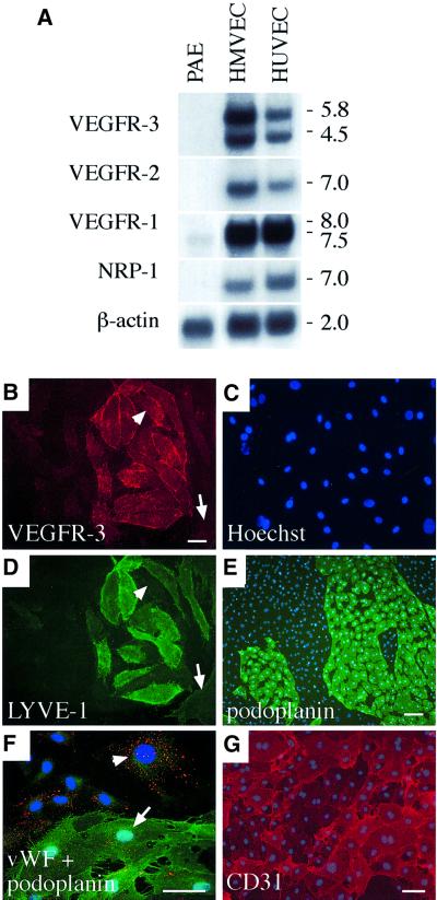
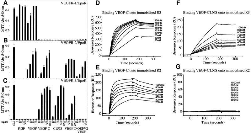
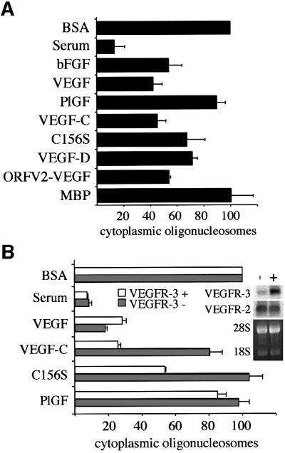
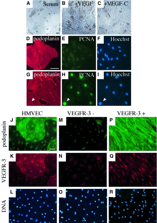
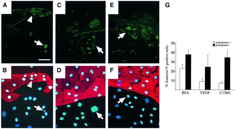
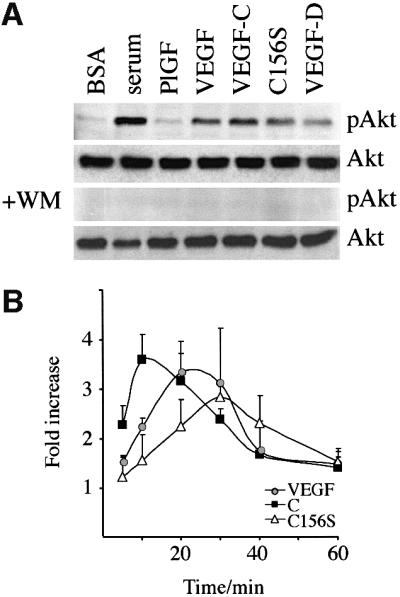
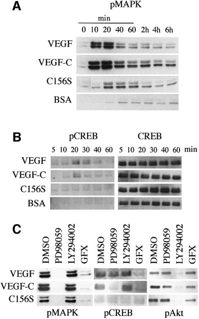
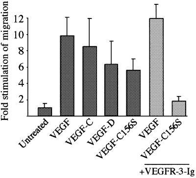
Vascular endothelial growth factor receptor-3 (VEGFR-3/Flt4) binds two known members of the VEGF ligand family, VEGF-C and VEGF-D, and has a critical function in the remodelling of the primary capillary vasculature of midgestation embryos. Later during development, VEGFR-3 regulates the growth and maintenance of the lymphatic vessels. In the present study, we have isolated and cultured stable lineages of blood vascular and lymphatic endothelial cells from human primary microvascular endothelium by using antibodies against the extracellular domain of VEGFR-3. We show that VEGFR-3 stimulation alone protects the lymphatic endothelial cells from serum deprivation-induced apoptosis and induces their growth and migration. At least some of these signals are transduced via a protein kinase C-dependent activation of the p42/p44 MAPK signalling cascade and via a wortmannin-sensitive induction of Akt phosphorylation. These results define the critical role of VEGF-C/VEGFR-3 signalling in the growth and survival of lymphatic endothelial cells. The culture of isolated lymphatic endothelial cells should now allow further studies of the molecular properties of these cells.


Figures

None
Fig. 1. (A) Expression of VEGFR mRNAs in primary human dermal microvascular endothelial cells (HMVECs), human umbilical vein endothelial cells (HUVECs) and in the porcine aortic endothelial (PAE) cell line. A northern blot containing 8 µg of the mRNAs was probed with radiolabelled cDNA fragments of human VEGF receptors and with β-actin for the control of equal loading. Numbers to the right denote the sizes of the transcripts (kb). (BG) HMVECs consist of two distinct populations of blood vascular and lymphatic endothelial cells. Immunofluorescence double staining using antibodies against VEGFR-3 (B) and LYVE-1 (D) with conterstaining of the nuclei by Hoechst fluorochrome (C). Note that LYVE-1 expression is not detected in all VEGFR-3-positive cells (arrowhead in B and D) while some VEGFR-3-negative cells are also stained weakly with LYVE-1 antibodies (arrow in B and D). Immunolabelling with antibodies against podoplanin (green in E and F), vWF (red, F) and CD31 (G). The nuclei were stained with the Hoechst fluorochrome (E–G). vWF expression occurs primarily in the podoplanin-negative cells (arrowhead in F), but weaker expression is also detected on podoplanin-positive cells (arrow in F). Stainings in (B–E) were carried out using live cells on ice, and in (F) and (G) after PFA fixation. Scale bars in (B–D), (F) and (G), 50 µm; in (E), 100 µm.
None
Fig. 2. (A–C) Analysis of the receptor specificities of different VEGFs using the Ba/F3 bioassay. Measurement of the viability of Ba/F3 cells expressing the chimeric receptors VEGFR-1/EpoR (A), VEGFR-2/EpoR (B) or VEGFR-3/EpoR (C) in the presence of different VEGFs at the indicated concentrations. Cell viability was determined using the MTT assay. Data represent the mean values from triplicate assays (mean ± SD). (DG) Biosensor analysis of the interaction of VEGF-C (D and E) and VEGF-C156S (F and G) with VEGFR-3 (D and F) and VEGFR-2 (E and G). Chimeric receptor proteins were immobilized onto a carboxymethylated dextran surface. Growth factors were injected over the surface at a flow rate of 20 µl/min at the indicated concentrations. The sensorgrams shown have been subtracted with the corresponding signal obtained when the same sample was passed over a blank control channel. Kinetic data derived from the biosensor analysis are shown in Table I.
None
Fig. 3. VEGFR-2- and VEGFR-3-, but not VEGFR-1-activating ligands inhibit apoptosis of serum-deprived HMVECs. Measurement of the cytoplasmic histone-associated DNA fragments (mono- and oligo nucleosomes) in serum-starved HMVECs consisting of two cell populations of blood vascular and lymphatic endothelial cells (A) or in the isolated cell populations after magnetic cell sorting using VEGFR-3 antibodies (B). The enrichment factor of cytoplasmic oligonucleosomes in the apoptotic cells grown for 24 h in serum-free medium (BSA) was chosen as 100%. Data represent mean values from three independent experiments (mean ± SD). Grey bars in (B) represent blood vascular endothelial cells and white bars represent VEGFR-3-expressing lymphatic endothelial cells. The inset in (B) shows a northern blot containing 4 µg of total RNAs extracted from blood vascular (–) and lymphatic endothelial cells (+). The blot was probed with radiolabelled cDNA fragments of human VEGFR-2 or VEGFR-3, and the 28S and 18S rRNAs were visualized by ethidium bromide staining of the gel for the control of equal loading. The following concentrations of growth factors were used in the apoptosis assays: bFGF, 10 ng/ml; PlGF, 500 ng/ml; VEGF, 50 ng/ml; VEGF-C, 100 ng/ml; VEGF- C156S, 500 ng/ml; VEGF-D, 500 ng/ml; ORFV2-VEGF, 500 ng/ml; and myelin basic protein (MBP) as an irrelevant control protein, 500 ng/ml.
None
Fig. 4. Isolation of VEGFR-3/podoplanin-positive and -negative endothelial cells using magnetic microbeads. (A–I) Culture of VEGFR-3-expressing lymphatic endothelial cells in complete medium containing 5% serum (A) and supplemented with VEGF (10 ng/ml, B) or VEGF-C (100 ng/ml, C). Staining of the VEGFR-3-positive cells grown for 5 days after sorting in serum (DF) or supplemented with VEGF-C (GI) for podoplanin (red; D and G) or PCNA (green; E and H). The nuclei were stained with the Hoechst fluorochrome (F and I). Note that if supplemented with VEGF-C, the cells are stained for PCNA (arrowhead in G–I). Immunofluorescence double-staining of non-sorted cells (JL) or VEGFR-3-negative (MO) and VEGFR-3-positive (PR) cell populations with antibodies against podoplanin (green; J, M and P) or VEGFR-3 (red; K, N and Q). The nuclei were stained with the Hoechst fluorochrome (L, O and R). The VEGFR-3+ cells were cultured in the presence of VEGF-C (100 ng/ml). Scale bars, 50 µm.
None
Fig. 5. (A–F) Annexin-V staining of HMVECs after 72 h of culture in serum-free medium alone (BSA, A) or with stimulation of VEGF (C) or VEGF-C (E). Simultaneous staining using antibodies against podoplanin (B, D and F) was used to distinguish lymphatic and blood vascular endothelial cells. Arrows indicate apoptotic, annexin-V-positive lymphatic endothelial cells, and arrowheads indicate apoptotic blood vascular endothelial cells. The nuclei were conterstained with Hoechst fluorochrome. (G) Quantitation of the annexin-V-positive cells (% of adherent cells) in the podoplanin-positive and -negative cell populations after 72 h of serum starvation. Note that detached cells were not included in the quantitation. Data represent mean values from five counted areas (×400) (mean ± SD). Scale bar in (A–F), 50 µm.
None
Fig. 6. VEGFR-2 or VEGFR-3 stimulation leads to PI3-kinase-dependent Akt phosphorylation. (A) The indicated ligands were used to stimulate HMVECs for 25 min, and Akt-Ser473 phosphorylation was analysed. Note that 30 nM wortmannin (WM) abolished the Akt activation in response to all VEGFs studied. (B) VEGF (grey circles), VEGF-C (black boxes) and VEGF-C156S (open triangles) induced phosphorylation of Akt with different kinetics. The data represent quantitations of optical densities of the signals from phosphorylated versus total Akt protein from three independent experiments (mean ± SD).
None
Fig. 7. (A and B) Simultaneous signalling via VEGFR-2 and VEGFR-3 upon VEGF-C stimulation leads to sustained p42/p44 MAPK activation in HMVECs. The p42/p44 MAPK activation was detected by western blotting using phospho-Thr202/Tyr204-MAPK-specific antibodies (A) and CREB phosphorylation using phospho-Ser133-specific antibodies (B). The growth factor concentrations used are: VEGF, 10 ng/ml; VEGF-C, 100 ng/ml; and VEGF-C156S, 500 ng/ml. (C) The VEGFR- 3-induced p42/p44 MAPK activation is mediated via PKC in HMVECs. Effects of inhibition of PKC by GF109203X (GFX), MEK1 by PD98059, and PI-3 kinase by LY294002 on p42/p44 MAPK Thr202/Tyr204 phosphorylation, CREB Ser133 phosphorylation and Akt Ser473 phosphorylation in HMVECs. The growth factor concentrations used are: VEGF, 1 ng/ml; VEGF-C, 10 ng/ml; and VEGF-C156S, 500 ng/ml.
None
Fig. 8. VEGFR-3 mediates endothelial cell migration. The migration of HMVECs in the presence of different VEGFs in a Boyden chamber assay. VEGF-C156S-, but not VEGF-stimulated migration was blocked by pre-incubating VEGF-C156S with a 10-fold molar excess of soluble VEGFR-3 (light grey bars). Data represent mean values from three independent experiments (mean ± SD). The growth factor concentrations used are: VEGF, 10 ng/ml; VEGF-C, 500 ng/ml; VEGF-D, 500 ng/ml; and VEGF-C156S, 3 µg/ml.

References

    1. Achen M.G., Jeltsch,M., Kukk,E., Makinen,T., Vitali,A., Wilks,A.F., Alitalo,K. and Stacker,S.A. (1998) Vascular endothelial growth factor D (VEGF-D) is a ligand for the tyrosine kinases VEGF receptor 2 (Flk1) and VEGF receptor 3 (Flt4). Proc. Natl Acad. Sci. USA, 95, 548–553. - PMC - PubMed
    1. Achen M.G. et al. (2000) Monoclonal antibodies to vascular endothelial growth factor-D block its interactions with both VEGF receptor-2 and VEGF receptor-3. Eur. J. Biochem., 267, 2505–2515. - PubMed
    1. Aiello L.P., Pierce,E.A., Foley,E.D., Takagi,H., Chen,H., Riddle,L., Ferrara,N., King,G.L. and Smith,L.E. (1995) Suppression of retinal neovascularization in vivo by inhibition of vascular endothelial growth factor (VEGF) using soluble VEGF-receptor chimeric proteins. Proc. Natl Acad. Sci. USA, 92, 10457–10461. - PMC - PubMed
    1. Baldwin M.E. et al. (2001) The specificity of receptor binding by vascular endothelial growth factor-D is different in mouse and man. J. Biol. Chem., 276, 19166–19171. - PubMed
    1. Banerji S., Ni,J., Wang,S.X., Clasper,S., Su,J., Tammi,R., Jones,M. and Jackson,D.G. (1999) LYVE-1, a new homologue of the CD44 glycoprotein, is a lymph-specific receptor for hyaluronan. J. Cell Biol., 144, 789–801. - PMC - PubMed

Publication types

MeSH terms

Substances