. 2001 Sep 3;20(17):4814-25.
doi: 10.1093/emboj/20.17.4814.

The FK506-binding protein 25 functionally associates with histone deacetylases and with transcription factor YY1

Affiliations

The FK506-binding protein 25 functionally associates with histone deacetylases and with transcription factor YY1

W M Yang et al. EMBO J. .

Abstract

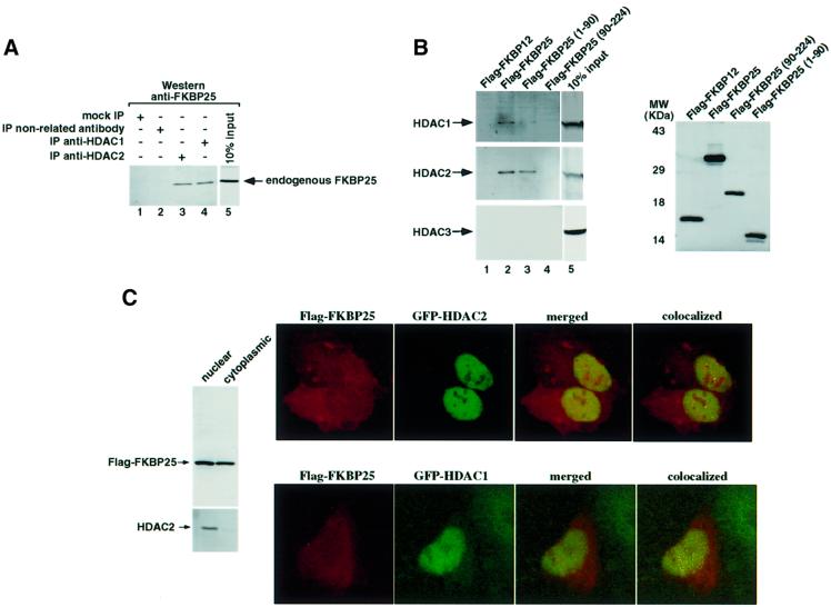
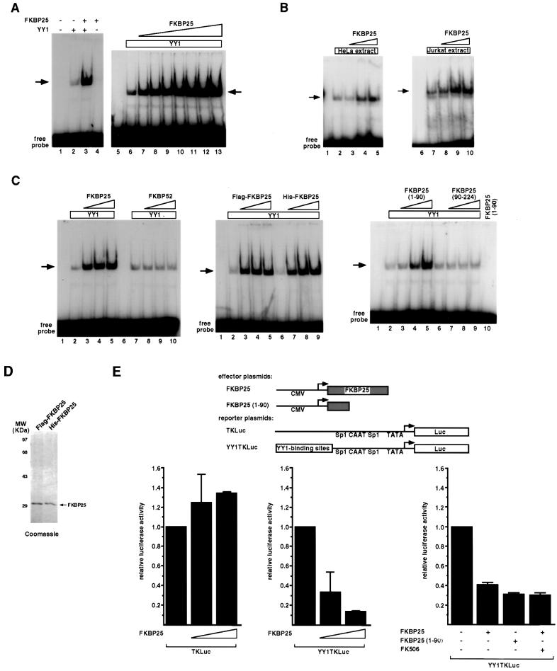
FK506-binding proteins (FKBPs) are cellular receptors for immunosuppressants that belong to a subgroup of proteins, known as immunophilins, with peptidylprolyl cis-trans isomerase (PPIase) activity. Sequence comparison suggested that the HD2-type histone deacetylases and the FKBP-type PPIases may have evolved from a common ancestor enzyme. Here we show that FKBP25 physically associates with the histone deacetylases HDAC1 and HDAC2 and with the HDAC-binding transcriptional regulator YY1. An FKBP25 immunoprecipitated complex contains deacetylase activity, and this activity is associated with the N-terminus of FKBP25, distinct from the FK506/rapamycin-binding domain. Furthermore, FKBP25 can alter the DNA-binding activity of YY1. Together, our data firmly establish a relationship between histone deacetylases and the FKBP enzymes and provide a novel and critical function for the FKBPs.


Figures

None
Fig. 1. The FKBP25 protein. Schematic drawing of the FKBP25 protein. Locations of the helix–loop–helix (HLH) motif and the conserved FK506/rapamycin-binding domains are indicated. Numbers indicate the position of amino acid residues.
None
Fig. 2. Association of FKBP25 with histone deacetylase enzymatic activity. (A) Anti-FKBP25 immunoprecipitates from HeLa whole-cell extracts were assayed for histone deacetylase activity as described in Materials and methods. (BD) HeLa cells were transfected with plasmids encoding different Flag epitope-tagged proteins, and histone deacetylase activity was assayed from anti-Flag immunoprecipitates. ‘+ competitor’ indicates the addition of excess Flag peptide and ‘+ TSA’ indicates treatment with 400 nM TSA (Wako). The amount of released [3H]acetic acid represents histone deacetylase activity. Each assay was performed in duplicate from two to four independent samples, and the values shown are the averages ± SD. The different background level (vector) reflects the different specific activity of the labeled input materials.
None
Fig. 3. Physical interaction between HDAC1/2 and FKBP25. (A) Anti-HDAC1 and anti-HDAC2 immunoprecipitates from HeLa whole-cell extracts were separated by SDS–PAGE, transferred onto a membrane and probed with an anti-FKBP25 antibody. ‘Mock IP’ indicates a reaction carried out identically but without the primary antibody. The input lane was loaded with one-tenth the amount of extract used in the immunoprecipitation reactions. (B) Western blots were performed on anti-Flag immunoprecipitates using anti-HDAC1 (top left), anti-HDAC2 (middle left) or anti-HDAC3 (bottom left) antibodies. A separate western blot was performed on the same anti-Flag immunoprecipitates using an anti-Flag antibody to demonstrate equivalent immunoprecipitation of the targeted proteins. (C) Co-localization of FKBP25 with HDAC1/2. HeLa cells were transfected with equal amounts of Flag-FKBP25 and GFP–HDAC1 or GFP–HDAC2 expression constructs, fixed, stained with anti-Flag antibodies and analyzed by confocal microscopy. The white dots indicate areas where two signals are within 0.02 µm. Left panel: western blot analysis to confirm the dual nuclear and cytoplasmic localization of FKBP25 by subcellular fractionation. Immunoblotting with the anti-HDAC2 antibody is shown as a control.
None
Fig. 4. HDAC-dependent transcriptional repression by FKBP25. (A) Schematic drawing of plasmids used in transient transfection assays. SV40 indicates SV40 early promoter/enhancer. Sp1, CAAT and TATA represent some of the cis-acting regulatory elements located in the TK promoter. Bent arrows indicate the direction of transcription from each plasmid. (B) Transfection assay results show that Gal4-FKBP25 and Gal4-FKBP25 (1–90) fusions repress transcription when targeted to promoters, and that the repression is reversed by TSA. All transfections were normalized to equal amounts of DNA with parental expression vectors. The results are the mean ± SD from at least three separate transfections.
None
Fig. 5. In vivo interaction between FKBP25 and YY1. (AC) Western blots were performed on anti-Flag or anti-YY1 immunoprecipitates using anti-YY1, anti-FKBP25 or anti-Flag antibodies. Immunoprecipitated products were obtained from a HeLa or Jurkat whole-cell extract either untreated or transfected with plasmids expressing the indicated Flag-tagged proteins. ‘Mock IP’ indicates a reaction carried out identically but without the primary antibody. In each set of experiment, a separate western blot was performed on the same immunoprecipitates to confirm equivalent immunoprecipitation of the targeted proteins.
None
Fig. 6. In vitro interaction between FKBP25 and YY1. Representative autoradiograms of in vitro translated YY1 proteins (A and E) or Gal4-YY1 fusion proteins (F) captured by GST fusion proteins. Several independent experiments yielded consistent results. The input lanes were loaded with one-tenth the amount of 35S-labeled proteins used in the binding reactions. Gels were stained with Coomassie Blue prior to autoradiography to show approximately equal amounts of GST fusion proteins in each lane. (B and C) A highly purified sample of YY1 protein expressed from E.coli together with purified GST–FKBP25 fusion proteins were used in pull-down assays to demonstrate a direct interaction between the two proteins. Western blots were performed to determine the amount of purified YY1 captured by GST–FKBP25. (D) Schematic drawing of YY1 and YY1 deletion constructs. Acidic, acidic domain; His, histidine cluster; GA, glycine/alanine-rich region; GK, glycine/lysine-rich region; Zn Fingers, zinc finger region. For simplicity, the Gal4 portion of 260–333 and 300–333 is not shown here. The ability of each YY1 protein to bind GST–FKBP25 is indicated (+ or –). The FKBP25-binding domain was localized to residues 300–333 of YY1.
None
Fig. 7. FKBP25 augments YY1’s DNA-binding and transcriptional repression activity. (AC) EMSA. The arrows indicate protein–DNA complexes specifically inhibited by the addition of excess YY1-binding DNA but not by the addition of an unrelated DNA (data not shown). Amounts of FKBP proteins added: 5 ng (A, lanes 3 and 4); 1–7 ng (A, lanes 7–13); 1, 3 and 5 ng (B, lanes 3–5 and 8–10; C, left panel, lanes 3–5 and 8–10; C, middle panel, lanes 3–5 and 7–9; C, right panel, lanes 3–5 and 7–10). (D) Coomassie Blue staining of an SDS–polyacrylamide gel containing highly purified FKBP25 proteins used in the EMSA. (E) Top panel: schematic drawing of plasmids used in transient transfection assays. CMV indicates the cytomegalovirus promoter/enhancer. Bottom panel: transfection assay results show that FKBP25 represses transcription through YY1. All transfections were normalized to equal amounts of DNA with parental expression vectors. The results are the mean ± SD from at least two separate transfections. ‘+ FK506’ indicates treatment with a final concentration of 1 mg/ml of FK506 (Fujisawa, USA).

Similar articles

Cited by

References

    1. Aravind L. and Koonin,E.V. (1998) Second family of histone deacetylases. Science, 280, 1167.
    1. Arevalo-Rodriguez M., Cardenas,M.E., Wu,X., Hanes,S.D. and Heitman,J. (2000) Cyclophilin A and Ess1 interact with and regulate silencing by the Sin3-Rpd3 histone deacetylase. EMBO J., 19, 3739–3749. - PMC - PubMed
    1. Benton B.M., Zang,J.H. and Thorner,J. (1994) A novel FK506- and rapamycin-binding protein (FPR3 gene product) in the yeast Saccharomyces cerevisiae is a proline rotamase localized to the nucleolus. J. Cell Biol., 127, 623–639. - PMC - PubMed
    1. Carmen A.A., Rundlett,S.E. and Grunstein,M. (1996) HDA1 and HDA3 are components of a yeast histone deacetylase (HDA) complex. J. Biol. Chem., 271, 15837–15844. - PubMed
    1. Cress W.D. and Seto,E. (2000) Histone deacetylases, transcriptional control, and cancer. J. Cell Physiol., 184, 1–16. - PubMed

Publication types

MeSH terms