. 2001 Sep 3;20(17):4826-35.
doi: 10.1093/emboj/20.17.4826.

Invertebrate connectin spans as much as 3.5 microm in the giant sarcomeres of crayfish claw muscle

Affiliations

Invertebrate connectin spans as much as 3.5 microm in the giant sarcomeres of crayfish claw muscle

A Fukuzawa et al. EMBO J. .

Abstract

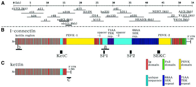
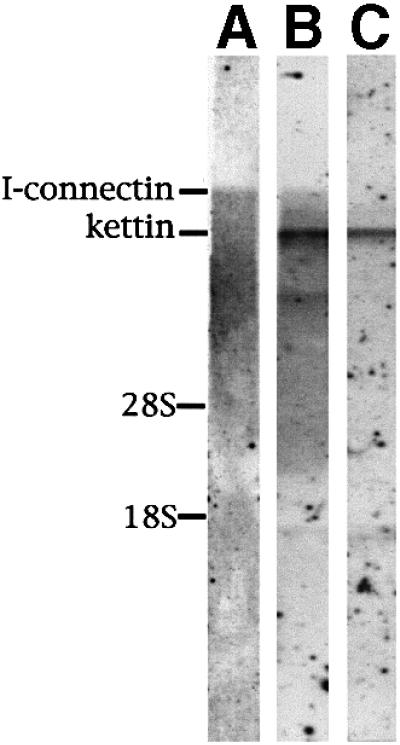
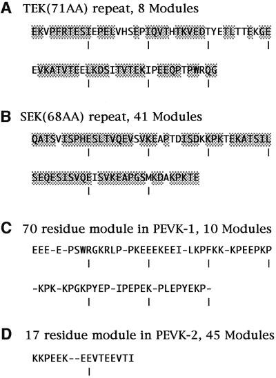
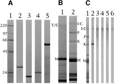
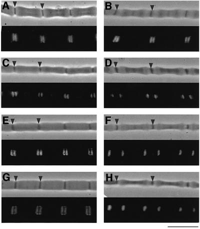
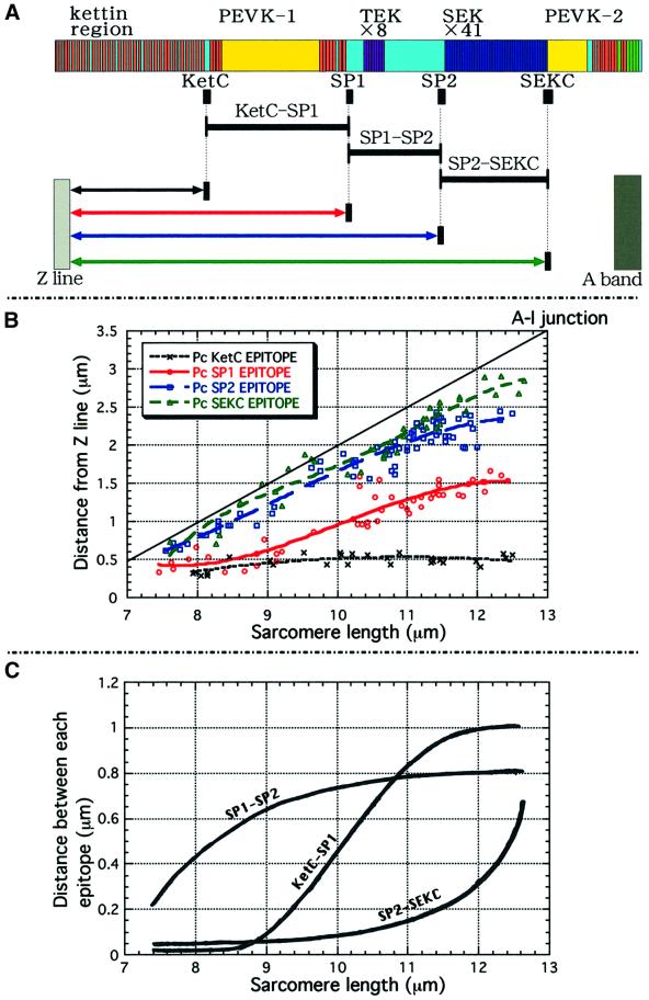
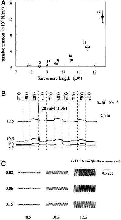
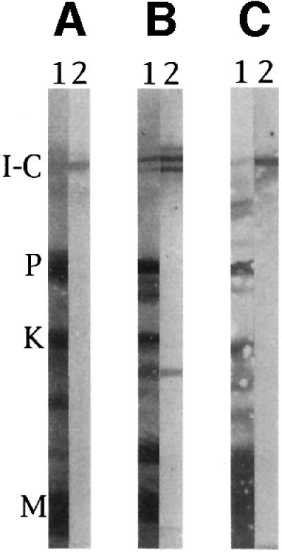
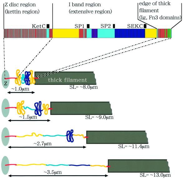
In crayfish claw closer muscle, the giant sarcomeres are 8.3 microm long at rest, four times longer than vertebrate striated muscle sarcomeres, and they are extensible up to 13 microm upon stretch. Invertebrate connectin (I-connectin) is an elastic protein which holds the A band at the center of the sarcomere. The entire sequence of crayfish I-connectin was predicted from cDNA sequences of 53 424 bp (17 352 residues; 1960 kDa). Crayfish I-connectin contains two novel 68- and 71-residue repeats, and also two PEVK domains and one kettin region. Kettin is a small isoform of I-connectin. Immunoblot tests using antibody to the 68-residue repeats revealed the presence of I-connectin also in long sarcomeres of insect leg muscle and barnacle ventral muscle. Immunofluorescence microscopy demonstrated that the two repeats, the long spacer and the two PEVK domains contribute to sarcomere extension. These regions rich in charged amino acids, occupying 63% of the crayfish I-connectin molecule, may allow a span of a 3.5 microm distance as a new class of composite spring.


Figures

None
Fig. 1. The domain structure of crayfish I-connectin predicted from the cloned cDNAs. (A) cDNAs sequenced to reconstruct the cDNA contig encoding I-connectin. (B) The domain structure of I-connectin. Bars a and b, cDNAs used for northern blotting; KetC, SP1, SP2 and SEKC (black boxes), portions bacterially expressed and used for raising antibodies. (C) The domain structure of kettin cDNA. Bar c, 1.3 kb cDNA corresponding to the 3′-untranslated region (UTR) used for northern blotting.
None
Fig. 2. Northern blot tests to detect the expression of kettin and I-connectin mRNAs in crayfish claw closer muscle. (A) Detectionof I-connectin mRNA by probe a in Figure 1B. (B) Detection of I-connectin and kettin mRNAs by probe b in Figure 1B. Note that kettin was expressed much more than I-connectin. (C) Detection of kettin mRNA by probe c in Figure 1C. 28S, vertebrate 28S rRNA; 18S, vertebrate 18S rRNA.
None
Fig. 3. Amino acid sequences characteristic of crayfish I-connectin. (A) TEK repeats. Consensus sequences conserved in more than seven repeats out of eight are shaded. (B) SEK repeats. Consensus sequences conserved in >29 repeats out of 41 are shaded. (C) PEVK-1 module. Consensus sequences are >50% conserved. (D) PEVK-2 module. Consensus sequences are as in (C).
None
Fig. 4. Recombinant peptides used for raising antibodies and immunoblot tests of the reactions of the antibodies with crayfish claw closer muscle proteins. (A) SDS–PAGE patterns of purified recombinant peptides: lane 1, molecular weight markers (sizes shown in kDa); lanes 2–5, KetC, SP1, SP2 and SEKC recombinant peptides shown in Figure 1B. Electrophoresis was on 10% polyacrylamide gels. (B) SDS–PAGE patterns of rabbit cardiac muscle and crayfish claw closer muscle: lane 1, rabbit cardiac muscle; lane 2, crayfish claw closer muscle. T/C, titin/connectin; I-C, I-connectin; P, projectin; K, kettin; M, myosin heavy chain. Electrophoresis was on 2–6% polyacrylamide gels. (C) Immunoblot tests: lane 1, amido black stain of crayfish claw closer muscle; lanes 2–6, treated with antibodies PcKetC, PcSP1, PcSP2, PcSEKC and Pc3000K, respectively. Electrophoresis was on 2–6% polyacrylamide gels.
None
Fig. 5. Immunofluorescence microscopic observations of giant sarcomeres of crayfish claw closer muscle using antisera against recombinant peptides of crayfish I-connectin. (A and B) Treated with PcKetC: sarcomere length (SL), (A) 9.2 µm; (B) 11.8 µm. (C and D) Treated with PcSP1: SL, (C) 8.7 µm; (D) 11.8 µm. (E and F) Treated with PcSP2: SL, (E) 8.9 µm; (F) 12.0 µm. (G and H) Treated with PcSEKC: SL, (G) 8.5 µm; (H) 12.0 µm. Upper, phase- contrast image; lower, fluorescence image. Arrowheads indicate the Z line. Bar, 10 µm.
None
Fig. 6. Extensibility of various regions of the crayfish I-connectin molecule linking the Z line and the A band. (A) Positions of the four epitopes in the entire sequence of I-connectin. (B) Changes in the distance between each epitope and the Z line at increasing SL. Crosses, KetC; open circles, SP1; open squares, SP2; open triangles, SEKC. (C) Changes in the distance between each epitope at increasing SL.
None
Fig. 7. (A) The relationship between passive tension and sarcomere length. Horizontal and vertical bars attached to each symbol indicate the standard error of means and the number of measurements. (B) Traces of passive tension showing a reversible effect of ionic strength. Sarcomere length (SL) is shown on the left in micrometers. The dotted lines indicate the changes in the incubating solution, the ionic strength of which is shown by the vertical numbers. BDM (20 mM) was added to the solution during the period shown by the rectangle. The cross-sectional area of the fiber was 40 000, 33 000, 76 000 and 56 000 µm2 for the traces at SL of 12.5, 10.5, 9.5 and 8.5 µm, respectively. (C) Representative traces of tension oscillation in response to 20 Hz sinusoidal length perturbation of 2% fiber length amplitude. Ionic strength is shown on the left and SL is shown below the traces in micrometers. For the traces at SL 12.5 µm, those obtained in the presence of 20 mM BDM were superimposed on the last three-quarters of the traces in the absence of BDM at ionic strength 0.02 and 0.06. All the traces in the figure were obtained from a specimen with 17 000 µm2 cross-sectional area.
None
Fig. 8. Immunoblot detection of invertebrate connectin (I-connectin) in several arthropod striated muscles. SDS–PAGE was carried out using 2.3–4% polyacrylamide gels. (A) Barnacle (Dolichopus nitidus) ventral muscle; (B) beetle (Allomyrina dichotoma) leg muscle; (C) fly (Calliphora lata) larva muscle. Lane 1, amido black stain; lane 2, treated with Pc SEKC (cf. Figure 1B). I-C, I-connectin; P, projectin; K, kettin; M, myosin heavy chain.
None
Fig. 9. Scheme of structural changes of the I-connectin molecule in a giant sarcomere of crayfish claw muscle at varied sarcomere length. The N-terminal kettin domain and the C-terminal Ig and Fn3 domains are practically inextensible. At rest, all the extensible regions (PEVK-1 to PEVK-2) are largely retracted. At SL ∼9 µm, the region spacer-1–TEK repeats–spacer-2 (SP1–SP2) is greatly extended. At SL ∼11.4 µm, PEVK-1 is almost entirely extended. At SL ∼13 µm, SEK repeats (SP2–SEKC) are nearly completely elongated (cf. Figure 6C).

References

    1. Benian G.M., Ayme-Southgate,A. and Tinley,T.L. (1999) The genetics and molecular biology of the titin/connectin-like proteins of invertebrates. Rev. Physiol. Biochem. Pharmacol., 138, 235–268. - PubMed
    1. Daley J., Southgate,R. and Ayme-Southgate,A. (1998) Structure of the Drosophila projectin protein: isoforms and implication for projectin filament assembly. J. Mol. Biol., 279, 201–210. - PubMed
    1. Endo T. and Nadal-Ginard,B. (1987) Three types of muscle-specific gene expression in fusion-blocked rat skeletal muscle cells: translational control in EGTA-treated cells. Cell, 49, 515–526. - PubMed
    1. Freiburg A. et al. (2000) Series of exon-skipping events in the elastic spring region of titin as the structural basis for myofibrillar elastic diversity. Circ. Res., 86, 1114–1121. - PubMed
    1. Gautel M., Mues,A. and Young,P. (1999) Control of sarcomeric assembly: the flow of information on titin. Rev. Physiol. Biochem. Pharmacol., 138, 97–137. - PubMed

Publication types

MeSH terms

Associated data