. 2001 Sep 3;20(17):4987-97.
doi: 10.1093/emboj/20.17.4987.

The exon-exon junction complex provides a binding platform for factors involved in mRNA export and nonsense-mediated mRNA decay

Affiliations

The exon-exon junction complex provides a binding platform for factors involved in mRNA export and nonsense-mediated mRNA decay

H Le Hir et al. EMBO J. .

Abstract

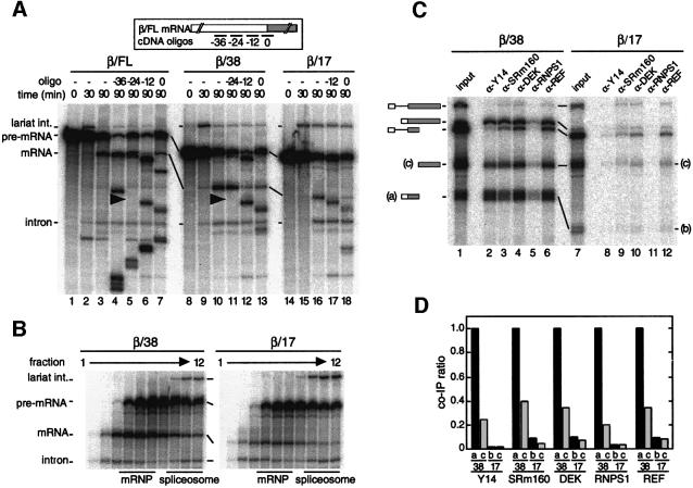
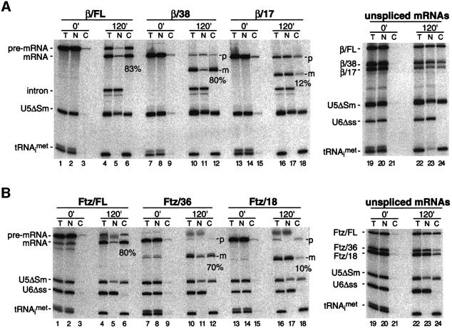
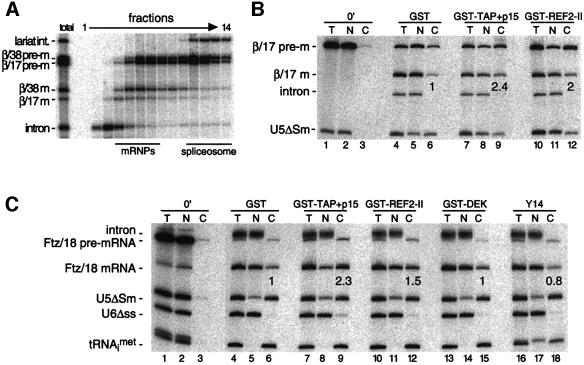
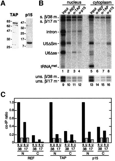
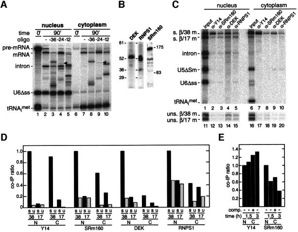
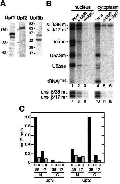
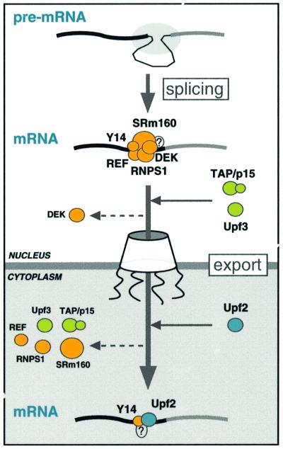
We recently reported that spliceosomes alter messenger ribonucleoprotein particle (mRNP) composition by depositing several proteins 20-24 nucleotides upstream of mRNA exon-exon junctions. When assembled in vitro, this so-called 'exon-exon junction complex' (EJC) contains at least five proteins: SRm160, DEK, RNPS1, Y14 and REF. To better investigate its functional attributes, we now describe a method for generating spliced mRNAs both in vitro and in vivo that either do or do not carry the EJC. Analysis of these mRNAs in Xenopus laevis oocytes revealed that this complex is the species responsible for enhancing nucleocytoplasmic export of spliced mRNAs. It does so by providing a strong binding site for the mRNA export factors REF and TAP/p15. Moreover, by serving as an anchoring point for the factors Upf2 and Upf3, the EJC provides a direct link between splicing and nonsense-mediated mRNA decay. Finally, we show that the composition of the EJC is dynamic in vivo and is subject to significant evolution upon mRNA export to the cytoplasm.


Figures

None
Fig. 1. The EJC is not deposited on spliced β/17 mRNA. (A) Labeled β/FL (lanes 1–7), β/38 (lanes 8–13) or β/17 pre-mRNA (lanes 14–18) was incubated under splicing conditions in HeLa cell nuclear extract for the times indicated. Aliquots from 90 min reactions were further incubated with the indicated cDNA oligos (short bars underneath the mRNA schematic at top; named according to the center position of the oligo relative to the exon–exon junction, which was defined as 0). Splicing substrates, intermediates and products are indicated to the left. (B) Glycerol gradient fractionation of 90 min splicing reactions containing β/38 (left) or β/17 pre-mRNA (right). Bars indicate positions of mRNP and spliceosome.(C) Co-immunoprecipitation of β/38 (lanes 1–6) and β/17 (lanes 7–12) RNA species after splicing (2 h), separation of mRNPs from spliceosomes by glycerol gradient fractionation. 5′ (a or b) and 3′ (c) mRNA fragments resulted from subsequent RNase H cleavage with a single cDNA oligo centered 49 nt downstream of the exon–exon junction. Reactions were then subjected to immunoprecipitation with the antibodies indicated. Lanes 1 and 7 correspond to 1/15th of input RNA. (D) Co-immunoprecipitation ratios of fragments a, b and c from β/38 and β/17 mRNAs. Ratios shown were determined by dividing the absolute co-immunoprecipitation efficiency for each RNA with the indicated antibody by the absolute co-immunoprecipitation efficiency of β/38 fragment ‘a’ with that same antibody. In all experiments, RNAs were separated by 10% denaturing PAGE.
None
Fig. 2. The EJC promotes efficient export of spliced mRNAs. (AXenopus oocyte nuclei were injected with control RNAs plus the indicated pre-mRNA (lanes 1–18) or unspliced control mRNAs (lanes 19–24). RNA samples from total oocyte (T), nuclear (N) and cytoplasmic (C) fractions were collected immediately (0 min) or 120 min after injection and analyzed by 10% denaturing PAGE. One oocyte equivalent of RNA, from a pool of 10 oocytes, was loaded per lane in all panels. Splicing substrates, products and control RNAs are identified to the left of each gel. (B) Same as (A), with the corresponding Ftz-derived RNAs. Percentages indicate the proportion of each spliced mRNA in the cytoplasm 120 min after injection.
None
Fig. 3. (A) Glycerol gradient fractionation of splicing substrates and products present in Xenopus oocyte nuclei 30 min after injection of β/38 and β/17 pre-mRNAs shows that both spliced mRNAs are released from spliceosomes in vivo. RNAs were extracted and analyzed as in Figure 1B. Bars indicate positions of mRNPs and spliceosomes. (B and C) Stimulation of spliced β/17 and Ftz/18 mRNA export by co-injection of recombinant proteins. Xenopus oocyte nuclei were injected with indicated control RNAs (at left) and either β/17 pre-mRNA (B) or Ftz/18 pre-mRNA (C). Where indicated, purified recombinant proteins (25 nl of 1 mg/ml stocks) were co-injected with RNAs (lanes ≥7) and co-injection of purified GST served as a control (lanes 4–6). RNA samples from total oocytes (T), nuclear (N) and cytoplasmic (C) fractions were collected immediately after injection (0 min; lanes 1–3) or 3 h after injection (lanes 4–18) and analyzed as in Figure 2. Export efficiencies of β/17 and Ftz/18 mRNAs in the presence of test proteins are indicated relative to their export efficiencies with GST alone.
None
Fig. 4. The EJC promotes recruitment of TAP/p15 heterodimers to spliced mRNAs. (A) Protein samples from Xenopus oocyte nuclei were analyzed by western blotting with anti-TAP and anti-p15 antibodies. (B) Control RNAs plus either β/38 and β/17 pre-mRNAs (upper panel, lanes 1–8) or unspliced β/38 and β/17 mRNAs (lower panel, lanes 9–16) were co-injected in Xenopus oocyte nuclei. Two hours after injection, RNA samples from nuclear and cytoplasmic fractions were subjected to immunoprecipitation with the antibodies indicated. Lanes 1, 5, 9 and 13 correspond to 1/15th of input RNA. RNAs were analyzed as in Figure 2. (C) Co-immunoprecipitation ratios of spliced (s) or unspliced (u) β/38 and β/17 mRNAs isolated from nuclear (N) or cytoplasmic (C) compartments with antibodies against the species indicated. For each antibody, ratios were calculated by dividing the absolute co-immunoprecipitation efficiency of the species indicated by the absolute co-immunoprecipitation efficiency of nuclear spliced β/38 mRNA with that antibody.
None
Fig. 5. Y14 and SRm160 remain associated with spliced mRNAs after export. (A) Spliced β/FL mRNA exhibits a similar protection to RNase H digestion in both nucleus and cytoplasm. Following injection of β/FL pre-mRNA into oocyte nuclei, nuclear (lanes 1–5) and cytoplasmic (lanes 6–10) fractions were collected immediately (lanes 1 and 6) or after 90 min (lanes 2–5 and 7–10). Aliquots of 90 min samples were further incubated alone (lanes 2 and 7) or with the cDNA oligos indicated (lanes 3–5 and 8–10). RNAs were separated by 8% denaturing PAGE. (B) Protein samples from Xenopus oocyte nuclei were analyzed by western blotting with anti-DEK, anti-RNPS1 and anti-SRm160 antibodies. (C and D) Same as Figure 4B and C, respectively, except that experiments were performed with antisera raised against Y14, SRm160, RNPS1 and DEK. (E) Co-immunoprecipitation ratios of spliced β/38 mRNA isolated from nuclear (N) or cytoplasmic (C) fractions with antibodies against the species indicated.Co-immunoprecipitations were performed after 1.5 or 3 h incubation and in the absence (–) or presence (+) of competitor unspliced β/38 mRNA (comp.) in the cytoplasm. In (D) and (E), co-immunoprecipitation ratios were calculated as in Figure 4C.
None
Fig. 6. The EJC promotes efficient recruitment of hUpf3 and hUpf2 to spliced mRNAs. (A) Protein samples from total Xenopus oocyte lysates were analyzed by western blotting using anti- hUpf3b, hUpf2 and hUpf1 antibodies. (B and C) Same as Figure 4B and C except that experiments were performed with antisera raised against hUpf3b and hUpf2.
None
Fig. 7. A model for the evolution of the EJC upon mRNA export. Pre-mRNA splicing deposits the EJC upstream of the splice junction. At this stage, the complex contains at least five proteins, SRm160, DEK, RNPS1, REF and Y14. After splicing, the TAP/p15 heterodimer and Upf3 join the complex in the nucleus. DEK probably dissociates (dashed arrow) before mRNA export to the cytoplasm, whereas SRm160 and the nucleocytoplasmic shuttling proteins RNPS1, REF, TAP/p15 and Upf3 are all likely to leave after mRNA export. Y14 remains stably associated with spliced mRNA in both compartments, while Upf2 only joins the complex in the cytoplasm. Question marks symbolize potential unidentified components.

References

    1. Applequist S.E., Selg,M., Raman,C. and Jack,H.M. (1997) Cloning and characterization of HUPF1, a human homolog of the Saccharomyces cerevisiae nonsense mRNA-reducing UPF1 protein. Nucleic Acids Res., 25, 814–821. - PMC - PubMed
    1. Bachi A. et al. (2000) The C-terminal domain of TAP interacts with the nuclear pore complex and promotes export of specific CTE-bearing RNA substrates. RNA, 6, 136–158. - PMC - PubMed
    1. Blencowe B.J., Issner,R., Nickerson,J.A. and Sharp,P.A. (1998) A coactivator of pre-mRNA splicing. Genes Dev., 12, 996–1009. - PMC - PubMed
    1. Braun I.C., Rohrbach,E., Schmitt,C. and Izaurralde,E. (1999) TAP binds to the constitutive transport element (CTE) through a novel RNA-binding motif that is sufficient to promote CTE-dependent RNA export from the nucleus. EMBO J., 18, 1953–1965. - PMC - PubMed
    1. Braun I.C., Herold,A., Rode,M., Conti,E. and Izaurralde,E. (2001) Overexpression of TAP/p15 heterodimers bypasses nuclear retention and stimulates nuclear mRNA export. J. Biol. Chem., 276, 20536–20543. - PubMed

MeSH terms