. 2001 Sep 3;20(17):5022-31.
doi: 10.1093/emboj/20.17.5022.

The Usf-1 transcription factor is a novel target for the stress-responsive p38 kinase and mediates UV-induced Tyrosinase expression

Affiliations

The Usf-1 transcription factor is a novel target for the stress-responsive p38 kinase and mediates UV-induced Tyrosinase expression

M D Galibert et al. EMBO J. .

Abstract

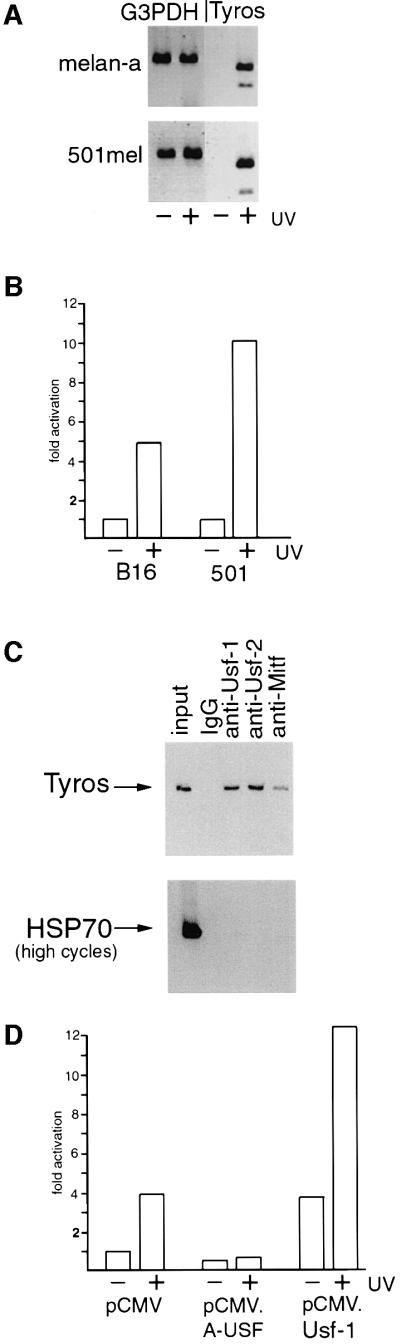
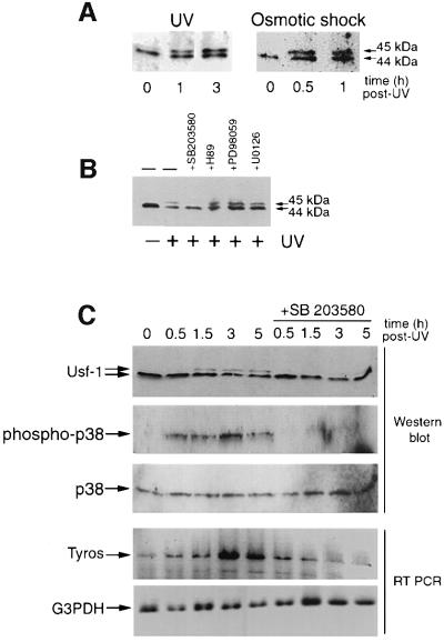
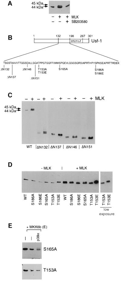
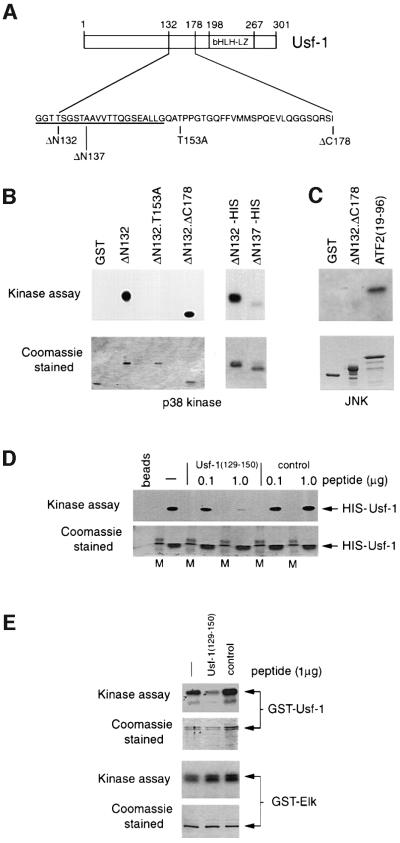
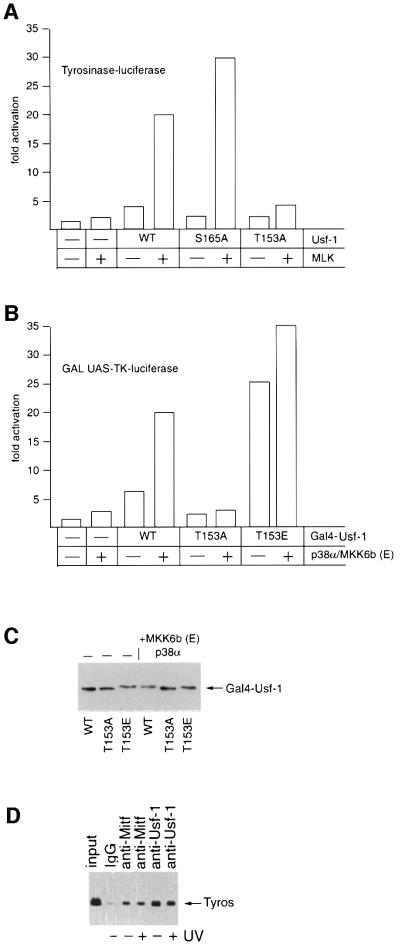
The stress-activated signalling cascade leading to phosphorylation of the p38 family of kinases plays a crucial role during development and in the cellular response to a wide variety of stress-inducing agents. Although alterations in gene expression characteristic of the stress response require the regulation of key transcription factors by the p38 family, few downstream targets for this signalling pathway have been identified. By examining the ability of pigment cells to respond to UV irradiation as part of the UV-induced tanning response, we show that while the microphthalmia-associated transcription factor Mitf regulates basal Tyrosinase expression, it is the ubiquitous basic helix-loop-helix-leucine zipper transcription factor Usf-1 that is required for the UV activation of the Tyrosinase promoter. Consistent with this we demonstrate that Usf-1 is phosphorylated and activated by the stress-responsive p38 kinase. The results suggest that activation of Usf-1 by p38 at a wide variety of viral and cellular promoters will provide a link between stimuli as diverse as UV irradiation, glucose, viral infection and pro-inflammatory cytokines, and the changes in gene expression associated with the stress response.


Figures

None
Fig. 1. UV-inducibility of the Tyrosinase promoter is USF dependent. (A) The Tyrosinase gene is UV inducible. RT–PCR using mRNA derived from mouse melanocytes (melan-a) or a human melanoma cell line (501mel) treated or untreated with UV (254 nm, 40 J/m2). Primers were specific for the Tyrosinase gene and G3PDH was used as a control. RNA was prepared 3 h post-irradiation. (B) The Tyrosinase promoter is UV responsive. A Tyrosinase promoter-luciferase reporter (100 ng) extending from –300 to +80 was transfected into either mouse (B16) or human (501mel) melanoma, and luciferase activity determined. UV irradiation was performed 24 h post-transfection where indicated. (C) Usf-1 and Usf-2 bind the Tyrosinase promoter in vivo. Chromatin immunoprecipitations were performed on 501mel cells using the indicated antibodies or control non-specific IgG. Recovered DNA was subject to PCR using primers specific for either Tyrosinase or the HSP70 promoter. Controls (not shown) confirmed that the PCR reactions were in the logarithmic phase. (D) UV-inducibility of the Tyrosinase promoter is inhibited by dominant-negative USF. The Tyrosinase promoter-luciferase reporter (100 ng) was co-transfected into B16 melanoma cells together with either a Usf-1 expression vector (pCMV.Usf-1, 500 ng), a dominant-negative USF expression vector (pCMV.A-USF, 500 ng), or a control vector (pCMV), and luciferase activity determined. Where indicated, cells were treated with UV.
None
Fig. 2. Stress-induced phosphorylation of Usf-1. (A) Western blotting analysis using anti-Usf-1 Ab (Santa Cruz) of B16 melanoma cells, before and after UV irradiation (254 nm, 40 J/m2) or osmotic shock (0.5 M sorbitol, 20 min), at indicated time post-treatment. The 45 kDa band corresponds to the phosphorylated form of Usf-1 described previously (Galibert et al., 1997). (B) Stress-induced phosphorylation of Usf-1 is abolished when B16 melanoma cells are pre-treated with the SB 203580 compound (10 µM, 20 min prior to UVC stimulation), a highly specific inhibitor of the p38 family kinases, but not with the MEK inhibitors PD98059 (50 µM) or U0126 (10 µM) or the H89 (10 µM) inhibitor of MSK1. (C) A time course of induction of Tyrosinase expression in 501mel cells in response to UV irradiation in the presence or absence of the SB 203580 compound. Tyrosinase mRNA was detected by RT–PCR and compared with the levels of a G3PDH control. Protein extracts from the corresponding time points were western blotted and probed for Usf-1, total p38 or double-phospho-p38. Similar results have been obtained using B16 cells.
None
Fig. 3. Requirements within Usf-1 for stress-induced phosphorylation in vivo. (A) MLK-mediated phosphorylation of Usf-1. B16 melanoma cells were transfected with a vector expressing SV5 epitope-tagged Usf-1 (800 ng) either alone or together with 10 ng of an MLK expression vector (pCMV.MLK). Cells were pre-treated with the SB 203580 compound (10 µM) where indicated. (B) Schematic representation of the Usf-1 protein and various point and deletion mutations expressed in the co-transfection assays with MLK shown in (C) and (D). (C and D) Vectors expressing SV5 epitope-tagged WT or the indicated Usf-1 deletion or point mutants were transfected into COS7 cells either alone or together with the MLK expression vector. Cell extracts were assayed by SDS–PAGE and western blotting using the anti-SV5 antibody. Similar results were obtained in B16 melanoma cells. (E) SV5 epitope-tagged Usf-1 (pCMV.SV5-Usf-1) or the S165A or T153A mutants were expressed in B16 melanoma cells either alone or together with MKK6b(E) and p38α as indicated.
None
Fig. 4. Requirements within Usf-1 for stress-induced phosphorylation in vitro. (A) Schematic of Usf-1 proteins used in the in vitro kinase assays. (B) Usf-1 is phosphorylated by p38α in vitro. In vitro kinase assays were performed using either purified GST–Usf-1 fusion protein or His6-tagged Usf-1 proteins in the presence of recombinant activated p38α kinase [γ-32P]ATP. Kinase assays using the indicated Usf-1 derivatives are shown in the upper panel after SDS–PAGE and autoradiography. The lower panel shows the same gel stained with Coomassie Blue indicating that equivalent amounts of protein were used in each assay. (C) JNK does not efficiently phosphorylate Usf-1. The indicated GST fusion proteins were incubated with purified active JNK and [γ-32P]ATP. Efficient phosphorylation of ATF2 was observed but not of either GST alone or the GST-Usf-1 fusion protein. (D) A competitor peptide can inhibit the phosphorylation of His-tagged Usf-1 by p38α in vitro. Active recombinant p38α kinase was pre-incubated with either Usf-1 competitor peptide corresponding to Usf-1 amino acids 129–150, or a control peptide (0.1–1 µg) derived from the Pho4 activation domain, for 30 min prior to the kinase reaction in the presence of His-tagged Usf-1 recombinant protein as indicated. M indicates a molecular weight marker. (E) The Usf-1 peptide inhibits phosphorylation of Usf-1 but not Elk by p38. Kinase assays were performed as in (D) using the indicated GST fusion proteins in the presence or absence of the Usf-1 peptide or the control Pho4 peptide.
None
Fig. 5. Thr153 and active p38α are required for efficient transcription activation by Usf-1. (A) The Tyrosinase promoter-luciferase reporter (300 ng) was transfected into B16 melanoma cells either alone or together with the MLK and Usf-1 expression vectors (50 ng) in the indicated combinations and luciferase activity determined. (B) A GAL UAS-luciferase reporter (200 ng) was transfected into COS7 cells either alone or together with vectors expressing WT or mutant Usf-1 fused to the Gal4 DNA-binding domain in the presence or absence of co-expressed p38α/MKK6b(E) kinases as indicated. (C) Western blot of the indicated Gal4–Usf-1 chimeric proteins expressed in the transfected cells using anti-Gal4 DNA-binding domain antibody (Clontech). (D) Chromatin immunoprecipitation assay at the Tyrosinase promoter using the indicated antibodies before or after UV irradiation (254 nm, 40 J/m2) of B16 melanoma cells.

References

    1. Abdel-Malek Z., Suzuki,I., Tada,A., Im,S. and Akcali,C. (1999) The melanocortin-1 receptor and human pigmentation. Ann. NY Acad. Sci., 885, 117–133. - PubMed
    1. Aksan I. and Goding,C.R. (1998) Targeting the microphthalmia basic helix-loop-helix-leucine zipper transcription factor to a subset of E-Box elements in vitro and in vivo. Mol. Cell. Biol., 18, 6930–6938. - PMC - PubMed
    1. Alpert D., Schwenger,P., Han,J. and Vilcek,J. (1999) Cell stress and MKK6b-mediated p38 MAP kinase activation inhibit tumor necrosis factor-induced IκB phosphorylation and NF-κB activation. J. Biol. Chem., 274, 22176–22183. - PubMed
    1. Aquilla E., Whelchel,A., Knot,H.J., Nelson,M. and Posada,J. (1996) Activation of multiple mitogen-activated protein kinase signal transduction pathways by the endothelin B receptor requires the cytoplasmic tail. J. Biol. Chem., 271, 31572–31579. - PubMed
    1. Armstrong B.K., Kricker,A. and English,D.R. (1997) Sun exposure and skin cancer. Australas. J. Dermatol., 38, S1–S6. - PubMed

Publication types

MeSH terms