. 2001 Sep 1;535(Pt 2):591-600.
doi: 10.1111/j.1469-7793.2001.00591.x.

Force and power output of fast and slow skeletal muscles from mdx mice 6-28 months old

Affiliations

Force and power output of fast and slow skeletal muscles from mdx mice 6-28 months old

G S Lynch et al. J Physiol. .

Abstract

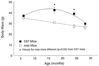
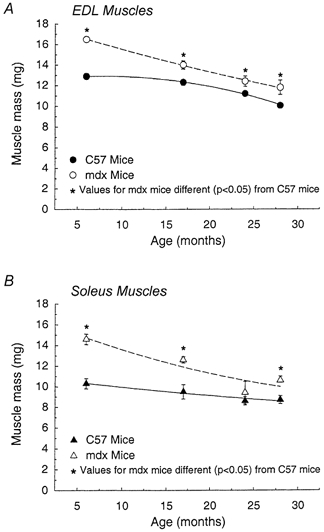
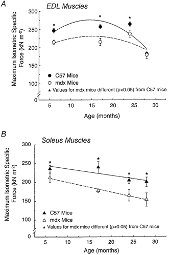
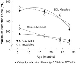
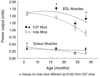
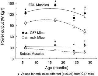
1. Differences in the effect of age on structure-function relationships of limb muscles of mdx (dystrophin null) and control mice have not been resolved. We tested the hypotheses that, compared with limb muscles from age-matched control mice, limb muscles of 6- to 17-month-old mdx mice are larger but weaker, with lower normalised force and power, whereas those from 24- to 28-month-old mdx mice are smaller and weaker. 2. The maximum isometric tetanic force (P(o)) and power output of limb muscles from 6-, 17-, 24- and 28-month-old mdx and control mice were measured in vitro at 25 degrees C and normalised with respect to cross-sectional area and muscle mass, respectively. 3. Body mass at 6 and 28 months was not significantly different in mdx and control mice, but that of control mice increased 16 % by 17 months and then declined 32 % by 28 months. The body masses of mdx mice declined linearly with age with a decrease of 25 % by 28 months. From 6 to 28 months of age, the range in the decline in the masses of EDL and soleus muscles of mdx and control mice was from 16 to 28 %. The muscle masses of mdx mice ranged from 9 % to 42 % greater than those of control mice at each of the four ages and, even at 28 months, the masses of EDL and soleus muscles of mdx mice were 17 % and 22 % greater than control values. 4. For mdx mice of all ages, muscle hypertrophy was highly effective in the maintenance of control values for absolute force for both EDL and soleus muscles and for absolute power of soleus muscles. Throughout their lifespan, muscles of mdx mice displayed significant weakness with values for specific P(o) and normalised power approximately 20 % lower than values for control mice at each age. For muscles of both strains, normalised force and power decreased approximately 28 % with age, and consequently weakness was more severe in muscles of old mdx than in those of old control mice.


Figures

Figure 1
Figure 1. Cross-sections of EDL (A-D) and soleus (E-H) muscles from 6- (A, B, E and F) and 24- (C, D, G and H) month-old mdx and control mice stained with haematoxylin and eosin (H&E)
Note in cross-sections of muscles from mdx mice: (i) the variability in cross-sectional area of fibres, (ii) the large number of small atrophic fibres, (iii) the large number of central myonuclei, 50-70 %, in muscles of mdx mice.
Figure 2
Figure 2. Body mass of mdx and control C57BL male mice plotted at 6, 17, 24 and 28 months old
The effect of age (P < 0.001) and the strain of mouse (P < 0.001) were both significant. The effect of age was different for mdx compared with control mice with significant (P < 0.001) interactions. Here and in subsequent figures symbols and error bars indicate means ± 1 s.e.m; where no error bars are apparent they are masked by the symbol. Asterisks indicate significant differences between data for mdx and control C57 mice (P < 0.05).
Figure 3
Figure 3. Mass of EDL (A) and soleus (B) muscles of mdx and control male mice plotted against age
For both EDL and soleus muscles, the effect of age (P < 0.001) and the strain of mouse (P < 0.001) were significant. For both muscles the effect of age was different for mdx compared with control mice; significant interactions were observed for the EDL muscles (P < 0.013) and for the soleus muscles (P < 0.034). Asterisks indicate significant differences between data for mdx and control C57 mice (P < 0.05).
Figure 5
Figure 5. Maximum isometric specific force (Po) of EDL muscles (A) and soleus muscles (B) plotted against age
For EDL muscles, the specific Po of both mdx and control mice decline with age (P < 0.05). Effect of age was not significacntly different for the two strains (P < 0.13 for interactions). For soleus muscles, the specific Po of both mdx and control mice declined with age (P < 0.05). Effect of age was not signifcantly different for the two strains (P < 0.64 for interactions). Asterisks indicate significant differences between data for mdx and control C57 mice (P < 0.05).
Figure 4
Figure 4. Maximum isometric tetanic force (Po) of EDL and soleus muscles of mdx and control mice plotted against age
The effect of age was significant (P < 0.001) for each muscle, but the strain of mouse had no significant effect for EDL (P < 0.41) or for soleus (P < 0.86) muscles. No significant interactions were observed for the EDL muscles (P < 0.15), but the effect of age was significantly greater (P < 0.031) for soleus muscles of mdx mice with a significant difference by 24 months old, whereas absolute Po of soleus muscles of control mice showed no significant decrease with age. Asterisks indicate significant differences between data for mdx and control C57 mice (P < 0.05).
Figure 6
Figure 6. Absolute power output of EDL and soleus muscles of mdx and control mice plotted against age
The effect of age was significant (P < 0.001) for both muscles and the effect of the strain of mouse was significant for EDL muscles (P < 0.001), but not for soleus muscles (P < 0.19). Significant interactions between age and strain were observed for the EDL (P < 0.028) and soleus (P < 0.02) muscles. Asterisks indicate significant differences between data for mdx and control C57 mice (P < 0.05).
Figure 7
Figure 7. Normalised power output of EDL and soleus muscles of mdx and control mice plotted against age
The effect of age (P < 0.001) and the strain of mouse (P < 0.001) were each significant for both muscles. The interaction between age and strain was not significant for EDL muscles (P < 0.15) with the normalised power of EDL muscles of both strains decreasing after 17 months of age. For soleus muscles a significant (P < 0.037) interaction between age and strain was observed. Asterisks indicate significant differences between data for mdx and control C57 mice (P < 0.05).

References

    1. Anderson JE, Bressler BH, Ovalle WK. Functional regeneration in the hindlimb skeletal muscle of the mdx mouse. Journal of Muscle Research and Cell Motility. 1988;9:499–515. - PubMed
    1. Anderson JE, McIntosh LM, Moor AN, Yablonka-Reuveni Z. Levels of MyoD protein expression following injury of mdx and normal limb muscle are modified by thyroid hormone. Journal of Histochemistry and Cytochemistry. 1998;46:59–67. - PubMed
    1. Beilharz M, Lareu R, Garrett KL, Grounds MD, Fletcher S. Quantitation of muscle precursor cell activity in skeletal muscle by Northern analysis of MyoD and myogenin probes: application to dystrophic (mdx) mouse muscles. Molecular and Cellular Neuroscience. 1992;3:326–333. - PubMed
    1. Brooks SV. Rapid recovery following contraction-induced injury to in situ skeletal muscles in mdx mice. Journal of Muscle Research and Cell Motility. 1998;19:179–187. - PubMed
    1. Brooks SV, Faulkner JA. Contractile properties of skeletal muscles from young, adult and aged mice. Journal of Physiology. 1988;404:71–82. - PMC - PubMed

Publication types