. 2001 Oct;75(19):8957-67.
doi: 10.1128/JVI.75.19.8957-8967.2001.

Antigenically distinct conformations of CXCR4

Affiliations

Antigenically distinct conformations of CXCR4

F Baribaud et al. J Virol. 2001 Oct.

Abstract

The major human immunodeficiency virus type 1 (HIV-1) coreceptors are the chemokine receptors CCR5 and CXCR4. The patterns of expression of the major coreceptors and their use by HIV-1 strains largely explain viral tropism at the level of entry. However, while virus infection is dependent upon the presence of CD4 and an appropriate coreceptor, it can be influenced by a number of factors, including receptor concentration, affinity between envelope gp120 and receptors, and potentially receptor conformation. Indeed, seven-transmembrane domain receptors, such as CCR5, can exhibit conformational heterogeneity, although the significance for virus infection is uncertain. Using a panel of monoclonal antibodies (MAbs) to CXCR4, we found that CXCR4 on both primary and transformed T cells as well as on primary B cells exhibited considerable conformational heterogeneity. The conformational heterogeneity of CXCR4 explains the cell-type-dependent ability of CXCR4 antibodies to block chemotaxis to stromal cell-derived factor 1 alpha and to inhibit HIV-1 infection. In addition, the MAb most commonly used to study CXCR4 expression, 12G5, recognizes only a subpopulation of CXCR4 molecules on all primary cell types analyzed. As a result, CXCR4 concentrations on these important cell types have been underestimated to date. Finally, while the factors responsible for altering CXCR4 conformation are not known, we found that they do not involve CXCR4 glycosylation, sulfation of the N-terminal domain of CXCR4, or pertussis toxin-sensitive G-protein coupling. The fact that this important HIV-1 coreceptor exists in multiple conformations could have implications for viral entry and for the development of receptor antagonists.


Figures
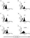
FIG. 1
FIG. 1
Binding of CXCR4 MAbs to Jurkat CD4+ cells is inhibited by T22. Binding of MAbs 12G5, 1, 2, 17, 18, and Leu3A to Jurkat CD4+ cells in the presence (thick line) or in the absence (solid black area) of T22 (40) was examined. The background binding measured with mIgGs (thin line) is also shown. The percentages of inhibition obtained in the presence of T22 in this experiment were 81.2, 88.4, 85.2, 86.5, 86.8, and 0% for 12G5, 1, 2, 17, 18, and Leu3A, respectively. FL2-H, staining obtained with the indicated MAb. A representative experiment out of three done is shown.
FIG. 2
FIG. 2
Equilibrium binding of anti-CXCR4 MAbs. (A) Jurkat CD4+ cells were stained with the indicated concentrations of CXCR4 MAbs 12G5, 1, 2, 8, 12, 16, 17, and 18 for 1 h followed by PE-coupled anti-mouse antibody and analyzed by flow cytometry as described in Materials and Methods. A representative experiment out of three done is shown. (B) Relative affinity for each antibody presented as normalized EC50 (nEC50). The EC50 of each antibody was the concentration of antibody that gave a half-maximal GMCF value, based upon flow cytometric analysis using serial dilutions of the MAbs on Jurkat CD4+ and SupT1 cells. The results are the mean and standard error of the mean obtained from three experiments with each cell type.
FIG. 3
FIG. 3
CXCR4 heterogeneity on lymphoid cell lines. The indicated cell lines were incubated with saturating levels of CXCR4 MAbs 12G5, 1, 2, 8, 12, 16, 17, and 18 for 1 h and then processed for flow cytometry. The cell lines are grouped into three panels (A, B, and C) for convenience. All results have been normalized to the GMCF value obtained for 12G5, which was set at 100%. The results are representative of five different experiments and are reported as the mean and standard error of the mean for each cell line.
FIG. 4
FIG. 4
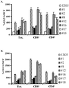
CXCR4 heterogeneity on PBLs. Human PBLs treated for 4 days with IL-2 alone (A) or with IL-2–PHA (B) were stained with the panel of CXCR4 MAbs and analyzed by flow cytometry as described in Materials and Methods. Antibodies to CD8 and CD4 were also used to examine CXCR4 heterogeneity on these T-cell subsets. Tot., total. The results shown are the mean and standard error of the mean obtained for two or three donors in five different experiments.
FIG. 5
FIG. 5
CXCR4 heterogeneity on B cells. Fresh blood was stained with the panel of CXCR4 MAbs as described in Materials and Methods. All results have been normalized to the GMCF value obtained for 12G5, which was set at 100%. A representative experiment out of three done with eight donors each is shown. The mean value is indicated by a horizontal bar.
FIG. 6
FIG. 6
Reactivity of the CXCR4 MAbs to human, rat, and feline CXCR4. (A) Amino acid sequence comparison of the amino-terminal domain (N-term.) and the first, second, and third extracellular loops (ECL1, ECL2, and ECL3) of human, rat, and feline CXCR4. Human CXCR4 is used as the reference sequence, with different amino acids in the rat or feline sequence indicated in bold. A dash indicates amino acid identity, and a dot indicates a gap created to maximize the alignment. (B) 293T cells were transfected with human, rat, or feline CXCR4, and the reactivities of MAbs 12G5, 1, 2, 8, 12, 16, 17, 18, and 4G10 were analyzed by flow cytometry. The mean and standard error of the mean for one representative experiment out of three done in triplicate are shown.
FIG. 7
FIG. 7
Chemotaxis inhibition of T cells correlates with differential antibody reactivity. HS-Sultan cells were incubated with CXCR4 MAbs 12G5, 2, 1, 18, and 4G10. A fraction of the cells was analyzed by flow cytometry (white columns), and the rest was used for a chemotaxis assay (black columns). The mean and standard error of the mean for one representative experiment out of three done in triplicate are shown. The chemotaxis index is defined as the ratio of the number of cells that migrate under a given condition to the number of cells that migrate in the absence of the chemoattractant. NEG., chemotaxis index with no chemoattractant in the lower chamber; POS., chemotaxis in response to SDF-1α. The column labeled SDF is the chemotaxis control and shows the chemotaxis index when the concentration of SDF-1α in the upper chamber is equivalent to that in the lower chamber.
FIG. 8
FIG. 8
Sulfation and glycosylation do not influence antibody reactivity. 293T cells were transfected with CXCR4 or a CXCR4 construct lacking the first 12 (Δ12-CXCR4), 15 (Δ15-CXCR4), or 23 (Δ23-CXCR4) N-terminal amino acids. At 48 h later, the cells were stained with MAbs 12G5, 1, 2, and 18 and analyzed by flow cytometry. A representative experiment out of three done is shown.
FIG. 9
FIG. 9
Pertussis toxin treatment does not influence antibody reactivity. Jurkat cells were incubated overnight in the presence or absence (−) of 100 ng of pertussis toxin/ml. The cells were then stained with MAbs 12G5, 1, 2, 8, 12, 16, 17, and 18 and analyzed by flow cytometry. One experiment out of two done is shown.

References

    1. Baba M, Nishimura O, Kanzaki N, Okamoto M, Sawada H, Iizawa Y, Shiraishi M, Aramaki Y, Okonogi K, Ogawa Y, Meguro K, Fujino M. A small-molecule, nonpeptide CCR5 antagonist with highly potent and selective anti-HIV-1 activity. Proc Natl Acad Sci USA. 1999;96:5698–5703. - PMC - PubMed
    1. Berger E A, Murphy P M, Farber J M. Chemokine receptors as HIV-1 coreceptors: roles in viral entry, tropism, and disease. Annu Rev Immunol. 1999;17:657–700. - PubMed
    1. Berson J F, Long D, Doranz B J, Rucker J, Jirik F R, Doms R W. A seven-transmembrane domain receptor involved in fusion and entry of T-cell-tropic human immunodeficiency virus type 1 strains. J Virol. 1996;70:6288–6295. - PMC - PubMed
    1. Bleul C C, Farazan M, Choe H, Parolin C, Clark-Lewis I, Sodroski J, Springer T A. The lymphocyte chemoattractant SDF-1 is a ligand for LESTR/fusin and blocks HIV-1 entry. Nature. 1996;382:829–833. - PubMed
    1. Brelot A, Heveker N, Montes M, Alizon M. Identification of residues of CXCR4 critical for human immunodeficiency virus coreceptor and chemokine receptor activities. J Biol Chem. 2000;275:23736–23744. - PubMed

Publication types