. 2001 Oct;75(19):9029-36.
doi: 10.1128/JVI.75.19.9029-9036.2001.

Recombinant herpes simplex virus type 1 expressing murine interleukin-4 is less virulent than wild-type virus in mice

Affiliations

Recombinant herpes simplex virus type 1 expressing murine interleukin-4 is less virulent than wild-type virus in mice

H Ghiasi et al. J Virol. 2001 Oct.

Abstract

The effect of interleukin-4 (IL-4) on herpes simplex virus type 1 (HSV-1) infection in mice was evaluated by construction of a recombinant HSV-1 expressing the gene for murine IL-4 in place of the latency-associated transcript (LAT). The mutant virus (HSV-IL-4) expressed high levels of IL-4 in cultured cells. The replication of HSV-IL-4 in tissue culture and in trigeminal ganglia was similar to that of wild-type virus. In contrast, HSV-IL-4 appeared to replicate less well in mouse eyes and brains. Although BALB/c mice are highly susceptible to HSV-1 infection, ocular infection with HSV-IL-4 resulted in 100% survival. Furthermore, 57% of the mice survived coinfection with a mixture of HSV-IL-4 and a lethal dose of wild-type McKrae, compared with only 10% survival following infection with McKrae alone. Similar to wild-type BALB/c mice, 100% of IL-4(-/-) mice also survived HSV-IL-4 infection. T-cell depletion studies suggested that protection against HSV-IL-4 infection was mediated by a CD4(+)-T-cell response.


Figures
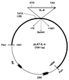
FIG. 1
FIG. 1
Construction of the pHSV-IL-4 plasmid containing the murine IL-4 gene under control of the LAT promoter. Details of the preparation of the recombinant transfer vector are given in Materials and Methods. The plasmid contains LAT nucleotides −1041 to +4656 (thick line) with a deletion from nucleotides 76 to 1667. The complete gene for mouse IL-4, including the stop codon and its poly(A), site is inserted into the deleted region. The IL-4 gene is under the control of the LAT promoter. Insertion of enhanced green fluorescent protein into HSV-1 in the same location resulted in high-level long-term expression throughout both acute and latent infection (36).
FIG. 2
FIG. 2
Construction and structure of the HSV-IL-4 mutant virus. (A) The top schematic shows the HSV-1 McKrae genome in the prototypic orientation. TRL and IRL represent the long terminal and internal (or inverted) repeats, TRS and IRS represent the short terminal and internal (or inverted) repeats. UL and US represent the long and short unique regions, respectively. The solid rectangle represents the very stable 2-kb LAT. The start site for LAT transcription is indicated by the arrow at +1, the relative location of the LAT promoter TATA box 28 nucleotides upstream of the start of transcription. (B) dLAT2903 has a deletion from LAT nucleotides −161 to +1667 (XXXXXX) in both copies of LAT and makes no LAT RNA. (C) HSV-IL-4 was constructed from dLAT2903 by homologous recombination between dLAT2903 DNA and a plasmid containing the complete LAT promoter and the entire structural IL-4 gene [including its 3′ poly(A) signal] as described in Materials and Methods.
FIG. 3
FIG. 3
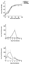
Replication of HSV-IL-4 in tissue culture and mouse tears. (A) HSV-IL-4 replication in tissue culture. Subconfluent RS cell monolayers in triplicate from two separate experiments were infected with 0.01 PFU of HSV-IL-4, dLAT2903, or McKrae per cell as described in Materials and Methods. Total virus was harvested at the indicated times postinfection by two cycles of freeze-thawing. The amount of virus at each time for each virus was determined by standard plaque assays on RS cells. Each point represent the mean ± the standard error of the mean (n = 6). (B) Virus titers in BALB/c mouse tears. BALB/c mice were ocularly infected with 2 × 10⁵ PFU of HSV-IL-4 or dLAT2903 per eye. Tear films were collected from days 1 to 7, and virus titers were determined by standard plaque assays. Each point represents the mean ± the standard error of the mean of titers from 10 eyes. (C) Virus titers in C57BL/6 mouse tears. C57BL/6 mice were ocularly infected with 2 × 10⁶ PFU per eye of HSV-IL-4 or dLAT2903. Virus titers were determined as for panel B. Each point represents the mean + the standard error of the mean of the titers from 10 eyes.
FIG. 4
FIG. 4
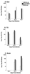
Virus titers in whole eyes, TG, and brain. BALB/c mice were ocularly infected with 2 × 10⁵ PFU of HSV-IL-4, dLAT2903, or McKrae per eye as described in Materials and Methods. The mice were euthanized on the indicated days. Eyes (A), TG (B), and brain (C) were removed and homogenized, and virus titers were determined as described in Materials and Methods. Each bar represents the mean ± the standard error of the mean of 10 eyes, 10 TG, or 5 brains.
FIG. 5
FIG. 5
Survival following infection with HSV-IL-4. (A) Survival of BALB/c mice coinfected with HSV-IL-4 and McKrae. Mice were coinfected ocularly with 2 × 10⁴ PFU of HSV-IL-4/eye and 2 × 10⁴ PFU of wt McKrae/eye. Survival was determined 28 days after infection. (B) Survival of BALB/c IL-4−/− mice infected with HSV-IL-4. IL-4−/− mice were inoculated ocularly with 2 × 10⁵ PFU of HSV-IL-4 or dLAT2903 as described in Materials and Methods. Survival was measured 28 days after infection. (C) Survival of CD4+-depleted or CD8+-depleted BALB/c mice after ocular infection with HSV-IL-4. CD4+ or CD8+ T-cell depletions were done as described in Materials and Methods. Mice were inoculated ocularly with 2 × 10⁵ PFU of HSV-IL-4/eye, and survival was measured 28 days after infection.

Similar articles

Cited by

References

    1. Abehsira-Amar O, Gibert M, Joliy M, Theze J, Jankovic D L. IL-4 plays a dominant role in the differential development of Tho into Th1 and Th2 cells. J Immunol. 1992;148:3820–3829. - PubMed
    1. Bachmann M F, Schorle H, Kuhn R, Muller W, Hengartner H, Zinkernagel R M, Horak I. Antiviral immune responses in mice deficient for both interleukin-2 and interleukin-4. J Virol. 1995;69:4842–4846. - PMC - PubMed
    1. Biron C A. Cytokines in the generation of immune responses to, and resolution of, virus infection. Curr Opin Immunol. 1994;6:530–538. - PubMed
    1. Cheers C, Janas M, Ramsay A, Ramshaw I. Use of recombinant viruses to deliver cytokines influencing the course of experimental bacterial infection. Immunol Cell Biol. 1999;77:324–330. - PubMed
    1. Cherwinski H M, Schumacher J H, Brown K D, Mosmann T R. Two types of mouse helper T cell clone. III. Further differences in lymphokine synthesis between Th1 and Th2 clones revealed by RNA hybridization, functionally monospecific bioassays, and monoclonal antibodies. J Exp Med. 1987;166:1229–1244. - PMC - PubMed

Publication types