

Cooperation of an RNA packaging signal and a viral envelope protein in coronavirus RNA packaging
- PMID: 11533169
- PMCID: PMC114474
- DOI: 10.1128/JVI.75.19.9059-9067.2001
Cooperation of an RNA packaging signal and a viral envelope protein in coronavirus RNA packaging
Abstract
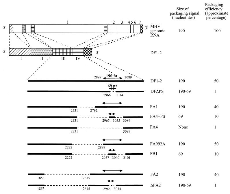
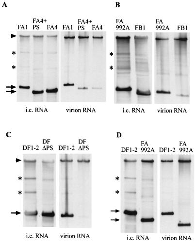
Murine coronavirus mouse hepatitis virus (MHV) produces a genome-length mRNA, mRNA 1, and six or seven species of subgenomic mRNAs in infected cells. Among these mRNAs, only mRNA 1 is efficiently packaged into MHV particles. MHV N protein binds to all MHV mRNAs, whereas envelope M protein interacts only with mRNA 1. This M protein-mRNA 1 interaction most probably determines the selective packaging of mRNA 1 into MHV particles. A short cis-acting MHV RNA packaging signal is necessary and sufficient for packaging RNA into MHV particles. The present study tested the possibility that the selective M protein-mRNA 1 interaction is due to the packaging signal in mRNA 1. Regardless of the presence or absence of the packaging signal, N protein bound to MHV defective interfering RNAs and intracellularly expressed non-MHV RNA transcripts to form ribonucleoprotein complexes; M protein, however, interacted selectively with RNAs containing the packaging signal. Moreover, only the RNA that interacted selectively with M protein was efficiently packaged into MHV particles. Thus, it was the packaging signal that mediated the selective interaction between M protein and viral RNA to drive the specific packaging of RNA into virus particles. This is the first example for any RNA virus in which a viral envelope protein and a known viral RNA packaging signal have been shown to determine the specificity and selectivity of RNA packaging into virions.
Figures
Similar articles
-
Characterization of N protein self-association in coronavirus ribonucleoprotein complexes.Virus Res. 2003 Dec;98(2):131-40. doi: 10.1016/j.virusres.2003.08.021. Virus Res. 2003. PMID: 14659560 Free PMC article.
-
Murine coronavirus packaging signal confers packaging to nonviral RNA.J Virol. 1997 Jan;71(1):824-7. doi: 10.1128/JVI.71.1.824-827.1997. J Virol. 1997. PMID: 8985424 Free PMC article.
-
Characterization of the coronavirus M protein and nucleocapsid interaction in infected cells.J Virol. 2000 Sep;74(17):8127-34. doi: 10.1128/jvi.74.17.8127-8134.2000. J Virol. 2000. PMID: 10933723 Free PMC article.
-
Coronavirus genomic RNA packaging.Virology. 2019 Nov;537:198-207. doi: 10.1016/j.virol.2019.08.031. Epub 2019 Aug 30. Virology. 2019. PMID: 31505321 Free PMC article. Review.
-
Molecular Basis of Encapsidation of Hepatitis C Virus Genome.Front Microbiol. 2018 Mar 7;9:396. doi: 10.3389/fmicb.2018.00396. eCollection 2018. Front Microbiol. 2018. PMID: 29563905 Free PMC article. Review.
Cited by
-
Ribonucleocapsid assembly/packaging signals in the genomes of the coronaviruses SARS-CoV and SARS-CoV-2: detection, comparison and implications for therapeutic targeting.J Biomol Struct Dyn. 2022 Jan;40(1):508-522. doi: 10.1080/07391102.2020.1815581. Epub 2020 Sep 9. J Biomol Struct Dyn. 2022. PMID: 32901577 Free PMC article.
-
Rift Valley fever virus NSs mRNA is transcribed from an incoming anti-viral-sense S RNA segment.J Virol. 2005 Sep;79(18):12106-11. doi: 10.1128/JVI.79.18.12106-12111.2005. J Virol. 2005. PMID: 16140788 Free PMC article.
-
Chimeric coronavirus-like particles carrying severe acute respiratory syndrome coronavirus (SCoV) S protein protect mice against challenge with SCoV.Vaccine. 2008 Feb 6;26(6):797-808. doi: 10.1016/j.vaccine.2007.11.092. Epub 2007 Dec 26. Vaccine. 2008. PMID: 18191004 Free PMC article.
-
Multiple Roles of SARS-CoV-2 N Protein Facilitated by Proteoform-Specific Interactions with RNA, Host Proteins, and Convalescent Antibodies.JACS Au. 2021 Jun 15;1(8):1147-1157. doi: 10.1021/jacsau.1c00139. eCollection 2021 Aug 23. JACS Au. 2021. PMID: 34462738 Free PMC article.
-
The molecular biology of coronaviruses.Adv Virus Res. 2006;66:193-292. doi: 10.1016/S0065-3527(06)66005-3. Adv Virus Res. 2006. PMID: 16877062 Free PMC article. Review.
References
Publication types
MeSH terms
Substances
Grants and funding
LinkOut - more resources
Full Text Sources
