. 2001 Oct;75(19):9096-105.
doi: 10.1128/JVI.75.19.9096-9105.2001.

Receptor binding transforms the surface subunit of the mammalian C-type retrovirus envelope protein from an inhibitor to an activator of fusion

Affiliations

Receptor binding transforms the surface subunit of the mammalian C-type retrovirus envelope protein from an inhibitor to an activator of fusion

A L Barnett et al. J Virol. 2001 Oct.

Abstract

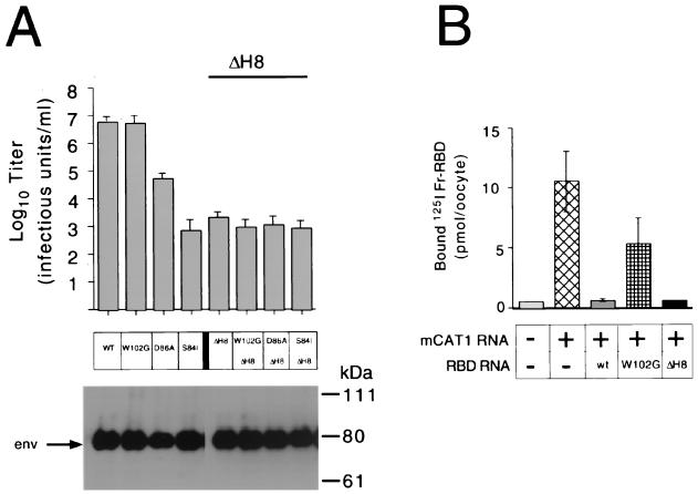
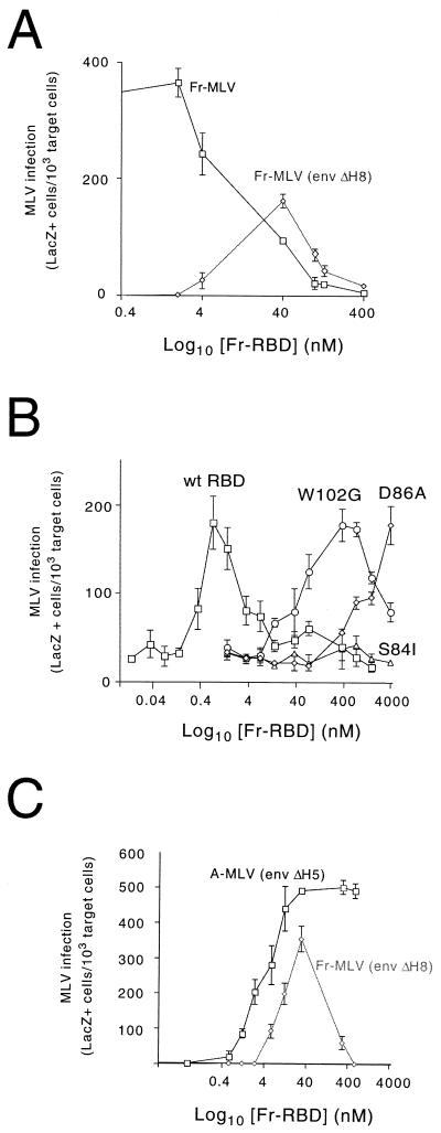
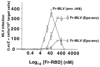
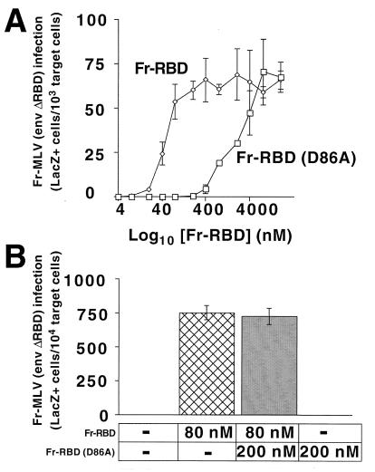
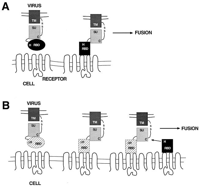
The envelope protein (Env) of murine leukemia viruses (MLVs) is composed of a surface subunit (SU) and a transmembrane subunit (TM), which mediates membrane fusion, resulting in infection. SU contains a discrete N-terminal receptor binding domain (RBD) that is connected to the remainder of Env by a short, proline-rich segment. Previous studies suggest that after receptor binding, the RBD interacts directly with the remainder of Env to trigger fusion (A. L. Barnett, R. A. Davey, and J. M. Cunningham, Proc. Natl. Acad. Sci. USA 98:4113-4118, 2001). To investigate the role of the RBD in activating fusion, we compared infection by several MLVs that are defective unless rescued in trans by the addition of soluble RBD to the culture medium. Infection by MLV lacking a critical histidine residue near the N terminus of the viral RBD is dependent on the expression of receptors for both the RBD in the viral Env and the soluble RBD supplied in trans. However, infection by MLVs in which the RBD has been deleted or replaced by the ligand erythropoietin are dependent only on expression of the receptor for the soluble RBD. We were able to expand the host range of xenotropic MLV to nonpermissive murine fibroblasts only if the RBD was deleted from the xenotropic viral envelope and the soluble RBD from ecotropic Friend MLV was supplied to the culture medium. These findings indicate that receptor binding transforms the RBD from an inhibitor to an activator of the viral fusion mechanism and that viruses lacking the critical histidine residue at the N terminus of the RBD are impaired at the activation step.


Figures

FIG. 1
FIG. 1
The effect of deletion of HIS8 (ΔH8) in Fr-MLV SU on receptor binding and infection in comparison with the effect of substitutions for residues in the receptor binding pocket that reduce binding affinity and titer. (A) Infection. End point dilution of virus on NIH 3T3 fibroblasts was used to measure the titers of Fr-MLVs with ΔH8 and/or substitutions for one of three residues (W102G, D86A, or S84I) in the receptor binding pocket of SU that reduce receptor binding affinity (13). Each titer was determined in triplicate by end point dilution from independent stocks of virus, and standard errors are indicated. Incorporation of Env into virions was monitored by immunoblotting with anti-gp70 antibody (below). (B) Binding. Binding of exogenous 125I-Fr-RBD to Xenopus oocytes was measured 2 days after injection with capped mRNA encoding mCAT1 alone or in combination with a capped mRNA encoding either Fr-RBD, Fr-RBD (W102G), or Fr-RBD (ΔH8). Each measurement is the mean of the counts per minute from five oocytes ± 1 standard error. +, present; −, absent; WT or wt, wild type.
FIG. 2
FIG. 2
(A) Relationship between the concentration of soluble Fr-RBD in the medium and infection by Fr-MLV or Fr-MLV (env ΔH8). Fr-MLV or Fr-MLV (env ΔH8) infection of human 293 mCAT1 cells as a function of the concentration of purified Fr-RBD in the medium (0 to 400 nM) was determined. Infection was assessed 2 days postexposure to a 1:10 dilution of virus by recording the proportion of target cells that were stained blue by 5-bromo-4-chloro-3-indolyl-β-d-galactopyranoside. The results for each RBD concentration were determined in triplicate wells of a six-well plate. Standard errors are indicated. (B) Effect of substitutions for residues in the binding pocket on the capacity of soluble Fr-RBD to rescue Fr-MLV (env ΔH8) infection. Fr-MLV (env ΔH8) infection of NIH 3T3 cells as a function of the soluble RBD concentration (0 to 4,000 nM) was determined using either purified wild-type (wt) Fr-RBD (squares), Fr-RBD (W102G) (circles), Fr-RBD (D86A) (diamonds), or Fr-RBD (S84I) (triangles). LacZ-positive cells were counted 2 days postexposure to a 1:10 dilution of virus. The effect of each RBD concentration was assessed in triplicate on a six-well plate. Standard errors are indicated. (C) Relationship between Fr-RBD concentration and infection by Fr-MLV (env ΔH8) and A-MLV (env ΔH5). Human 293 mCAT1 cells were exposed to 1:10 dilutions of Fr-MLV (env ΔH8) or A-MLV (env ΔH5) in the presence of the indicated concentration of purified Fr-RBD protein. The infection level was determined as described above.
FIG. 3
FIG. 3
Relationship between Fr-RBD concentration and infection by Fr-MLV (Epo-env). Infection on 293 cells that express mCAT1 alone (triangles) or in combination with EpoR (squares) of a 1:10 dilution of Fr-MLV (Epo-env) supernatant was measured as a function of the concentration of Fr-RBD (0 to 2,000 nM). The cells were assayed for acquired β-galactosidase expression 48 h postinfection. The effect of each concentration of Fr-RBD on infection was assessed in triplicate wells of a six-well plate; standard errors are indicated. As a reference, the effect of Fr-RBD on Fr-MLV (env ΔH8) infection was replotted from Fig. 2C (circles). In a separate experiment, we observed no detectable difference in the relationship between Fr-RBD concentration and Fr-MLV (env ΔH8) infection on human 293 mCAT1 and human 293 mCAT1-EpoR cells (data not shown).
FIG. 4
FIG. 4
Infection of NIH 3T3 cells (A) and 293 mCAT1 cells (B) by X-MLV, X-MLV (env ΔH7), and X-MLV (env ΔRBD) was measured in the presence (100 nM) or absence of soluble Fr-RBD. The NIH 3T3 cells (CL-13) used in this experiment express fourfold more mCAT1 than normal NIH 3T3 cells (12). It should be noted that the deletion of the conserved histidine residue (His7) in X-MLV reduces the titer of X-MLV by 10-fold to 104 IU/ml compared to a 104-fold reduction in titer caused by the deletion of the equivalent His residue in A-MLV or Fr-MLV. +, present; −, absent.
FIG. 5
FIG. 5
(A) RBD-dependent infection by Fr-MLV (env ΔRBD) as a function of the concentration of Fr-RBD or Fr-RBD (D86A). Human 293 mCAT1 cells were exposed to Fr-MLV (env ΔRBD) carrying lacZ in the presence of Fr-RBD (diamonds) or Fr-RBD (D86A) (squares) over a concentration range of 0 to 8,000 nM. Two days after exposure to virus, the cells were stained with 5-bromo-4-chloro-3-indolyl-β-d-galactopyranoside, and blue cells were counted. The experiments were performed in triplicate wells of a six-well plate. (B) Effect of an excess of Fr-RBD (D86A) on Fr-RBD-dependent infection by Fr-MLV (env ΔRBD). Human 293 mCAT1 cells were exposed to Fr-MLV (env ΔRBD) encoding lacZ alone or in the presence of either Fr-RBD (80 nM) or Fr-RBD (D86A) (200 nM) or both. Infection was assessed as described above. −, absent. The error bars represent ±1 standard error.
FIG. 6
FIG. 6
Schematic diagram to illustrate the proposed mechanism of soluble-RBD-dependent activation of fusion. The viral membrane containing the Env protein is at the top. Env is depicted with the RBD (oval) connected by the proline-rich region (curved line) to the C-terminal segment (light shaded rectangle). The transmembrane domain (dark shaded rectangle) is connected to the C-terminal segment by a disulfide bond. The cell membrane containing the virus receptor is at the bottom. (A) Proposed steps leading to the activation of fusion, assuming a mechanism in which the viral RBD and the C-terminal segment of SU interact in cis. The viral RBD is depicted as an oval that, on receptor contact, undergoes a conformational change (depicted as a rectangle). In this situation, fusion is triggered by a specific interaction between the bound conformation of the RBD and the C-terminal segment and is dependent upon the conserved histidine residue. (B) Proposed steps for RBD-dependent Fr-MLV (env ΔH8) infection in trans. Receptor binding to viral RBD (ΔH) results in exposure of the C-terminal segment of SU, enabling a productive interaction with soluble RBD bound to a distinct receptor. As in cis, the interaction between soluble RBD and the C-terminal segment in trans is strongly dependent on the conserved histidine residue.

References

    1. Albritton L M, Tseng L, Scadden D, Cunningham J M. A putative murine ecotropic retrovirus receptor gene encodes a multiple membrane-spanning protein and confers susceptibility to virus infection. Cell. 1989;57:659–666. - PubMed
    1. Anderson M M, Lauring A S, Burns C C, Overbaugh J. Identification of a cellular cofactor required for infection by feline leukemia virus. Science. 2000;287:1828–1830. - PubMed
    1. Bae Y, Kingsman S M, Kingsman A J. Functional dissection of the Moloney murine leukemia virus envelope protein gp70. J Virol. 1997;71:2092–2099. - PMC - PubMed
    1. Barnett A L, Davey R A, Cunningham J M. Modular organization of the Friend murine leukemia virus envelope protein underlies the mechanism of infection. Proc Natl Acad Sci USA. 2001;98:4113–4118. - PMC - PubMed
    1. Battini J L, Danos O, Heard J M. Receptor choice determinants in the envelope glycoproteins of amphotropic, xenotropic, and polytropic murine leukemia viruses. J Virol. 1992;66:1468–1475. - PMC - PubMed

Publication types

LinkOut - more resources