. 2001 Oct;75(19):9262-73.
doi: 10.1128/JVI.75.19.9262-9273.2001.

Function of Rta is essential for lytic replication of murine gammaherpesvirus 68

Affiliations

Function of Rta is essential for lytic replication of murine gammaherpesvirus 68

T T Wu et al. J Virol. 2001 Oct.

Abstract

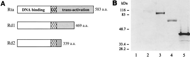
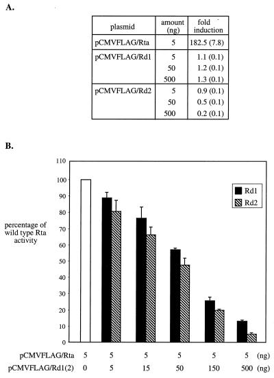
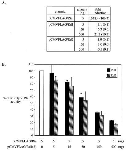
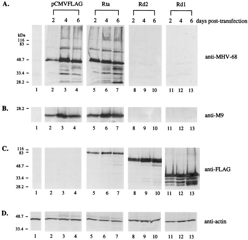
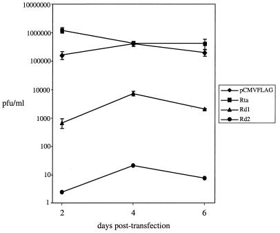
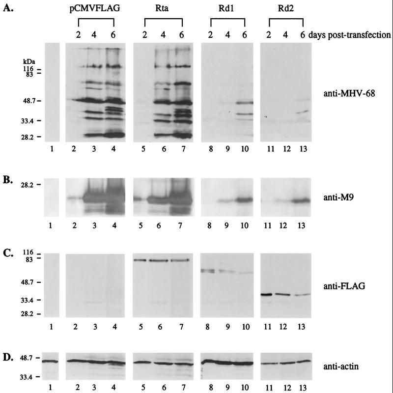
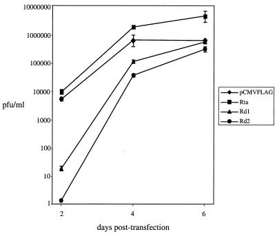
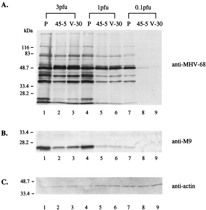
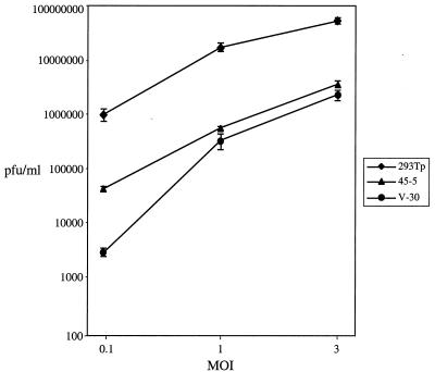
Rta, encoded primarily by open reading frame 50, is well conserved among gammaherpesviruses. It has been shown that the Rta proteins of Epstein Barr virus (EBV), Kaposi's sarcoma-associated herpesvirus (KSHV, or HHV-8), and murine gammaherpesvirus 68 (MHV-68; also referred to as gamma HV68) play an important role in viral reactivation from latency. However, the role of Rta during productive de novo infection has not been characterized in gammaherpesviruses. Since there are cell lines that can support efficient productive de novo infection by MHV-68 but not EBV or KSHV, we examined whether MHV-68 Rta plays a role in initiating viral lytic replication in productively infected cells. Rta, functioning as a transcriptional activator, can activate the viral promoter of early lytic genes. The amino acid sequence alignments of the Rta homologues suggest that the organizations of their functional domains are similar, with the DNA binding and dimerization domains at the N terminus and the trans-activation domain at the C terminus. We constructed two mutants of MHV-68 Rta, Rd1 and Rd2, with deletions of 112 and 243 amino acids from the C terminus, respectively. Rd1 and Rd2 could no longer trans-activate the promoter of MHV-68 gene 57, consistent with the deletions of their trans-activation domains at the C terminus. Furthermore, Rd1 and Rd2 were able to function as dominant-negative mutants, inhibiting trans-activation of wild-type Rta. To study whether Rd1 and Rd2 blocked viral lytic replication, purified virion DNA was cotransfected with Rd1 or Rd2 into fibroblasts. Expression of viral lytic proteins was greatly suppressed, and the yield of infectious viruses was reduced up to 10(4)-fold. Stable cell lines constitutively expressing Rd2 were established and infected with MHV-68. Transcription of the immediate-early gene, rta, and the early gene, tk, of the virus was reduced in these cell lines. The presence of Rd2 also led to attenuation of viral lytic protein expression and virion production. The ability of Rta dominant-negative mutants to inhibit productive infection suggests that the trans-activation function of Rta is essential for MHV-68 lytic replication. We propose that a single viral protein, Rta, governs the initiation of MHV-68 lytic replication during both reactivation and productive de novo infection.


Figures

FIG. 1
FIG. 1
Construction and expression of wild-type and mutant Rta proteins. (A) The structures of the wild-type and mutant Rta proteins are shown, with the open boxes representing the DNA binding domains, the hatched boxes representing the dimerization domains (DZ), and the shaded boxes representing the activation domains. The size of each protein is indicated at the right. (B) Wild-type and mutant Rta proteins are expressed in 293T cells. The coding sequences of Rta, Rd1, and Rd2 were individualy cloned into pCMVFLAG. The cells were transfected with the empty vector, pCMVFLAG (lane 1), pCMVFLAG/Rta/KSHV (lane 2), pCMVFLAG/Rta (lane 3), pCMVFLAG/Rd1 (lane 4), or pCMVFLAG /Rd2 (lane 5). Ten percent of the total cell lysates was loaded onto a 10% denaturing polyacrylamide gel. Western blot analysis was carried out using the monoclonal antibody against the FLAG epitope. The masses of individual proteins in the molecular weight standard are indicated at the left.
FIG. 2
FIG. 2
Rd1 and Rd2 function as dominant-negative mutants of Rta in 293T cells. (A) Rd1 and Rd2 do not activate the ORF57 promoter. 293T cells were cotransfected with 50 ng of the reporter construct, p57Luc, and 5 ng of pCMVFLAG/Rta, or 5 to 500 ng of pCMVFLAG/Rd1, or 5 to 500 ng of pCMVFLAG/Rd2. The total amounts of plasmid DNA were brought up to 600 ng with pCMVFLAG. The control transfection was carried out with 550 ng of pCMVFLAG and 50 ng of p57Luc. Each transfection included 1 ng of pRLCMV containing the Renilla luciferase gene driven by the constitutively active CMV promoter for nomalization of variations among transfections. At 48 h posttransfection, total cell lysates were harvested for analysis of luciferase activity. Normalized luciferase activity was calculated by dividing the level of firefly luciferase activity by the level of Renilla luciferase activity in each transfection. The fold induction was then calculated by dividing the level of normalized luciferase activity by that of the control transfection. Standard deviations derived from four experiments are shown in parentheses. (B) Rd1 and Rd2 inhibit wild-type Rta trans-activation of the ORF57 promoter. 293T cells were transfected with 50 ng of p57Luc and 5 ng of pCMVFLAG/Rta alone or with 5 to 500 ng of pCMVFLAG/Rd1 or 5 to 500 ng of pCMVFLAG/ Rd2. Total cell lysates were harvested at 48 h posttransfection, and the luciferase activity in each transfection was measured. The fold induction was calculated as described above. Wild-type Rta activity was expressed as the percentage of the fold induction relative to cotransfection of p57Luc and pCMVFLAG/Rta. Standard deviations derived from four experiments are expressed as error bars.
FIG. 3
FIG. 3
Rd1 and Rd2 function as dominant-negative mutants of Rta in BHK-21 cells. The transfections described in the legend to Fig. 2 were repeated in BHK-21 cells. (A) Rd1 and Rd2 do not activate the ORF57 promoter. (B) Rd1 and Rd2 inhibit wild-type Rta trans-activation of the ORF57 promoter.
FIG. 4
FIG. 4
Rd1 and Rd2 inhibit MHV-68 lytic protein expression from transfected virion DNA in 293T cells. pCMVFLAG (0.2 μg; lanes 2 to 4), pCMVFLAG/Rta (0.2 μg; lanes 5 to 7), pCMVFLAG/Rd1 (0.2 μg; lanes 8 to 10), or pCMVFLAG/Rd2 (0.2 μg; lanes 11 to 13) was cotransfected into 293T cells with 0.2 μg of virion DNA derived from the recombinant green fluorescent protein-expressing virus tw25. Total cell lysates were harvested at 2, 4, and 6 days posttransfection (as indicated above the lanes), and 10% of each lysate, including a negative control of untransfected cells (lanes 1), was used for Western blot analysis. (A) Expression of MHV-68 lytic proteins is suppressed by Rd1 and Rd2. The membrane was probed with the polyclonal rabbit serum against the MHV-68-infected cell lysates (anti-MHV-68). (B) Expression of MHV-68 M9 protein is suppressed by Rd1 and Rd2. The membrane was probed with anti-M9 polyclonal rabbit serum. (C) Wild-type and mutant Rta proteins are expressed in 293T cells. The membrane was probed with the monoclonal antibody against the FLAG epitope (anti-FLAG). (D) The levels of cellular β-actin were examined (anti-actin). The membrane was probed with monoclonal antibody against cellular β-actin.
FIG. 5
FIG. 5
Rd1 and Rd2 inhibit the production of infectious viruses after transfection of virion DNA in 293T cells. The supernatants from the transfections described in the legend to Fig. 4 were harvested, and the viral titers were determined using plaque assays. The assays were repeated three times for each transfection. Standard deviations are expressed as error bars.
FIG. 6
FIG. 6
Rd1 and Rd2 inhibit MHV-68 lytic protein expression from transfected virion DNA in BHK-21 cells. The transfections described in the legend to Fig. 4 were repeated in BHK-21 cells. Total cell lysates were harvested at 2, 4, and 6 days posttransfection (as indicated above the lanes), and 10% of each lysate, including a negative control of untransfected cells (lanes 1), was used for Western blot analysis. (A) Expression of MHV-68 lytic proteins is suppressed by Rd1 and Rd2. The membrane was probed with the polyclonal serum against MHV-68-infected cell lysates (anti-MHV-68). (B) Expression of MHV-68 M9 protein is suppressed by Rd1 and Rd2. The membrane was probed with a rabbit serum against the MHV-68 M9 protein (anti-M9). (C) Wild-type and mutant Rta proteins are expressed in BHK-21 cells. The membrane was probed with the monoclonal antibody against the FLAG epitope (anti-FLAG). (D) The levels of cellular β-actin were examined. The membrane was probed with monoclonal antibody against cellular β-actin (anti-actin).
FIG. 7
FIG. 7
Rd1 and Rd2 inhibit the production of infectious viruses after transfection of virion DNA in BHK-21 cells. The supernatants from the transfections described in the legend to Fig. 6 were harvested, and the viral titers were determined using plaque assays. The assays were repeated three times for each transfection. Standard deviations are expressed as error bars.
FIG. 8
FIG. 8
Transcription of tk and rta genes is suppressed in stable cell lines expressing Rd2. Two cell lines, 45-5 and V-30, stably transfected with pCMVFLAG/Rd2 and the parental 293T cells (P) were infected with wild-type MHV-68 (3 PFU/cell). Total RNA from uninfected (U) or infected cells was harvested at 4 or 15 h postinfection, and 30% of each RNA sample was used for Northern blot analysis. (A) Transcription of rta is reduced in 45-5 and V-30 cells. The probe was derived from the rta gene (nt 68651 to 69378). (B) Transcription of tk is reduced in 45-5 and V-30 cells. The same membrane was stripped and rehybridized with a probe derived from the tk gene (nt 32879 to 34813). (C) The RNA loadings were examined by rehybridizing with a probe derived from the cellular GAPDH (glyceraldehyde-3-phosphate dehydrogenase) gene.
FIG. 9
FIG. 9
Viral protein expression is inhibited in stable cell lines expressing Rd2. 45-5, V-30, and the parental 293T (P) cells were infected with wild-type MHV-68 at the different MOIs (PFU/cell) indicated above the panel. Total cell lysates were harvested at day 2 postinfection, and 10% of each lysate was used for Western blot analyses. (A) Expression of viral lytic proteins is reduced in 45-5 and V-30 cells. The membrane was probed with polyclonal serum against MHV-68-infected cell lysates. (B) Expression of M9 (a viral late protein) is reduced in 45-5 and V-30 cells. The membrane was probed with polyclonal antibody against the recombinant MHV-68 M9 protein. (C) The protein loadings were examined by reprobing with monoclonal antibody against cellular β-actin.
FIG. 10
FIG. 10
The yield of infectious viruses is reduced in cell lines expressing Rd2. The supernatants were harvested from the infections described in the legend to Fig. 9, and viral titers were determined using plaque assays. The assays were performed three times for each infection, and standard deviations are expressed as error bars.

Similar articles

Cited by

References

    1. Buisson M, Manet E, Trescol-Biemont M C, Gruffat H, Durand B, Sergeant A. The Epstein-Barr virus (EBV) early protein EB2 is a posttranscriptional activator expressed under the control of EBV transcription factors EB1 and R. J Virol. 1989;63:5276–5284. - PMC - PubMed
    1. Chevallier-Greco A, Manet E, Chavrier P, Mosnier C, Daillie J, Sergeant A. Both Epstein-Barr virus (EBV)-encoded trans-acting factors, EB1 and EB2, are required to activate transcription from an EBV early promoter. EMBO J. 1986;5:3243–3249. - PMC - PubMed
    1. Chomczynski P, Sacchi N. Single-step method of RNA isolation by acid guanidinium thiocyanate-phenol-chloroform extraction. Anal Biochem. 1987;162:156–159. - PubMed
    1. Countryman J, Miller G. Activation of expression of latent Epstein-Barr herpesvirus after gene transfer with a small cloned subfragment of heterogeneous viral DNA. Proc Natl Acad Sci USA. 1985;82:4085–4089. - PMC - PubMed
    1. Cox M A, Leahy J, Hardwick J M. An enhancer within the divergent promoter of Epstein-Barr virus responds synergistically to the R and Z transactivators. J Virol. 1990;64:313–321. - PMC - PubMed

Publication types

LinkOut - more resources